Thursday, 13 June 201908:00 | Conference Registration, Materials Pick-Up, Morning Coffee and Pastries | |
Session Title: Conference Opening Session |
| | 08:30 |  | Conference Chair Chairman's Welcome, Introduction to the Conference and Technology Landscape of 3D-Culture
Terry Riss, Senior Product Manager, Cell Health, Promega Corporation, United States of America
|
| 09:00 |  | Keynote Presentation Generating Human Liver Spheres From Pluripotent Stem Cells and Their Application
David Hay, Chair of Tissue Engineering, MRC Centre for Regenerative Medicine, University of Edinburgh, United Kingdom
Liver disease is an escalating global health issue. While liver transplantation is an effective mode of therapy, there are a shortage of donor organs. Therefore, developing renewable sources of human liver tissue is important for the clinic. Pluripotent stem cell-derived liver tissue represents a potential alternative to cadaver derived hepatocytes and whole organ transplant. At present, two-dimensional differentiation procedures deliver tissue that is hepatocyte like, but lacks certain functions and long-term stability. Efforts to overcome these limiting factors have focussed on building three-dimensional (3D) cellular aggregates. Although enabling for the field, their widespread application has been limited due to cost and overreliance on undefined biological components. Our studies focused on the development of self assembled 3D liver tissue under defined conditions. In vitro generated 3D tissues exhibited stable phenotype, providing an attractive resource for the clinic and long-term in vitro modelling studies. Our most recent tissue engineering advances will be discussed at the meeting. |
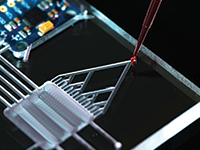
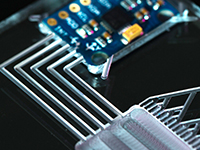
| 09:30 |  | Keynote Presentation Vascularized Tumor Spheroids for Drug Screening on Injection Molded IMPACT Platform
Noo Li Jeon, Professor, Seoul National University, Korea South
The field of microfluidics-based three-dimensional (3D) cell culture systems is rapidly progressing from academic proof-of-concept studies to valid solutions to real-world problems. Polydimethylsiloxane (PDMS)-based microfluidics platforms have been widely adopted for tumor-on-a-chip systems. However, due to the inherent material limitations that make it difficult to scale-up production, PDMS has not been widely used in standardized commercial applications for preclinical screening testing. In this presentation, injection-molded tumor spheroid chip made of polystyrene (PS) in a standardized 96-well plate format with a user-friendly design. Spontaneous liquid patterning is achievable with high repeatability. To demonstrate the feasibility of the device, we fabricated array of vascularized tumor spheroids and developed a 3D tumor angiogenesis model for drug screening. This model is easy- and ready-to-use, and suitable for mass-production systems, with the ability to deliver robust and reproducible results. |
| 10:00 | Morning Coffee Break and Networking in the Exhibit Hall | 10:45 |  | Keynote Presentation Utilizing Multi-Organ Human on a Chip Systems to Predict in vivo Outcomes For Efficacy and Toxicity
James Hickman, Professor, Nanoscience Technology, Chemistry, Biomolecular Science and Electrical Engineering, University of Central Florida; Chief Scientist, Hesperos, United States of America
The utilization of multi-organ human-on-a-chip or body-on-a-chip systems for toxicology and efficacy, that ultimately should lead to personalized medicine applications, is a topic that has received much attention recently for drug discovery and subsequent regulatory approval. Hesperos has been constructing these systems with up to 6 organs and have demonstrated long-term (>28 days) evaluation of drugs and compounds, that have shown similar response to results seen from clinical data or reports in the literature. Application of these systems for ALS, Alzheimer’s, rare diseases, diabetes and cardiac and skeletal muscle mechanistic toxicity will be reviewed. The development of an in vitro PDPK modeling that predicts in vivo results will also be presented. The system utilizes a pumpless platform with a serum free recirculating medium. This methodology integrates microsystems fabrication technology and surface modifications with protein and cellular components, for initiating and maintaining self-assembly and growth into biologically, mechanically and electronically interactive functional multi-component systems. Hesperos has received Phase II and Phase IIB SBIR grants from NCATS to apply Advanced Manufacturing Technologies and automation to these systems in collaboration with NIST in addition to support form pharmaceutical and cosmetic companies. This talk will also give results of six workshops held at NIH to explore what is needed for validation and qualification of these new systems. |
| 11:15 |  Complex 3D In-Vitro Liver-Disease Models For Fast, Automation-Compatible and Translational Drug Discovery Complex 3D In-Vitro Liver-Disease Models For Fast, Automation-Compatible and Translational Drug Discovery
Jan Lichtenberg, CEO and Co-Founder, InSphero AG
Scaffold-free 3D microtissues have evolved into the most widely used technology for highly predictive and most scalable cell-based assays in drug safety and discovery. While they provide a faithful in-vitro approximation of the in-vivo tissue microenvironment, they were not amenable for modeling microphysiological features up to now. Focusing on advantages and challenges in daily industry use, we’ll describe two case studies illustrating how a 3D microtissue can be used as a predictive surrogate for drug discovery. One study investigates a complex co-culture system recapitulating the hallmarks of nonalcoholic steatohepatitis (NASH) in a screening-compatible format. Consisting of primary human hepatocytes, NPCs and stellate cells, these 3D microtissues can be elastically driven into and out of specific disease states. As second application, we will describe the use of reconstituted, 3D primary human pancreatic islets for discovery of anti-diabetic drugs. Finally, we present a new multi-organ-on-a-chip system featuring microfluidic channels and chambers that were specifically engineered for culturing 3D microtissues and organoids under physiological flow conditions. The platform complies with the SBS plate standard and is made of polystyrene to prevent unwanted compound absorption. It allows for an automated and on-demand interconnection of up to 10 microtissues per channel in a highly flexible fashion. Enabling an automated removal and re-insertion of 3D microtissues from and into the device, this platform finally ads deep endpoints such as next-gen sequencing, lipidomics, FACS to the analytical toolbox for organ-on-a-chip applications. Summarizing, we will focus on the major challenges encountered when implementing human, primary cell based 3D and organ-chip assays – and our suggestions on how to address them in an industry-wide effort.
| 11:45 |  | Keynote Presentation Merging Organ-on-a-Chip with Organoids: The Best of Two Worlds?
Paul Vulto, Managing Director, MIMETAS, Netherlands
Organ-on-a-Chip and organoids are two fields of development that are rapidly redefining cell culture biology in the 21st century. Organ-on-a-Chip technology has demonstrated how microengineering technology can add aspects such as perfusion flow, patterning of tissues, precise control over gradients and application of mechanical strain to tissue culture. Organoid protocols have been carefully optimized to maintain the stem cell niche in developing and adult tissues, while at the same time permitting cell proliferation and differentiation.
In this lecture, I will show latest progress at MIMETAS integrating organoid protocols and Organ-on-a-Chip technology. I will show a range of tissue models comprising complex co-cultures, in a perfused system using state of the art stem cell and organoid protocols. Following this presentation, it will become clear that the two fields are merging together to aid cell culture of the 21st century. |
| 12:15 | Networking Lunch in the Exhibit Hall and Poster Viewing | |
Session Title: Organoids and 3D-Culture - Technologies and Applications |
| | |
Emerging Themes in Organoids and Organs-on-Chips: An Introduction | | Session Chair: Paul Vulto, Managing Director, MIMETAS, Netherlands |
| | 13:30 | Phenotypic Screening in 3D Culture Including ECM: Advantages and Challenges
Nathalie Maubon, CEO/CSO, HCS Pharma, France
Grégory Maubon, Digital Coordinator, HCS Pharma, France
Cellular assays in 3D culture have shown many advantages to better mimic the in vivo situation. 3D technologies for phenotypic screening have become simpler and more accessible, such as the use of Ultra Low Attachment (ULA) plates. However, we can now go even further using 3D technologies which allow us to reproduce the micro-environment of healthy or pathological organs of interest. During this talk, we will show how the cellular microenvironment impacts the proliferation and/or differentiation of cells. A few examples from the fields of oncology, CNS and metabolic disease will be presented. High-Content Screening (HCS) devices, such as our ImageXpress® Micro Confocal system, are now fast enough and sensitive enough to allow image acquisition in 3D cellular models. Nevertheless, to go further, perceptions and processes need to be changed. We will discuss cutting-edge new technologies, including virtual and augmented realities, deep learning and machine learning, and explain how these new technologies can be of benefit to phenotypic screening. | 14:00 | 3D Hydrogel as Clinically-Relevant Neuroblastoma Model Where to Test Novel Antibody-Mediated Therapies
Silvia Scaglione, Professor, National Council of Research (CNR), Genoa (Italy) and Head of Laboratory of Tissue Engineering, Italy
A 3D cell-laden alginate hydrogels was designed as in vitro neuroblastoma model. The system revealed a clinical-relevant constitutive and IFN-gamma inducible NB immunophenotype suggesting its potential as future platform for set up and validate immune based therapeutic approaches. | 14:30 | BrainSpheres to Study Developmental Neurotoxicity
David Pamies, Researcher, University of Lausanne, Switzerland
Developmental neurotoxicity is of high concern due to different reasons: 1) no routine testing for DNT is carried out in the U.S., in the EU, or elsewhere, as DNT testing is not required by law unless triggered by neurotoxic or endocrine effects in adult rodents, 2) DNT guidelines are expensive and time-consuming, 3) human brain complexity may not be completely tested by animal testing, and 4) brain defects can be difficult to detect. Experts in the field have suggested an in vitro testing battery to cover brain development key events (such as neural stem cell proliferation and differentiation, migration, neurite outgrowth, synaptogenesis, network formation, myelination, and apoptosis). Also, the use of more human-relevant models, using 3D organotypic iPSC derived systems, have been suggested as an alternative to classical in vitro models. Here we present a 3D brain human-derived iPSC model to study developmental neurotoxicity and different applications of the model. | 15:00 | Afternoon Coffee Break and Networking in the Exhibit Hall and Poster Viewing | 15:30 |  Magnetic 3D Bioprinting, from Spheroids to Fingerprinting Cells in 3D Using a 2D Workflow Magnetic 3D Bioprinting, from Spheroids to Fingerprinting Cells in 3D Using a 2D Workflow
Glauco Souza, Director of Global Business Development and Innovation, 3D Cell Culture at Greiner Bio-One; Adjunct Assistant Professor, Greiner Bio-One and University of Texas Health Science Center at Houston
The growing push for 3D cell culture models is limited by technical challenges in handling, processing, and scalability to high-throughput applications. To meet these challenges, we use our platform, magnetic 3D bioprinting, in which cells are individually magnetized and assembled with magnetic forces. In magnetizing cells, not only do we make routine cell culture and experiments feasible and scalable, but we also gain fine spatial control in the formation of spheroids and more complex structures. This presentation will focus on recent developments using this platform, particularly in cancer biology and immunology. Specifically, we will present a method for phenotypic profiling of cell types within spheroids using real-time high-throughput imaging. This label-free method allows for multiplexing with other assay endpoints for high-content screening. Overall, we use magnetic 3D bioprinting to create functionally and structurally representative spheroids for high-throughput screening.
| 16:00 | High Content Screening of Organoid Cultures to Visualize and Quantify Intestinal Toxicity
Bram Herpers, COO, OcellO B V, Netherlands
Establishing organoids from human material enables development of 3D cell culture models for the intestinal epithelium to investigate physiological and toxicological mechanisms. At OcellO we combine a high throughput 3D human intestinal organoid culture platform with high content phenotype-based analysis to visualize and quantify the effects of compounds and treatment conditions on the epithelial integrity, injury and inflammation in a diverse (healthy and diseased) patient-derived organoid population. | 16:30 | 3D Microtumor-based Combinatorial Drug Discovery
Jens Kelm, CEO and Co-Founder, PreComb Therapeutics AG, Switzerland
3D tissue culture technologies for drug discovery are becoming of age. However, new 3D technologies and models are primarily used to exchange existing models to improve individual aspects in the developmental process. However, 3D models do not only reflect tissue and organ disease biology better than 2D models they allow to create novel assays to build completely new drug development processes. To take full advantage of 3D we present a 3D-based discovery processes to develop novel drug combinations. | 17:00 |  | Keynote Presentation Organoids, A Preclinical Patient Population
Robert Vries, CEO, HUB Organoids, Netherlands
Key to the improvement of drug development and disease modeling is a preclinical model that represents patients. We previously developed an adult stem cells derived culture system that allowed for the genetically and phenotypically stable expansion of human epithelial cells. The Organoids derived from a variety of organs such as lung, liver, pancreas, breast, etc, are a powerful tool to study Cancer, Cystic Fibrosis and other diseases. Recent studies show that the in vitro response to drug treatments tested organoids correlate with the clinical outcome of the patient from which the organoid was derived. Therefore, the organoids appear to faithfully represent a patient in the lab. The scalability of the organoids potentially provides a novel approach to screening in early discovery, drug development as well as predictive diagnostics. |
| 17:30 | Industry Panel Discussion Focusing on 3D-Culture and Organoids
| 18:15 | Networking Reception with Beer and Wine -- Meet Colleagues and Network with New Acquaintances | 19:15 | Close of Day 1 of the Conference | 19:30 | 3D-Culture and Organoids Short Course Presented by Dr. Terry Riss [Separate Registration Required to Attend this Short Course] |
Friday, 14 June 201908:00 | Morning Coffee, Pastries and Networking in the Exhibit Hall | |
Session Title: Tox Screening and Enabling Technologies |
| | 08:30 | Using the Multi-Organ-Chip Technology For Predictive Safety and Efficacy Testing In Drug Development
Katharina Schimek, Senior Scientist and Project Manager, TissUse GmbH, Germany
Present in vitro and animal tests for drug development do not reliable predict the human outcomes of tested drugs and substances because they are failing to emulate the organ complexity of the human body, leading to high attrition rates in clinical studies. These systemic responses of applied substances are ignored in most in vitro tests.
Microphysiological systems have proven to be a powerful tool for recreating human tissue- and organ-like functions at research level, providing the basis for the establishment of qualified preclinical assays with improved predictive power. However, industrial adoption of microphysiological systems and respective assays is progressing slowly due to their complexity. In the first part of the presentation examples of established single-organ chip, two-organ and four-organ chip solutions are highlighted. The underlying universal microfluidic Multi-Organ-Chip (MOC) platform with the size of a microscopic slide consists of an on-chip micro-pump and is capable to interconnect different organ equivalents. The micro-pump ensures stable long-term circulation of media through the tissue culture compartments at variable flow rates, adjustable to near to physiological mechanical stresses of the respective tissues. Issues to ensure long-term performance and industrial acceptance of microphysiological systems, such as design criteria, tissue supply and on chip tissue homeostasis will be discussed. Finally, a roadmap into the future is outlined, to further bring these assays into regulatory-accepted drug testing on a global scale. | 09:00 |  | Keynote Presentation 3D Microphysiological Models of Neurological Function and Disease and Their Application to Drug Screening
Roger Kamm, Cecil and Ida Green Distinguished Professor of Biological and Mechanical Engineering, Massachusetts Institute of Technology (MIT), United States of America
The capability to produce in vitro models of normal physiological function and disease is rapidly expanding, and it is now becoming clear that these microphysiological systems (MPS) will soon find their place in the multi-step process of identifying and vali-dating new drugs, and testing for their potentially toxic side-effects. In order to gain acceptance by the pharma and biotech industries, however, these systems will need to be further developed, validated, and methods developed to fabricate them at high throughput and consistency. In this presentation, we focus on systems being developed to model neurological function, disease, and the process of transport of drugs across the tight blood-brain barrier (BBB) to treat neurological disorders and cancer. These MPS are each derived entirely from human cells, produced in microfluidic platforms of different design, and include models of the BBB, Alzheimer’s Disease, and amyotrophic lateral sclerosis (ALS). |
| 09:30 | Quantitative Assessment of Tissue Chip Technologies
Murat Cirit, Director at Translational Center of Tissue Chip Technologies, Massachusetts Institute of Technology (MIT), United States of America
A large percentage of drug candidates fail at the clinical trial stage due to a lack of efficacy and unacceptable toxicity, primarily because the in vitro cell culture models and in vivo animal models commonly used in preclinical studies provide limited information about how a drug will affect human physiology. The need for more physiologically relevant in vitro systems for preclinical efficacy and toxicity testing has led to a major effort to develop “Microphysiological Systems (MPS)”, aka tissue chips (TC), based on engineered human tissue constructs. Translational Center of Tissue Translational Center of Tissue Chip Technologies (TC2T) has been established to bridge between academic research and development and industrial application of MPS technologies via providing unbiased testing and validation of MPS technologies. We believe that the full impact of MPS technologies will be realized only when robust approaches for in vitro–in vivo (MPS-to-human) translation are developed and utilized. Therefore, TC2T takes a holistic approach—based on quantitative systems pharmacology (QSP)— to achieve quantitative characterization of these complex systems and translation of experimental insights to clinical outcomes. | 10:00 | Accurate Prediction of Organ-Specific Toxicities
Daniele Zink, Principal Research Scientist and Team Leader, Institute of Bioengineering and Nanotechology, Agency for Science Technology and Research (A*STAR), Singapore
Predicting toxicity to internal organs with alternative methods is currently a major challenge. We have developed the first methods that predict nephrotoxicity in humans with high accuracy. These methods include a high-throughput platform, which combines high-throughput screening (HTS) with phenotypic profiling and machine learning. With this technology toxicity to renal proximal tubular cells (PTC) could be predicted with 82% or 89% test balanced accuracy, depending on whether primary human PTC or a human PTC line were used. Based on the same technology we have developed a HTS platform for the prediction of hepatotoxicity in humans. This HTS platform has been validated with ~90 compounds that were separated into training and test sets, and both test sensitivity and specificity are in the range of 80%. In addition, we have developed a HTS platform for the prediction of vascular toxicity. Our predictive HTS methods are currently combined with our predictive approaches based on human induced pluripotent stem cells.
While the HTS platforms are robust, suitable for rapid compound screening and produce binary (yes/no) results with respect to toxicity prediction, we develop complementary microphysiological systems (MPS) for repeated dose testing. Results obtained with a kidney-specific MPS show a high correlation between IC50 values obtained in vitro and the lowest toxic doses in humans. It is expected that respective MPS can greatly facilitate in vitro to in vivo extrapolations (IVIVE). Current difficulties in this area are one of the major obstacles that prevent applications of in vitro methods in risk assessment. | 10:30 | Morning Coffee Break, Networking and Poster Viewing | 11:00 |  THE LIVE CELL SHIPPER - A Portable CO2 Incubator to Revolutionize Biopharmaceutical Supply Chain Programs THE LIVE CELL SHIPPER - A Portable CO2 Incubator to Revolutionize Biopharmaceutical Supply Chain Programs
Corné Swart, Head of Business Development, Cellbox Solutions GmbH
Are you tired of massive drawbacks due to inadequate logistics solutions for cell/tissue transport? The only thing you wish for is to get your cells or cell based products to the final destination safe and sound? #CELLBOX is the first self-sufficient transport incubator that maintains the optimal temperature and CO2 atmosphere conditions for living cells and tissues, in the same manner they would be cultivated in the laboratory. #CELLBOX reduces the lead time in your logistic chain by up to two-thirds: no need to freeze, thaw and regenerate samples any longer. Simultaneously, the device eliminates the loss of valuable and sensitive cells caused by the exposure to extreme conditions and toxic additives. Join the #CELLBOX technology presentation and learn why #CELLBOX is the perfect solution for safe and reliable shipping, regardless of whether the intended transport of your iPSC-derived cells, organoids, tissues and other cell based samples is by road, rail, sea or air.
| 11:30 | Exploiting Human Stem Cells to Assess Risk of Developmental Neurotoxicity from Psychotropic Drugs
Robert Halliwell, Professor of Neuroscience and Clinical Pharmacology, University of The Pacific, United States of America
The developing human nervous system is highly susceptible to damage from environmental agents including heavy metals, organophosphate pesticides and pharmaceutical agents including psychotropic drugs. Determining the risk of neurotoxicity in humans using animal models is not very reliable. Our lab has been pioneering the study of human stem cells as a model of neurogenesis to address development neurotoxicity from psychotropic agents in vitro. This presentation will describe the experiments we are conducting investigating a variety of human stem cells, including tissue (adult) and induced pluripotent stem (iPS) cells to derive functional neurons. I will present the morphological, immunocytochemical, electrophysiological and neuropharmacological properties of neurons derived from human stem cells. I will also describe the impact of a range of psychotropic agents, including a number of antiepileptic medicines on the differentiation and maturation of human stem cells into functional neurons in vitro. Finally, I will present data on the value of human stem cell-derived neurons in the study of neuroprotection. | 12:00 |  | Keynote Presentation Formation, Cultivation and Analysis of 3D Microtissues using a Microfluidic Multicellular Spheroid Array
Peter Ertl, Professor of Lab-on-a-Chip Systems, Vienna University of Technology, Austria
A long-standing trend in pharmaceutical development, toxicology and biomedicine has been the establishment of complex in vitro 3D cell cultures whose structures and function resemble human tissues. To meet the growing need to improve the predictive power of toxicological, pharmaceutical and preclinical studies, the multicellular spheroid array builds on the advancement of novel organ-on-a-chip technology that establishes, cultivates and supports a wide variety of multicellular spheroids (3D cell aggregates). This system thus closes an important technological gap, enabling rapid and easy production of spheroids of defined size and cell types.
|
| 12:30 | Networking Lunch, Exhibit and Poster Viewing | 13:15 |  Luncheon Technology Spotlight Presentation: BI/OND: Versatile Organ-on-Chip platform for 3D Tissues Luncheon Technology Spotlight Presentation: BI/OND: Versatile Organ-on-Chip platform for 3D Tissues
Nikolas Gaio, CTO, BI/OND Solutions B.V.
Nowadays, biologists must choose between two options when running pre-clinical models in the early phase of drug testing: in vivo studies conducted on animals or in vitro screening. However, in vitro model and animals have shown to be inadequate for testing new pharmaceuticals to treat the many diseases that afflict humans. To achieve better medicine, there is a considerable need for more accurate human-representative systems. Therefore, biologists developed high-content 3D tissue models (organoids, microtissues, patient-derived tissues, etc..). However, they found that maintaining these 3D tissue models alive it is extremely complex. In particular, it is fundamental to control oxygen gradients and nutrients using a dynamic system. BIOND Solutions (BI/OND) supports biologists working in pharmaceutical companies, biotech and academia to explore fundamental questions about human health and diseases by providing versatile, dynamic chips for 3D tissue models. The approach consists in combining human cells, or patient-derived tissue with the use of a microfluidic chip.
| 13:45 | Poster Awards | 14:00 |  3D Liver Spheroids for In Vitro Modeling 3D Liver Spheroids for In Vitro Modeling
Feng Li, Senior Scientist, Development, Corning Life Sciences
Spheroids made of primary human liver cells are being adapted to in vitro assays for drug discovery and development. We present work at Corning Life Sciences to facilitate researchers that are interested in implementing 3D liver models to their studies.
| 14:30 |  Prediction of Gastrointestinal Toxicity and Drug Absorption Using the Reconstructed 3D Model of Human Small Intestine Epithelium (EpiIntestinal) Prediction of Gastrointestinal Toxicity and Drug Absorption Using the Reconstructed 3D Model of Human Small Intestine Epithelium (EpiIntestinal)
Jan Markus, Production Manager/Senior Scientist, MatTek IVLSL
Small intestine is an important entry gateway for many nutrients, drugs and other substances. About 90% of orally administered drug absorption occurs in the small intestine. Thereby there is a need of good and reliable in vitro model capable to predict drug toxicity and absorption/metabolism patterns that would replace intestine models relying predominantly on the use of cell lines generated from the colon or kidney.
The reconstructed 3D human small intestine model – EpiIntestinal mimics morphology and cell-type composition of normal human small intestine. It is polarized, allows studying bidirectional drug penetration through intestinal wall and expresses proteins involved in active drug transport and metabolism at physiological level. This allows modelling of complex drug absorption profiles, including the permeation, metabolism, drug-drug interaction and adverse effects of drugs on epithelium. Our results revealed that EpiIntestinal mimics the in vivo drug absorption profile much closer than the cancer cell line-based model. EpiIntestinal was also shown to predict toxicity with much higher specificity and sensitivity than the animal model. All in all, this model represents a promising tool to model complex processes occurring in small intestine.
| 15:00 | High-Throughput Microfluidic Platform for Drug Screening of Vascularized 3D Tissues
Sara Previdi, Scientist, University Medical Center Leiden, Netherlands
3D tissues such as spheroids or organoids derived from human pluripotent stem cells (PSCs) represent a new type of three-dimensional in vitro model for understanding organ development, disease mechanism and drug testing. Despite the success in generating 3D cultures resembling different tissue types (brain, heart, intestine, liver, lung and kidney), these mini-organs show limited growth potential and an immature phenotype due to lack of vascularization. Several groups have attempted to improve vascularization of organoids by transplanting them into a host (i.e. mouse, chick). However, the low predictivity of animal models, boost the development of in vitro alternative strategies. In this regard, microfluidic techniques are increasingly recognized as important toolbox able to add physiologically relevant cues to traditional cell culture models. We recently described the use of the OrganoPlate® for generating 3D perusable angiogenic vessels. Here, we present the use of a high-throughput 'grafting' platform which allows vessels co-culture with 3D tissue aggregates and tissue vascularization. One unit of the Mimetas OrganoPlate® Graft is made of two microfluidic channels in which endothelial cells can be patterned against ECM through the use of the PhaseGuide® technology. Gradient of pro-angiogenic factors (VEGF, PMA, S1P and FGF-b) allows the formation of a perfused vascular bed on top of which tissue fragments (i.e. organoids or spheroids) can be added to enable vascularization. Tissue dependent vessels remodeling and stabilization can be monitored overtime by real time imaging and barrier integrity. When liver spheroids are used, vessels became leaktight to dextran 150 kDa after 14 days of co-culture. Moreover, expression of CD31+ cells around and in within the spheroids proves that endothelial cells migration and tissue envelopment occurred during co-culture. The high number of units (up to 64 chips in 384 well format) enables functionality studies and compound screening in a robust and automated way. We propose the use of the OrganoPlate® Graft as a vessels grafting platform for multiple 3D tissues allowing drug screening and disease modeling in a more physiological environment. | 15:30 | Establishment and Characterization of a Newly Generated Cell Line based on Primary Small Intestinal Tissue
Christina Fey, Researcher, Tissue Engineering and Regenerative Medicine, University hospital Würzburg, Germany
Newly generated immortalized intestinal cells based on human or mouse primary epithelial cells show properties comparable to the native gut and enable a much easier and cost-efficient handling compared to primary intestinal cells. | 16:00 | Human Induced Pluripotent Stem (iPS) Cells in Glaucoma Modeling with an Automated Platform and 3D Retinal Organoids
Maciej Daniszewski, Researcher, University of Melbourne, Australia
Glaucoma is a disease characterized by slow degeneration of retinal ganglion cells (RGCs) what leads to irreversible vision loss and may ultimately result in blindness. It is estimated that approximately 80 million people will be affected by the disease in 2020, and with aging populations, this number will only continue to grow, thus generating a significant financial burden on the healthcare system.
Nevertheless, its exact molecular mechanism remains elusive. The use of human pluripotent stem cells (hPSCs) and three-dimensional (3D) organoids may bring a long-awaited breakthrough. Organoids mimic the organization and physiology of the tissue and thus recapitulate the disease phenotype more closely than 2D models. Organoid-derived RGCs may help to extend our understanding of mechanisms that confer risk of developing the disease, but also those that govern its progression. | 16:30 | .png) | Keynote Presentation In vitro Safety Assays with Human Derived Cardiomyocytes…. .…Bridging the Translational Gap?
Gary Gintant, Senior Research Fellow, Abbvie, United States of America
Clever biological and engineering endeavors are providing numerous novel opportunities to address cardiac safety issues of drug candidates with the intent of bridging the “translational gap” between preclinical and clinical studies. These efforts involve both “generalized” and patient-specific cardiac preparations. The utility of such studies depend not only on the evolving preparations, but also on the appropriate questions and interpretations, both representing challenges for future efforts. |
| 17:00 | Closing Coffee Break and Networking Opportunities with Speakers and Delegates |
|


 Add to Calendar ▼2019-06-13 00:00:002019-06-14 00:00:00Europe/London3D-Culture, Organoids and Tox Screening Europe 20193D-Culture, Organoids and Tox Screening Europe 2019 in Rotterdam, The NetherlandsRotterdam, The NetherlandsSELECTBIOenquiries@selectbiosciences.com