Co-Located Conference AgendasExtracellular Vesicles (EVs): Technologies & Biological Investigations | Lab-on-a-Chip and Microfluidics 2021 | Organoids & Organs-on-Chips 2021 | Point-of-Care Diagnostics and Biosensors 2021 | 

Monday, 13 December 202113:00 | Conference Registration and Materials Pick-Up | |
Session Title: Conference Plenary Session |
| | |
Venue: Marriott Coronado Island Ballrooms C & D |
| | 14:00 |  | Conference Chair Welcome and Introduction by Conference Co-Chair -- Lab-on-a-Chip and Microfluidics -- Inputs and Outputs in Product Engineering
Leanna Levine, Founder & CEO, ALine, Inc., United States of America
A unique aspect to point-of-care product development is to determine if the biological or chemical test will perform in a microfluidic system often before the rest of the product concept is thoroughly fleshed out. This means the programmatic priorities are focused on translational research before developing design inputs for a regulated product with an instrument. ALine’s new partnership with Nectar Product Development will enable us to support not just the assay implementation, but also create the entire product development roadmap, including the path to regulatory approval. |
| 14:10 |  | Conference Chair Welcome introduction by Conference Co-Chair Lab-on-a-Chip, Microfluidics & Associated Fields – Current Status: The Two Pathways – Simple Standard Devices Versus Highly Individualized Integrated Devices
Claudia Gärtner, CEO, microfluidic ChipShop GmbH, Germany
This presentation will pinpoint two interesting completely different ways for the use and the style of Lab-on-a-Chip devices: namely the simpler standard devices and the highly specific multifunctional integrated lab-on-a-chip devices. Point-of-care diagnostic has been one of the most prominent fields for lab-on-a-chip devices in particular since the last years during the pandemic. These devices are rather specific and highly individualized from the different companies commercializing them. A different pathway are easier standard devices, less application specific that are provided off-the-shelf from various companies and are frequently used with existing non-specific-lab-on-a-chip laboratory equipment. This “Lab-on-a-Chip-Catalogue” of an ever-growing number of different devices from various suppliers round the globe is worth to be highlighted and the use of existing standards and the creation of new ones should be shown as “the second way” for microfluidics. |
| 14:20 |  | Conference Chair Welcome and Introduction by Conference Co-Chair -- Extracellular Vesicles and Cellular Communication
Lucia Languino, Professor of Cancer Biology, Thomas Jefferson University, United States of America
This conference is focused on current Technologies and Biological Investigations on Extracellular Vesicles. Extracellular vesicles are nano-sized membranous structures released by cells into the extracellular space, and easily detectable in body fluids. Extracellular vesicles are highly heterogenous; large extracellular vesicles are plasma membrane-derived extracellular vesicles, while small extracellular vesicles are of endosomal or non-endosomal origin and are secreted upon fusion with the plasma membrane. Extracellular vesicle biological functions contribute to cell-cell communications in physiological and pathological conditions by carrying unique cargo (proteins, lipids, mRNAs and miRNAs) that modifies the functional state of the recipient cells. Recent investigations have clearly demonstrated the therapeutic and diagnostic potential of extracellular vesicles. The therapeutic potential of extracellular vesicles is of great interest especially because these vesicles can be engineered to achieve specific targeting. Currently, more than 200 trials that utilize extracellular vesicles are already registered in clinicaltrials.gov in the US. In this conference, emerging biological topics and novel technologies will be presented to stimulate discussion between areas of research of microfluidics, organoids and extracellular vesicles. |
| 14:30 |  | Keynote Presentation Development of Novel Intestine-on-Chip Models
Nancy Allbritton, Frank and Julie Jungers Dean of the College of Engineering and Professor of Bioengineering, University of Washington in Seattle, United States of America
Organ-on-chips are miniaturized devices that arrange living cells to simulate functional subunits of tissues and organs. These microdevices provide exquisite control of the biochemical and biophysical microenvironment for the investigation of organ-level physiology and disease. 2D and 3D models displaying a polarized human colonic epithelium were developed to recapitulate gastrointestinal physiology. The 2D crypt mimic displays a spatially patterned monolayer of epithelium displaying a stem-cell niche and differentiated cell zone with cells migration between the two regions. This planar 2D format enables efficient image cytometry for high-throughput screening applications as well physiologic measurements difficult to perform in a 3D format e.g., calcium signaling measurements. The 3D model builds on the 2D model by providing the full architecture of the in vivo human crypt. These models support formation of gradients of growth factors, microbial metabolites, and gases. A thick impenetrable layer of mucus with biophysical parameters similar to that of a living human can be formed for epithelial cell-microbe studies. These bioanalytical platforms are envisioned as next-generation systems for assay of microbiome-behavior, drug-delivery, and toxin-interactions with normal and diseased intestinal epithelia. |
| 15:00 |  | Keynote Presentation Affinity Selection of Extracellular Vesicles using Plastic-based Microfluidic Devices for the Management of Different Diseases
Steve Soper, Foundation Distinguished Professor, Director, Center of BioModular Multi-Scale System for Precision Medicine, The University of Kansas, United States of America
We have been developing tools for the diagnosis of a variety of
diseases. The commonality in these tools is that they consist of
microfluidic devices made from plastics via injection molding. Thus, our
tools can be mass produced at low-cost to facilitate bench-to-bed side
transition and point-of-care testing (PoCT). We have also been
generating novel assays focused on using liquid biopsy samples that are
enabled using microfluidics. In this presentation, I will talk about the
evolution of our fabrication efforts of plastic-based microfluidic and
nanofluidic devices as well their surface modification to make the
devices biocompatible for in vitro diagnostics. One tool that we have
generated is a plastic device (38 × 42 mm) that consists of 1.5M
pillars, which are surface decorated with affinity agents targeting
certain disease-associated extracellular vesicles (EVs). The affinity
agents are covalently attached to the surface of the microfluidic device
using a bifunctional linker, which consists of a coumarin moiety to
allow for the photolytic release of the captured EVs using a blue-light
LED to minimize photodamage to the EVs’ molecular cargo. We have also
developed a high-throughput nano-Coulter counter (nCC) made from a
plastic via injection molding for the counting of captured EVs from
clinical samples to allow their enumeration. The nCC consists of
multiple pores that are ~350 nm to allow for high throughput counting
with exquisite LODs (500 EVs/mL). In this presentation, I will discuss
the utility of these microfluidic and nanofluidic devices in several
diseases, for example, using EVs as a source of mRNAs for molecular
sub-typing of breast cancer patients. EVs were affinity selected from
breast-cancer patients’ plasma by searching for both epithelial and
mesenchymal expressing EVs to allow for highly efficient sub-typing
using the PAM50 gene panel. In an addition, the microfluidic and
nanofluidic devices were integrated into a single platform
(modular-based system) for PoCT to screen for early stage ovarian
cancer. Affinity probes were used to target EVs specifically generated
from tumor cells that signal early-stage ovarian cancer disease with the
nCC used for enumerating the number of EVs captured. Finally, the
modular system was used for the detection of COVID-19 at the PoC by
affinity selecting SARS-CoV-2 viral particles. The integrated system
could process saliva samples to search for the viral particles and count
them in <20 min. |
| 15:30 | Coffee Break and Networking | 16:00 |  | Keynote Presentation From Droplet-based Microfluidics Developments to Droplet-based Digital PCR
Valérie Taly, CNRS Research Director, Professor and Group leader Translational Research and Microfluidics, Université Paris Cité, France
Droplet-based microfluidics has led to the development of highly
powerful systems that represent a new paradigm in High-Throughput
Screening where individual assays are compartmentalized within
microdroplet microreactors. The integration of such systems for series
of complex individual operations on droplets has offered a solution to
the necessary miniaturization and automation of individual biological
assays. By combining a decrease of assay volume and an increase of
throughput, this technology goes beyond the capacities of conventional
screening systems. We will show how by combining droplet-based
microfluidic systems and clinical advances in molecular diagnostic we
have developed an original method to perform millions of single molecule
PCR in parallel to detect and quantify a minority of mutant sequences
within a high quantity of non-mutated sequences in complex mixture of
DNA with a sensitivity and precision that was unreachable by
conventional procedures. |
| 16:30 |  | Keynote Presentation High-Purity and High-Sensitivity Rare-Cell Isolation with Aliquot Sorting
Daniel Chiu, A. Bruce Montgomery Professor of Chemistry, University of Washington, United States of America
This presentation describes the high-sensitivity isolation of rare
cells, including circulating tumor cells and fetal cells, using
fluorescence activated aliquot sorting. Here, a blood sample is divided
into nanoliter volumes or aliquots, then analyzed and sorted based on
the presence or absence of a rare cell. By performing two rapid, on the
millisecond timescale, back-to-back sorting, we also demonstrate
isolation of rare cells with high purity. Finally, we drastically
increased the number of nucleated cells sorted by first concentrating
peripheral blood mononuclear cells from whole blood, which translated
into an equivalent blood volume analyzed by over an order of magnitude,
from 8mL to over 100mL per experiment. |
| 17:00 |  | Keynote Presentation Extracellular Vesicles: Liquid Biopsies and Biological Nanocarriers For Therapy
Dominique PV de Kleijn, Professor Experimental Vascular Surgery, Professor Netherlands Heart Institute, University Medical Center Utrecht, The Netherlands, Netherlands
|
| 17:30 |  | Keynote Presentation Over-Engineering in the Life Sciences: Microfluidics and Microphysiological Systems Meet Artificial Intelligence
John Wikswo, Gordon A. Cain University Professor, A.B. Learned Professor of Living State Physics; Founding Director, Vanderbilt Institute for Integrative Biosystems, Vanderbilt University, United States of America
It is a worthwhile exercise to examine whether the addition of microfluidic pumps and valves to organ-chip systems represents either “over-engineering” or the means by which microphysiological systems can be made massively parallel. While several cited examples of over-engineering represent overly complicated or frivolous solutions to a problem, such as the $400 Juicero juice-pouch-squeezer or a $1,390 Porsche roof box rated at 200 km/hour, others are prescient and ultimately yield great value, for example the NASA Opportunity rover whose mission was planned to last 90 Mars sols (~93 Earth days) but in fact lasted 14 Earth years, and Mercedes Benz’s introduction in the 1970s of antilock braking systems and in 1980 the driver’s airbag and seat-belt tensioner. From the perspective of a biologist who uses an agar-filled Petri dish for microbial colony picking, a 1536 well plate and its associated high-throughput screening infrastructure are overkill; for big Pharma, it is a welcome tool that took 15 years to be refined and put into practical use. As we approach the 15th anniversary of Mike Shuler’s landmark 2007 patent “Devices and Methods for Pharmacokinetic-based Cell Culture System,” we realize that microfluidic tissue and organ chips have been perfused using gravity, pressurized reservoirs, external syringe or peristaltic pumps, or on-chip peristaltic pumps. Although the smallest devices abound with Quake-style pneumatic valves that also serve as pumps, very few organ-chip systems utilize valves. For over a decade, our group has been developing microfluidic pumps and valves, and we have long argued that multi-organ microphysiological systems will ultimately need automation of pumps and valves to control each chip, their closed-loop sensors, and organ-organ interconnections. Our Missing Organ MicroFormulator concept spawned an award-winning 96-channel MicroFormulator, which served as the foundation for CN Bio Innovations’ recently introduced PhysioMimixTM pharmacokinetic (PK) system that supports PK studies of oncological drugs. As our group develops its fourth generation of rotary microfluidic pumps and valves to enable our construction and Ross King’s coding of the machine-learning algorithms for the 1000-channel Genesis Self-Driving Chemostat for yeast systems biology, we realize that this hardware can be readily adapted to perfuse and control a dozen of Steve George’s and Scott Simon’s bone marrow and skin chips in an in vitro infection model, maintain and analyze multiple, coupled organ chips for six months or longer, support remote studies of select agents in a BSL-3 or BSL-4 facility, apply circadian rhythms to thousands of wells in dozens of well plates or hundreds of zebrafish or perfused organ chips, and accelerate and optimize the microbial production of antibodies, industrial feed stocks, food protein, and sequestered carbon dioxide. Is this over-engineering or the future of artificial intelligence in biology? |
| 18:00 | 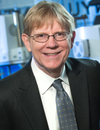 | Keynote Presentation Development and Deployment of ‘Smart Diagnostics’: Next Generation Point of Care Sensors with Capacity to Learn
John T McDevitt, Professor, Division of Biomaterials, New York University College of Dentistry Bioengineering Institute, United States of America
While COVID-19 has yielded devastating consequences over the past few years, the global pandemic also has opened the door for acceleration of development of core diagnostic capabilities that have the potential to lead to lasting impact for our society. With this vantage point in mind, in the recent past the McDevitt laboratory has launched a series of efforts that target the development and deployment of ‘smart diagnostics’ that serve as distributed point of care sensor nodes with capacity to learn. These mini-sensor ensembles with embedded artificial intelligence integrate programmable chip-based diagnostic systems capable of multiplexed measurements alongside clinical decision support tools that utilize strategically chosen nonclinical data elements that elicit signatures that can be used to capture diseases before they spiral out of control. As such, these efforts link for the first-time the following five key disciplines: i) lab-on-a-chip technologies, ii) in vitro diagnostics, iii) -omics research, iv) artificial intelligence, and v) digital healthcare delivery systems.
Importantly, the combination of point-of-care medical microdevices and machine learning has the potential transform the practice of medicine. In this area, scalable lab-on-a-chip devices have many advantages over standard laboratory methods including portability, faster analysis, reduced cost, lower power consumption, and higher levels of integration and automation. Despite significant advances in medical microdevice technologies over the years, several remaining obstacles are preventing clinical implementation and market penetration of these novel medical microdevices. Similarly, while machine learning has seen explosive growth in recent years and promises to shift the practice of medicine toward data-intensive and evidence-based decision making, its uptake has been hindered due to the lack of integration between clinical measurements and disease determinations.
In this talk, our recent advances in ‘smart diagnostics’ will be highlighted. These smart diagnostics include single-use microfluidic cartridges that serve as fully integrated, self-contained devices that contain aqueous buffers suitable for automated completion of all assay steps within nontraditional healthcare settings. Further, a portable analyzer instrument is fashioned to integrate fluid delivery, optical detection, image analysis, and user interface, representing a universal system for acquiring, processing, and managing clinical data while overcoming many of the challenges facing the widespread clinical adoption of lab-on-a-chip technologies. Intimate linkages between these medical microdevices and cloud connected databases allows for early disease detection algorithms to be used to impact clinical progress. |
| 18:30 | Networking Reception with Beer, Wine and a Light Dinner in the Exhibit Hall -- Meet Colleagues and Network with Sponsors and Exhibitors | 20:00 | Close of Day 1 of the Conference |
Tuesday, 14 December 202108:00 | Conference Registration and Continental Breakfast Served in the Exhibit Hall | 08:30 | Please Refer to the Lab-on-a-Chip and Microfluidics Agenda for Programming for the Morning Session of December 14, 2021 | 12:30 | Buffet Lunch and Networking in the Exhibit Hall with Exhibitors and Conference Sponsors | 13:30 | Please Refer to the Point-of-Care Diagnostics Agenda for Programming for the Afternoon Session of December 14, 2021 | 18:30 | Networking in the Exhibit Hall with Exhibitors and Conference Sponsors with Beer and Wine | 19:30 | Close of Conference Day 2 |
Wednesday, 15 December 202108:00 | Continental Breakfast Served in the Exhibit Hall | |
Session Title: Technologies and Themes in the Organs-on-Chips Space + Themes in Organoids Research |
| | |
Venue: Marriott Coronado Island Ballroom C |
| | 08:30 |  | Keynote Presentation The NIH Microphysiological Systems Program: In Vitro 3D Models for Safety and Efficacy Studies
Danilo Tagle, Director, Office of Special Initiatives, National Center for Advancing Translational Sciences at the NIH (NCATS), United States of America
Approximately 30% of drugs have failed in human clinical trials due to adverse reactions despite promising pre-clinical studies, and another 60% fail due to lack of efficacy. A number of these failures can be attributed to poor predictability of human response from animal and 2D in vitro mkodels currently being used in drug development. To address this challenges in drug development, the NIH Tissue Chips or Microphysiological Systems program is developing alternative innovative approaches for more predictive readouts of toxicity or efficacy of candidate drugs. Tissue chips are bioengineered 3D microfluidic platforms utilizing chip technology and human-derived cells and tissues that are intended to mimic tissue cytoarchitecture and functional units of human organs and systems. In addition to drug development, these microfabricated devices are useful for modeling human diseases, and for studies in precision medicine and environment exposures. Presenattion will elaborate in the development and utility of microphysiologicals sytems and in the partnerships with various stakeholders for its implementation. |
| 09:00 | 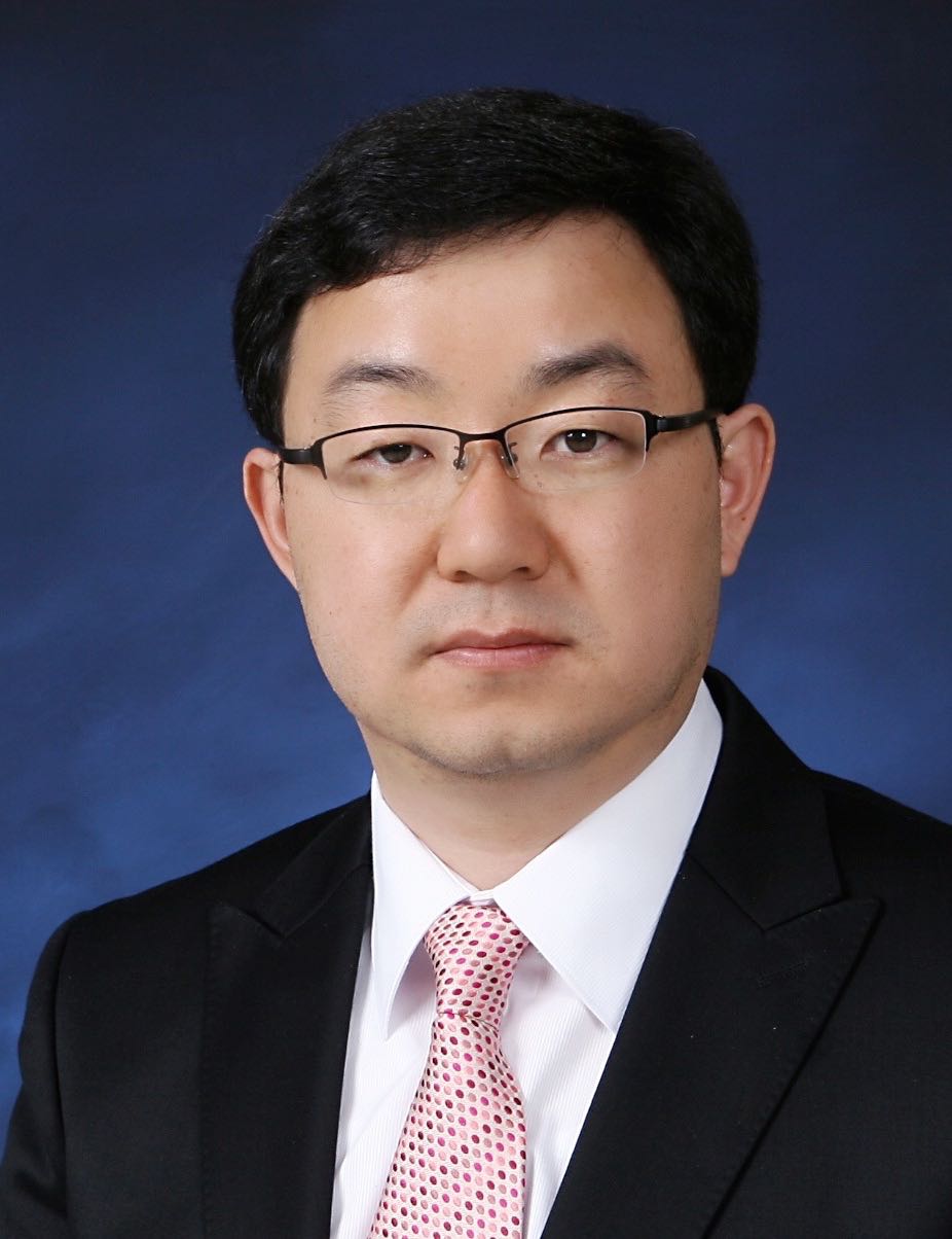 | Keynote Presentation High Throughput Vascularized Organoids For Drug Screening
Noo Li Jeon, Professor, Seoul National University, Korea South
Recent developments in microfluidics based organ-on-a-chip approaches have progressed rapidly in last few years. This presentation will describe recent work in our laboratory where new patterning method based on open microfluidics combined with injection molded devices enable formation of array of vascularized tumor spheroids and organoids that can be used for drug screening. Utilizing a microfluidic design with spontaneous capillary flow (SCF) based patterning mechanism, we present a novel in-vitro platform, Sphero-IMPACT and T-IMPACT (Injection-Molded Plastic Array 3D Culture platform). Drugs and immune cells can delivered via the perfusable vessel networks and live time-lapse imaging can be used to follow dynamic changes in the tumor microenvironment. Recent results on vascularized iPSCs derived neurosphere will also be described. The IMPACT platform is a versatile, high throughput platform with potential applications in organoid based drug screening with the form factor and throughput of standard microtiter plates. |
| 09:30 |  | Keynote Presentation Body on a Chip: Will It Transform Drug Development?
Michael Shuler, Samuel B. Eckert Professor of Engineering, Cornell University, President Hesperos, Inc., United States of America
A physiologically representative, multi-organ microphysiological systems (MPS) based on human tissues (also known as “human-on-a-chip”) may be a transformative technology to improve the selection of drug candidates most likely to earn regulatory approval from clinical trials. Such microscale systems combine organized human tissues with the techniques of microfabrication based on PBPK (Physiologically Based Pharmacokinetic) models. I will describe such systems being constructed at Hesperos and at Cornell. They are “self-contained” in that they can operate independently and do not require external pumps as is the case with many other microphysiological systems. They are “low cost”, in part, because of the simplicity and reliability of operation. They maintain a ratio of fluid (blood surrogate) to cells that is more physiologic than in many other in vitro systems allowing the observation of the effects of not only drugs but their metabolites. While systems can be sampled to measure the concentrations of drugs, metabolites, or biomarkers, they also can be interrogated in situ for functional responses such as electrical activity, force generation, or integrity of barrier function. Operation up to 28 days has been achieved allowing observation of both acute and chronic responses with serum free media. We have worked with various combinations of internal organ modules (liver, fat, neuromuscular junction, skeletal muscle, cardiac, bone marrow, blood vessels and brain) and barrier tissues (eg skin, GI tract, blood brain barrier, lung, and kidney). We have achieved unidirectional flow in a pumpless system which is important for mimicking the response of vascular tissues and constructed blood brain mimics with human in vitro like characteristics. The use of microelectrode arrays to monitor electrically active tissues (cardiac and neuronal) and micro cantilevers (muscle) have been demonstrated. While most systems use 5 or fewer organ modules, we have demonstrated that a 13 “organ” compartment device can be constructed. Most importantly these technical advances allow prediction of both a drug’s potential efficacy and toxicity (side-effects) in pre-clinical studies and have been applied to circulating immune cells, cancer cells, and rare diseases such as Myasthenia gravis. |
| 10:00 |  | Keynote Presentation Using Microfluidics for Immune Cell Trafficking and Capture
Steven C. George, Edward Teller Distinguished Professor and Chair, Department of Biomedical Engineering, University of California-Davis, United States of America
Microfluidic technology has played a leading role to advance our understanding of fundamental biological processes including cell separation and isolation, next generation sequencing, and cell trafficking. Over the past five years our lab has applied the basic principles of microfluidics to control fluid shear and flow to create simple microphysiological systems to better understand: 1) how to capture and isolate rare immune cells from the peripheral circulation, and 2) the principles which guide and control immune cell (lymphocytes, monocytes, and neutrophils) trafficking in complex tissue microenvironments. For the former, we leverage the ability to coat surfaces with antigens that are recognized by rare populations of B lymphocytes in the peripheral circulation. We then control the shear force at the surface and can capture and isolate these rare cell populations. Understanding how these rare cell populations evolve over time following viral (e.g., SARS-CoV-2, influenza) infection is central to understanding immunity following infection or immunization. For the latter, we are pursuing three projects. The first involves neutrophil trafficking into the cardiac muscle during COVID19-induced “cytokine storm”, including counterstrategies that limit binding of neutrophils to the inflamed endothelium. The second involves modeling myeloid cell-directed immunosuppression in the tumor microenvironment, and how counterstrategies such as inhibiting STAT3 signaling, can enhance CAR-T cell trafficking and effector function. The third project describes small tumor cell-derived extracellular vesicle (sEV) transport (convection and binding to the extracellular matrix) and how the sEV can establish spatial gradients to guide monocyte migration in the tumor microenvironment. This talk will provide an overview of our major results from each of these projects. |
| 10:30 | Injectable and Drug Deliverable Nano-Matrix for Cartilage-on-a-Chip
Yupeng Chen, Associate Professor, University of Connecticut, United States of America
Janus base nano-matrix is a novel injectable solid scaffold that can be applied in the microchannels of tissue chips to achieve localized drug delivery and improve cartilage cells anchorage and functions. The injectable nano-matrix may be a suitable tissue engineering scaffold for cartilage-on-a-chip. | 11:00 | Tissue Organoids For Disease Modeling and Therapy Development
Shay Soker, Professor of Regenerative Medicine and Chief Science Program Officer, Wake Forest Institute for Regenerative Medicine, United States of America
Three dimensional tissue equivalents (organoids) replicate native human tissue structure and function and can be studied in vitro for several weeks to allow intensive investigations. Besides their advantages in drug toxicity testing and for development of new drugs, the human tissue organoid platform serves as a model system to explore human tissue development and disease. Our recent research was focused on the use of tissue organoids to study human development and congenital diseases as well as other common diseases such as tissue fibrosis and cancer. Once a disease model is established, patient-specific (personalized) therapies can be realized as pre-clinical testing platform. | 11:30 | 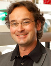 | Keynote Presentation Using Islet Spheroids to Model Beta Cell Targeted Interventions in Type 1 Diabetes
Matthias von Herrath, Vice President and Senior Medical Officer, Novo Nordisk, Professor, La Jolla Institute, United States of America
After investigating the human pathology of type 1 diabetes through organ donor pancreata (nPOD) it has become clear that beta cells become more recognizable by the immune system and that these beta cell Intrinsic events substantially contribute to their destruction. As immune reactivity to beta cells is even present to a low degree in healthy individuals, this ‘unmasking’ of the beta cell appears critical for T1D to develop. Upregulation of MHC is one of the key pathogenetic events. Thus, interventions should not only target the immune system, but should also be geared towards beta cells health and their recognizability. Here we have built, in collaboration with InSphero (Zurich, CH) a human islet spheroid immune attack model that replicates key events of human islet recognition and allows for the testing of drugs that can preserve beta cell function. |
| 12:00 | Pancreas Cancer Organoids for Research and Precision Medicine
Hervé Tiriac, Assistant Researcher, University of California-San Diego, United States of America
| 12:30 | Buffet Lunch and Networking in the Exhibit Hall with Exhibitors and Conference Sponsors | 13:00 |  | Keynote Presentation COVID-19 Therapeutics Safety ‘Clinical Trial’ with Cardiac Microphysiological Systems
Kevin Healy, Jan Fandrianto and Selfia Halim Distinguished Professorship in Engineering, University of California, Berkeley, United States of America
Three-dimensional (3D) microphysiological systems (MPSs) mimicking human organ function in vitro are an emerging alternative to conventional monolayer cell culture and animal models for myriad applications. Human induced pluripotent stem cells (hiPSCs) have the potential to capture the diversity of human genetics and provide an unlimited supply of cells. Combining hiPSCs with microfluidics technology in MPSs offers new perspectives for applications as diverse as drug development and organ preservation. This talk will address the use of our heart muscle MPS to assess the cardiac liabilities associated with repurposed therapeutics that treat SARS-CoV-2 infection. We propose that this MPS can help clinicians design their trials, rapidly project cardiac outcomes, and define new monitoring biomarkers to accelerate access of patients to safe COVID-19 therapeutics. |
| |
Session Title: 3D-Bioprinting Technologies, Tools and Emerging Themes |
| | |
Venue: Marriott Coronado Island Ballroom C |
| | 13:30 |  State-of-the-art Extrusion Bioprinting Technologies and Applications State-of-the-art Extrusion Bioprinting Technologies and Applications
Taci Pereira, Vice President and General Manager of Bioprinting, 3D Systems
In this talk, Taci Pereira, Vice President and General Manager of
Bioprinting at 3D Systems, will review key technologies from the Allevi
by 3D Systems bioprinting platform. These include hardware, software,
and material products for a wide range of research applications, such as
tissue engineering, regenerative medicine, disease modeling,
organs-on-chips, and bioink development.
| 14:00 |  Balancing Cost, Throughput and In Vivo Relevancy for In Vitro Liver Studies Balancing Cost, Throughput and In Vivo Relevancy for In Vitro Liver Studies
Peter Worthington, Director of Drug Discovery, Visikol
While we can generate ever more complex advanced cell culture models for use in our in vitro programs, not all research questions require this level of complexity. Through this presentation, Dr. Worthington will discuss how to balance cost, throughput and in vivo relevancy in the context of drug induced liver injury (DILI) and modeling liver disease (NASH, NAFLD). The presentation will review the use cases and applications for 2D cell culture models, 3D spheroid models and ex vivo precision cut tissue slice models.
| 14:30 |  Rapid Bioprinting for 3D Tissue Models Rapid Bioprinting for 3D Tissue Models
Shaochen Chen, Professor and Chair, NanoEngineering Department, University of California-San Diego
In this talk, I will present our laboratory’s recent research efforts in developing digital light processing (DLP) based rapid 3D bioprinting methods to create 3D tissue constructs using a variety of biomaterials and cells. These 3D printed scaffolds are functionalized with precise control of micro-architecture, mechanical (e.g. stiffness), chemical, and biological properties. Such functional scaffolds allow us to investigate cell-microenvironment interactions at nano- and micro-scales in response to integrated mechanical and chemical stimuli. From these fundamental studies we have been creating both in vitro and in vivo precision tissues for tissue regeneration, disease modeling, and drug discovery. Examples including 3D bioprinted liver and heart models will be discussed. I will also showcase 3D printed biomimetic scaffolds for treating spinal cord injury. Throughout the presentation, I will discuss engineer’s perspectives in terms of design innovation, biomaterials, mechanics, and scalable biomanufacturing.
| 15:00 |  Applications for Combining Extrusion and Light-based Bioprinting Techniques Applications for Combining Extrusion and Light-based Bioprinting Techniques
Nicole Diamantides, Bioprinting Field Application Scientist, CELLINK
Extrusion and light-based bioprinting are different 3D fabrication techniques that have been used for a wide variety of applications such as tissue engineering, 3D cell culture models for drug discovery, and creating models to study disease progression. Extrusion bioprinting is compatible with a wide variety of biomaterials and allows for the creation of multimaterial constructs that aid in mimicking the native tissue environment. Light-based printing, such as digital light processing, utilizes biomaterials that can be crosslinked upon exposure to light allowing for the creation of 3D structures with small features, such as channels which can act as vasculature. This talk will highlight how these two bioprinting techniques can be used in conjunction to create more complex and physiologically-accurate 3D models.
| 15:30 |  3D Printing is Allowing Users to Iterate a Wide Range of Microfluidic Devices Faster 3D Printing is Allowing Users to Iterate a Wide Range of Microfluidic Devices Faster
Hemdeep Patel, President, Co-Founder, CADworks3D
Over the last decade, 3D printing has changed the way designs are created, evaluated and iterated in all industries and in every facet of life. Since 2016, CADworks3D has been an active player in the world of microfluidics & bio-engineering by showcasing how 3D printers can radically improve the cycle of design, evaluation and iteration. The CADworks3D line of microfluidic 3D printers and 3D materials has users to test a wide range of devices from clear microfluidic encapsulated chips to master molds used for casting PDMS devices.
| 16:00 | Biochemical Compatibility of Stereolithographic Resins
Noah Malmstadt, Professor, Mork Family Dept. of Chemical Engineering & Materials Science, University of Southern California, United States of America
While 3D printing offers a promising route to rapidly prototyping and manufacturing microfluidic systems, it relies on materials that have yet to be fully tested in a biological context. Stereolithographic 3D printing, for instance, requires polymer precursors that can be cured via free radical chain reaction polymerization, as well as associated initiators and dyes. We have examined the ability of devices printed with a variety of common SLA resin formulations to support common reactions used in molecular biology laboratories, including PCR, translation, transcription, and reverse transcription. We found that all reactions are inhibited by all materials; some to the point where there are no products present at the limit of detection. This inhibition occurs not only when the material is incubated with the reaction, but if the reaction is performed in buffer that had previously been exposed to the material, suggesting that inhibitory molecules are leached from the cured resin and into buffer. In most cases, significant activity can be recovered by performing the reaction in the presence of BSA, which potentially acts to adsorb inhibitory molecules. | |
15-Minute Oral Presentations of Selected Posters in the OOAC Track -- Venue: Coronado Ballroom C |
| | 16:30 | Generation of iPSC-derived 3D Neurospheres for Modeling Retroviral Infection and EV-mediated Repair
Heather Branscome, Senior Scientist, ATCC and Research Assistant, George Mason University, United States of America
We report the generation of iPSC-derived neurospheres and show that these cultures are permissive to HIV-1 infection. In addition, we examine the functional effects of stem cell extracellular vesicles (EVs) on HIV-1 infected neurospheres. Our data demonstrates the feasibility of iPSC-derived neurospheres for modeling HIV-1 infection and highlights the potential of stem cell EVs for rescuing cellular damage. | 16:45 | Establishing a Microfluidic in vitro Model of Remote Ischaemic Damage in Connected Neuronal Cultures
Jennifer Knight, Medical Student at the University of Oxford, University of Oxford, United Kingdom
This project consisted of designing a microfluidic device in which we fluidically isolated the somas, axons, and dendrites of stem-cell derived neurons. This allowed us to create a novel in-vitro model where we can study remote damage in ischaemic stroke. | 17:00 | Anaerobic Microbiota Cultivation by Gut on a Chip Developed from Highly Transparent Thermoplastic and Off-Stoichiometry Thiol-ene Polymer
Artis Galvanovskis, Student, Latvian Biomedical Research and Study Centre, Latvia
The gut microbiota and its products play a critical role in human health and in many physiological processes. An altered microbiota can lead to various diseases like autoimmune disorders, metabolic disorders and cancer. Therefore, there is an urgent need for in vitro test platforms for human microbiota, but the current research methods are limited. A promising, new method currently in microbiota research is the gut on-chip (GOC). However, the usual chips from polydimethylsiloxane are not suited for research of anaerobic microbiota due to their gas permeability and small molecular absorption. To tackle this problem, we developed GOC from highly transparent thermoplastic and Off-stoichiometry thiol-ene polymer, both materials have low gas permeability and low small molecule absorption. This approach allows us to create an oxygen gradient from an aerobic endothelial channel to the anaerobic channel representing the gut lumen. Therefore, this study aims to use alternative materials for GOC microfabrication and test this GOC for anaerobic microbiota cultivation. Additionally, we established anaerobic microbiota isolation from stool samples using GutAlive kit. Currently, we are optimizing the cultivation process of isolated microbiota from stool samples in anaerobic conditions in our GOC with CACO2 and HUVEC cells. | 17:15 |  | Keynote Presentation Closing Keynote Presentation: Developing a Neurovascular Microphysiological System to Model Metastasis to the Brain and Alzheimer’s Disease
Roger Kamm, Cecil and Ida Green Distinguished Professor of Biological and Mechanical Engineering, Massachusetts Institute of Technology (MIT), United States of America
System complexity is always an important consideration in the context of model design to recapitulate disease processes or for drug development, and few systems pose the array of potentially important cell types and system parameters as are found in the brain. Over the past several years, we have sought to create models for a variety of applications in neurological function and disease starting with the blood brain barrier and cancer metastasis to the brain, and progressing to various manifestations of Alzheimer’s disease including cerebral amyloid angiopathy, amyloid beta plaque formation, and amyloid clearance from the brain via the meningeal lymphatics. In this talk, I will present the various stages of model development in terms of the essential cell types, the challenges/barriers that arise when adding complexity, and the ways in which each stage of the model is assessed. The emphasis will be on models that draw upon natural self-organization and self-assembly of a diverse cell population into a model with the morphological and functional characteristics of the healthy and diseased brain. |
| 18:00 | Close of Conference |
|


 Add to Calendar ▼2021-12-13 00:00:002021-12-15 00:00:00Europe/LondonOrganoids and Organs-on-Chips 2021Organoids and Organs-on-Chips 2021 in Coronado Island, CaliforniaCoronado Island, CaliforniaSELECTBIOenquiries@selectbiosciences.com