Other Track AgendasCirculating Nucleic Acids and Circulating Rare Cells: Liquid Biopsy for Early Cancer Detection | Extracellular Vesicles (EVs: Exosomes and Microvesicles): Research, Diagnostics and Therapeutics Applications |

Wednesday, 28 March 201808:00 | Conference Registration, Materials Pick-Up, Morning Coffee and Breakfast Pastries | |
Session Title: Conference Opening Plenary Session |
| | 09:00 |  | Keynote Presentation Aspirations For and the Current Status of Blood-based Cancer Diagnostics
Walter Koch, Vice President, Roche Molecular Systems, United States of America
Dozens of published research studies have reported that quantitative analysis of somatic mutations in blood is associated with dynamic responses of tumors to therapy, development of drug resistance during therapy and relapse after treatment, as well as genomic evolution of primary tumors and metastases. To date, only one “liquid biopsy” test has gained FDA approval; the cobas® EGFR Mutation Test v2 is used to genotype NSCLC for dozens of EGFR activating and resistance mutations used for selection of tyrosine kinase inhibitors Erlotinib or Osimertinib. Beyond genotyping tumors for therapy selection or clinical trial enrollment, there is an unmet medical need for better noninvasive biomarkers to monitor solid tumor therapy response, recurrence, and residual disease after surgical removal of early stage tumors. This talk will explore the clinical development needed for liquid biopsy tests to be broadly included in cancer diagnosis and treatment guidelines, and to be used in routine clinical care with reimbursement. |
| 09:30 |  | Keynote Presentation Subgroups of Extracellular Vesicles from Tumor Tissues for Biomarker Discovery and Liquid Biopsy Development in Malignant Disease
Jan Lötvall, Professor Krefting Research Centre, University of Gothenburg, Chief Scientist, Codiak BioSciences; Founding President of ISEV, United States of America
Extracellular vesicles (EVs) have the capacity to shuttle both proteins,
lipids and nucleotides such as RNA between cells, leading to an array
of functional changes in a recipient cell. Importantly, the EV secretome
changes significantly in disease, especially in cancer. We have
recently developed a process to isolate EVs specifically from tumor
tissues, and have utilized this technology to identify an array of
biomarker candidates in malignant melanoma, breast cancer and colon
cancer. Specifically, the technique identifies EV surface molecules from
tumor tissue EVs, which are not present on plasma EVs from healthy
individuals. Using this information, we have been able to develop assays
to specifically identify cancer-EVs in the circulation in cancer
patients compared to healthy individuals. This presentation will discuss
EV diversity, and will give several examples of circulating cancer EV
biomarkers that can function as liquid biopsies in cancer subgroup
identification, cancer monitoring and putatively cancer screening. |
| 10:00 |  | Keynote Presentation Carboxypeptidase E: A Biomarker for Multiple Cancers in Circulating Exosomes
Y. Peng Loh, Chief and Senior Investigator, Section on Cellular Neurobiology, Eunice Kennedy Shriver National Institute of Child health and Human Development, National Institutes of Health (NIH), United States of America
|
| 10:30 | Coffee Break and Networking in the Exhibit Hall | 11:15 |  | Keynote Presentation Extracellular Vesicles and Neuro-Oncologic Cancer
Bob Carter, Professor and Chief of Neurosurgery, Massachusetts General Hospital, Harvard Medical School, United States of America
Will discuss EVs as biomarkers in neuro-oncologic cancer. |
| 11:45 |  | Keynote Presentation Exosomal Integrins: Biomarkers for Liquid Biopsy
Lucia Languino, Professor of Cancer Biology, Thomas Jefferson University, United States of America
Cancer Extracellular Vesicles (EVs) found in blood are heterogeneous and show unique protein compositions. Our analysis shows that a prostate cancer EV subset, designated exosomes, carries a unique repertoire of integrins, which are surface receptors for extracellular matrix. Using iodixanol gradients, immunoblotting and size measurements we show that integrins are differentially expressed in cancer exosomes. Our recent studies demonstrate that exosomes from prostate cancer cells contribute to horizontal propagation of unique integrin-associated invasive phenotypes. We also show that exosomal integrins are transferred to cells in the tumor microenvironment and mediate response to therapy. This presentation will discuss the role of exosomal integrins in cancer progression and therapy response, and their potential use as clinical biomarkers for non-invasive diagnosis of cancer. |
| 12:15 |  | Keynote Presentation Extracellular Vesicles (EVs) and Cell Free DNA (cfDNA) as Blood-based Biomarkers: Plastic-based Microfluidics for their Enrichment and Analysis
Steve Soper, Foundation Distinguished Professor, Director, Center of BioModular Multi-Scale System for Precision Medicine, The University of Kansas, United States of America
While there are a plethora of different blood-based markers, EVs are generating significant interests due to their relatively high abundance (~1013 particles per mL of blood) and the information they carry. EVs contain a diverse array of nucleic acids, such as mRNA, lncRNA, and miRNA that can be used for disease management. In addition to EVs, cfDNA also are biomarkers that can be used to help manage different disease states using the mutations they possess that can have high diagnostic value. In spite of the relatively high abundance of cfDNA in diseased patients (~160 ng/mL), the extraction and enrichment of cfDNA has been inefficient, even by commercial kits, due to the low abundance of the tumor bearing DNA fragments (<0.01%) and the short nature of these fragments, especially cancer-related cfDNA (as small as 50 bp). In this presentation, we will discuss the design, fabrication and analytical figures-of-merit of a microfluidic device that can serve the dual purpose for the affinity-based selection of EVs and the solid phase extraction of cfDNA directly from plasma using the same device. The microfluidic is made from a plastic that can be injection molded to produce high quality devices at low cost. For EVs, the device is made cyclic olefin copolymer (COC) is UV/O3 activated to allow for the efficient immobilization of affinity agents to the surface of the device. In the case of cfDNA, the device is made from COC as well, but is only UV/O3 activated (i.e., no affinity agents used). Information will be provided as to the ability to molecularly profile the cargo contained within the affinity-selected EVs, in particular mRNA expression profiling. We will also discuss the use of this microfluidic to isolate with high recovery cfDNA from plasma samples with size selection capabilities. The isolated cfDNA could be queried for mutations using an allele-specific ligation detection reaction at a mutant to wild-type ratio <0.1%.
|
| 12:45 | Networking Lunch in the Exhibit Hall -- Meet the Exhibitors and View Posters | |
Session Title: Circulating Nucleic Acids (Cell-Free DNA and Circulating RNAs) as Circulating Biomarkers |
| | 14:30 | Low Coverage, Genome-Wide Sequencing of Cell-Free DNA Enables the Monitoring of Response to Immunotherapy in Cancer Patients
Taylor Jensen, Director, Research and Development, Sequenom (a LabCorp Company), United States of America
Inhibitors of the PD-1/PD-L1/CTLA4 immune checkpoint pathway have revolutionized cancer treatment with a subset of patients showing durable responses; however, challenges remain in the development of biomarkers to predict or monitor response to these therapies. The use of cell-free DNA (cfDNA) isolated from plasma, or liquid biopsy, provides a promising method for monitoring response. In contrast to methods that use ultra-deep (>30,000X) targeted sequencing, we will describe a recently completed a proof-of-concept study using low-coverage (~0.3X), genome-wide sequencing of cfDNA to detect tumor-specific copy number alterations. As part of this study, we have developed a novel metric, the Genome Instability Number (GIN), to monitor response to these drugs throughout treatment. In a series of case studies, we will describe how the GIN can be used to discriminate clinical response from progression, differentiate progression from pseudoprogression and identify hyperprogressive disease. In addition, we have utilized this metric to provide evidence for a delayed pharmacokinetic response for checkpoint inhibitors relative to targeted therapies. | 15:00 | Tracking Therapeutic Response Using an NGS-based ctDNA Assay
Abhijit Patel, Associate Professor, Yale University School of Medicine, United States of America
Our group has developed a simple, inexpensive, and ultra-sensitive NGS-based assay that enables highly multiplexed measurement of mutant ctDNA. Data will be presented from ongoing studies to establish the clinical utility of this technology, with a focus on monitoring of therapeutic response. | 15:30 | Coffee Break and Networking in the Exhibit Hall | |
Session Title: Afernoon Plenary Session -- Emerging Themes in Circulating Biomarkers |
| | 17:00 |  | Keynote Presentation New Tools for Liquid Biopsy: Rare Cells, Exosomes, and Circulating DNA
Daniel Chiu, A. Bruce Montgomery Professor of Chemistry, University of Washington, United States of America
This presentation will describe new technologies we developed for liquid
biopsy and precision medicine. The three new tools include a rare-cell
isolation instrument we call eDAR (ensemble decision aliquot ranking), a
nanofluidic technology for the high-sensitivity and high-purity
enrichment of exosomes, and a digital-nucleic-acid detection and
analysis platform based on our SD (self-digitization) chip. I will
outline the workings of these new tools, describe their performance, and
discuss the clinical questions we are addressing with these
next-generation technologies. |
| 17:30 |  | Keynote Presentation Utility and Challenges of ctDNA
J. Carl Barrett, Vice President, Translational Sciences Oncology, AstraZeneca, United States of America
ctDNA is rapidly becoming a major clinical tool for identifying patients for therapy, monitoring response of therapy and exploring mechanisms of resistance to therapies. Examples of all these applications will be presented. It is imperative that ctDNA assay are sensitive and specific. Data on the performance of different assays will be presented as well the basis for lack of concordance in certain situations. |
| 18:00 |  | Keynote Presentation Mt-SEA Pipeline: High Throughput Flow Cytometric Analysis of Exosomes in Clinical Biofluids
Jennifer Jones, NIH Stadtman Investigator, Head of Transnational Nanobiology, Laboratory of Pathology, Center for Cancer Research, National Cancer Institute, United States of America
Because Extracellular Vesicles (EVs) carry surface receptors that are
characteristic of their cells of origin, EVs have tremendous potential
as non-invasive biomarkers for diagnosis, risk-stratification, treatment
selection, and treatment monitoring. We developed a first-in-class
pipeline to characterize EV heterogeneity and provide high-sensitivity
quantification of informative EVs in biofluids before, during, and after
treatment. By combining multiplex assays with high-resolution, single
EV flow cytometric methods together into a Mutiplex-to-Single EV
Analysis (Mt-SEA) pipeline, we are able to characterize a broad range of
relevant EV subsets, while also accurately measuring the concentration
of specific EV populations. Detection of tumor-associated EVs and
detection of EV repertoire changes during treatment paves the way to
future evaluation of EVs as as biomarkers for use in personalized,
adaptive therapies. |
| 18:30 | Networking Reception with Beer, Wine and Appetizers in the Exhibit Hall. Engage with Colleagues and Visit the Exhibitors | 19:30 | Close of Day 1 of the Conference |
Thursday, 29 March 201807:00 | Morning Coffee, Breakfast Pastries and Networking in the Exhibit Hall | 07:40 | Hydrogel-based Optical and Electrical Sensors for Ultrasensitive Detection of Circulating Nucleic Acids in Liquid Biopsies
Dana Al Sulaiman, Postgraduate Researcher, Imperial College London, United Kingdom
Circulating cell-free Nucleic Acids (cfNAs) found in biofluids have received significant attention; however, their naturally low abundance, short length and high sequence homology represent a major challenge for quantitative and specific detection. Herein, we demonstrate how chemical and physical hydrogels can be exploited to redefine and overcome intrinsic limitations of current optical and electrical nucleic acid sensing techniques, thereby offering low-cost and easily tunable matrices for the next generation of cfNA sensors.
| |
Session Title: Evolution of Research in the Various Circulating Biomarker Classes -- Circulating Nucleic Acids and Circulating Vesicles |
| | 08:00 | 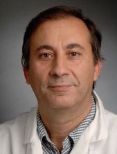 | Keynote Presentation Novel Technologies For the Detection of Clinically Relevant DNA Alterations in Circulating DNA
Mike Makrigiorgos, Professor of Radiation Oncology, Dana Farber Cancer Institute and Harvard Medical School, United States of America
With the increasing interest in treatment assessment using circulating DNA, sensitive and multiplexed detection of tumor-derived alterations in blood are desirable. We provide novel mutation enrichment-based methods that (a) enable several orders of magnitude improvement of microsatellite instability or mutations than currently possible; (b) are highly multiplex-able; (c) reduce the cost of both sample preparation and re-sequencing. Application in circulating DNA from clinical cancer samples will be presented. |
| |
Session Title: Evolution of Research in the Various Circulating Biomarker Classes -- Circulating Nucleic Acids and Circulating Vesicles |
| | 08:30 | 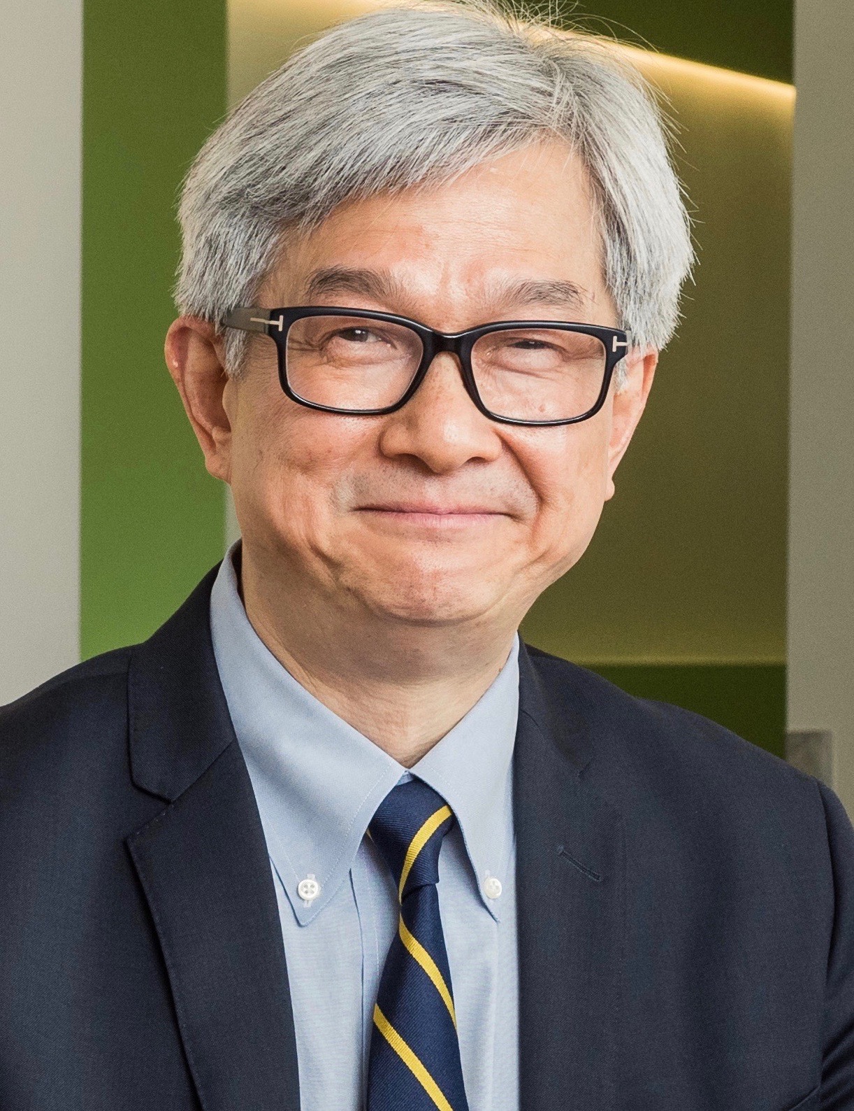 | Keynote Presentation EFIRM Liquid Biopsy (eLB)
David Wong, Felix and Mildred Yip Endowed Chair in Dentistry; Director for UCLA Center for Oral/Head & Neck Oncology Research, University of California-Los Angeles, United States of America
The advent of personalized medicine employing molecular targeted
therapies has markedly changed the treatment of cancer in the past
decade. Although tumor tissue biopsy-based genotyping is the current
clinical practice for guiding clinical management, biopsy procedures can
result in significant morbidity, limiting sampling to static snapshots
which are further limited in scope by the inherent sampling bias of the
analysis itself. To overcome these issues, technologies are needed for
rapid, cost-effective, and noninvasive identification of biomarkers at
various time points during the course of disease. Liquid biopsy is a
rapidly emerging field to address this unmet clinical need as
diagnostics based on cell-free circulating tumor DNA (ctDNA) can be a
surrogate for the entire tumor genome. The use of ctDNA via liquid
biopsy will facilitate analysis of tumor genomics that is urgently
needed for molecular targeted therapy. Currently, most targeted
approaches are based on PCR and/or next generation sequencing (NGS) for
liquid biopsy applications with performance concordance in the 60-80%
range with biopsy-based genotyping. We have developed a liquid biopsy
technology “Electric Field Induced Release and Measurement (EFIRM)-
Liquid Biopsy (eLB)” provides the most accurate targeted detection that
can assist clinical treatment decisions for the most common subtype of
lung cancer, non-small cell lung cancer (NSCLC), where tyrosine kinase
inhibitors (TKI) that can extend the disease progress free survival
period of these patients. eLB requires only 40 µl of sample volume, no
sample processing, reaction time is 15min and can be performed at the
point-of-care or high throughput reference lab using plasma or saliva.
eLB detects actionable EGFR mutations in NSCLC patients with >95%
concordance with biopsy-based genotyping. eLB is minimally (plasma)/
non-invasive (saliva) detecting the most common EGFR gene mutations that
are treatable with TKI such as Gefitinib or Erlotinib to effectively
extend the progression free survival of lung cancer patients. |
| 09:00 | 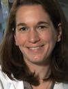 | Keynote Presentation Microfluidics for the Isolation of Biomarkers From Glioblastoma Patients
Shannon Stott, Assistant Professor, Massachusetts General Hospital & Harvard Medical School, United States of America
Glioblastoma is a highly fatal disease with few treatment options. Due to the location of the tumor, it is challenging to get dynamic, real-time information about the cancer. To address this need, we have developed microfluidic technologies to obtain information about these tumors by isolating rare cells (circulating tumor cells or “CTCs”) and tiny lipid particles, referred to as extracellular vesicles (EVs), from a simple blood draw. In this talk, data will be presented on our technological approach as well as our effort to interrogate their molecular content using next generation RNA sequencing and ddPCR. While the first application of this ‘liquid biopsy’ technology is in monitoring glioblastoma, it can be readily expanded to many different cancers and used to explore the underlying biology of metastasis. |
| 09:30 | 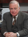 | Keynote Presentation Extracellular Vesicles as Mediators of Tissue Repair and Disease Reversal
Peter Quesenberry, Professor of Medicine, The Warren Alpert Medical School of Brown University, United States of America
Extracellular vesicle from marrow derived mesenchymal stem cells (MSCs) have been shown to have tissue repair characteristics. We have studied several models examining the capacity of MSC vesicles to promote tissue repair or disease reversal. In a murine model of monocrotaline induced pulmonary hypertension we have demonstrated that MSC derived vesicles will reverse or prevent the development of pulmonary hypertension as measured by vascular remodeling and right ventricular hypertrophy. In a similar vein we have demonstrated that MSC-derived vesicles mitigates radiation damage to murine bone marrow stem cells both in vitro and in vivo. This latter appears to be mediated by miRNA species. Lastly, MSC-derived vesicles have been shown to reverse the malignant phenotype of both colorectal and prostate cancer cells in vitro and in vivo.
The potential therapeutic applications of possible vesicle therapy are apparent and await appropriate progress in scale up approaches for harvest of vesicle types. |
| 10:00 | Challenges and Opportunities For Liquid Biopsies (ctDNA) in the Clinical Setting
Allison Welsh, Associate Director, Liquid Biopsy Development, Foundation Medicine, United States of America
| 10:30 | Coffee Break and Networking in the Exhibit Hall | 11:00 | Glioma Exosomes and Astrocytes: Conversion to the Dark Side
Michael Graner, Professor, Dept of Neurosurgery, University of Colorado Anschutz School of Medicine, United States of America
Glioblastomas (GBMs, WHO grade IV astrocytomas) are the worst of the
central nervous system tumors; despite maximum (and damaging)
therapeutic intervention, median survival time for patients is <15
months, and overall quality of life is poor. These abysmal outcomes have
changed little in 20 years. Clearly, our current therapies are
inadequate; we need innovative strides in understanding GBM biology to
rectify this situation. One “hot” research area is that of the impact of
tumor extracellular vesicles (EVs) on normal recipient cells. Tumor EVs
have extraordinary abilities to manipulate tumor microenvironments and
recipient cells both proximally and distally. Tumor EVs prepare the
“metastatic niche” for circulating tumor cells prior to colonization of a
target organ, deflect immune responses, and alter normal cells. Thus,
tumor EVs impact recipient cells to support tumor growth and
progression, which undoubtedly holds true for GBMs as well. However,
little is known about effects of GBM EVs on normal astrocytes—do GBM EVs
drive astrocyte phenotypic changes, potentially making the astrocytes
into tumor promotors? The answers could re-shape our paradigms on
gliomagenesis, particularly for recurrent tumors. Here we show that GBM
EVs activate cancer-type signaling pathways in recipient astrocytes,
promoting astrocyte migration towards the EVs, as well as astrocyte
anchorage-independent growth in soft agar. Astrocytes release of various
factors to generate a tumor-promoting milieu with increased tumor cell
growth, particularly in the areas of inflammatory responses to entities
that seem like viruses. We discuss the consequences of these phenomena
in the context of our current therapies with a view towards therapeutic
improvement. | 11:30 | Sequencing and Analysis of Patient Liquid Biopsies
Brian Dougherty, Executive Director, Translational Genomics, Oncology IMED, AstraZeneca R&D, United States of America
| 12:00 |  | Keynote Presentation About Noam Chomsky, DNA Motifs, Non-coding RNAs and Cancer Patients
George Calin, Professor and The Alan M. Gewirtz Leukemia & Lymphoma Society Scholar, University of Texas MD Anderson Cancer Center, United States of America
The newly discovered differential expression in numerous tissues, key cellular processes and multiple diseases for several families of long and short non-codingRNAs (ncRNAs, RNAs that do not codify for proteins but for RNAs with regulatory functions), including the already famous class of microRNAs (miRNAs) strongly suggest that the scientific and medical communities have significantly underestimated the spectrum of ncRNAs whose altered expression has significant consequences in diseases. MicroRNA and other short or long non-codingRNAs alterations are involved in the initiation, progression and metastases of human cancer. The main molecular alterations are represented by variations in gene expression, usually mild and with consequences for a vast number of target protein coding genes. The causes of the widespread differential expression of non-codingRNAs in malignant compared with normal cells can be explained by the location of these genes in cancer-associated genomic regions, by epigenetic mechanisms and by alterations in the processing machinery. MicroRNA and other short or long non-codingRNAs expression profiling of human tumors has identified signatures associated with diagnosis, staging, progression, prognosis and response to treatment. In addition, profiling has been exploited to identify non-codingRNAs that may represent downstream targets of activated oncogenic pathways or that are targeting protein coding genes involved in cancer. Recent studies proved that miRNAs and non-coding ultraconserved genes are main candidates for the elusive class of cancer predisposing genes and that other types of non-codingRNAs participate in the genetic puzzle giving rise to the malignant phenotype. Last, but not least, the shown expression correlations of these new ncRNAs with cancer metastatic potential and overall survival rates suggest that at least some member of these novel classes of molecules could potentially find use as biomarkers or novel therapeutics in cancers and other diseases. |
| 12:30 | Networking Lunch in the Exhibit Hall | |
Session Title: Transforming Circulating Biomarkers into Liquid Biopsies Delivering Actionable Clinical Information |
| | 13:00 | Abundant, Ultra-Short Trans-Renal Tumor DNA Fragments in Urine For Non-Invasive Cancer Detection and Monitoring
Qing Kang, Senior Scientist, Translational Medicine, Syros Pharmaceuticals, Inc., United States of America
Circulating tumor DNA (ctDNA) has shown great promise as a cancer biomarker in detecting tumor genotypes, monitoring cancer recurrence and resistance, as well as assessing treatment response. Compared to increased studies in plasma ctDNA, trans-renal tumor DNA (trDNA) exists as a largely untapped field. Here we present a proof-of-concept study to comprehensively understand the fundamentals of trDNA in different cohorts of cancer patients. | 13:30 |  | Keynote Presentation Circulating Circulome: A New Source of DNA For Liquid Biopsy?
Anindya Dutta, Harry F. Byrd Professor and Chair of Biochemistry and Molecular Genetics and Professor of Pathology, University of Virginia School of Medicine, United States of America
Cell-free circulating linear DNA is becoming very important for liquid biopsy. We have discovered small extrachromosomal circular DNA (eccDNA), called microDNA, in the nuclei of mammalian tissues and cell lines. Cell-free microDNAs from uniquely mapping regions of the genome are detected in plasma and serum from both mice and humans. The cell-free circulating circles are significantly longer (30%-60% >250 bases) than cell-free circulating linear DNA (~150 bases). Tumor-derived human microDNA is detected in the mouse circulation in a mouse xenograft model of human ovarian cancer. Human lung cancers contain longer microDNA than normal lung and these may be detected in the blood. Thus, circular DNA in the circulation is a previously unexplored pool of nucleic acids that could complement miRNAs and linear DNA in liquid biopsies. |
| 14:00 |  | Keynote Presentation Personalized Circulating Tumor DNA Technology
Cheng-Ho Jimmy Lin, Chief Scientific Officer, Oncology, Natera, United States of America
There is great promise in using circulating tumor DNA in biofluids and other human specimen in cancer care. I will present the different technologies for ctDNA; then we will the promises and challenges on how these can be applied for different indications such as diagnosis, early detection, treatment monitoring, minimal residual disease, and patient selection. |
| 14:30 | Non-Invasive Prenatal Screening for Genetic Disorders Using Cell-Free DNA in Maternal Plasma
Peter Benn, Professor Emeritus, Department of Genetics and Genome Sciences, University of Connecticut Health Center, United States of America
Methods to quantify fetal DNA copy number in maternal plasma has revolutionized prenatal screening and diagnosis of Down syndrome, other autosomal trisomies, sex chromosome abnormalities, and some microdeletion syndromes. This testing provides more accurate and screening than was previously possible and has thereby drastically reduced the number of chorionic villus biopsies and amniocenteses that are performed to confirm screening results. This non-invasive prenatal testing is being extended to include additional genetic conditions including single gene disorders. This talk will discuss the current status of this screening and comment on future prospects. | 15:00 |  | Keynote Presentation Development of Target Expression Assays in NSCLC Tumors and the CTC Compartment
Steven Pirie-Shepherd, Director, Pfizer, United States of America
Circulating tumor cells (CTCs) are a common focus of research, in part due to their relatively simple and non-invasive means of collection and their potential utility as biomarkers in cancer. They may also be studied to help further understand the metastatic process. In this presentation, we discuss development of an IHC assay to detect target expression in NSCLC tumors and an immunofluorescence assay to detect target expression in CTCs. We used these assays to profile matched samples obtained from treatment naïve NSCLC patients. We present data characterizing CTC enumeration as well as the detection and relative expression of target in CTCs and tumors from NSCLC patients and discuss the correlations between the presence of the target in CTCs and matched tumor resections. |
| 15:30 | Development of Predictive CTC Tests and Validation of their Clinical Utility for Therapeutic Benefit
Ryan Dittamore, Vice President, Translational Research and Clinical Affairs, Epic Sciences, United States of America
The development of liquid biopsy tests to inform clinical decisions in metastatic cancers is an unmet medical need. The presentation will overview novel test development, analytic validation, clinical development and demonstration of clinical utility in multiple clinical decision points in metastatic cancers. | 16:00 | Exploiting NGS and Liquid Biopsies For Clinical Individualized Neo-antigen Cancer Vaccines
John Castle, Executive Director, Vaccine Research and Translational Medicine, Agenus Switzerland Inc., Switzerland
We are treating cancer patients with individualized vaccines manufactured on-demand based on a NGS-profile of the patient tumor. We exploit NGS, bioinformatics, and computational immunology to generate the patient-specific vaccine blueprint. Our second-generation neoantigen vaccine platform, AutoSynVax™ (ASV™), entered the clinic in April and encodes mutation-containing peptides in a proprietary formulation and clinically tested adjuvant.
| 16:30 |  | Keynote Presentation The Droplet Biopsy Chip for Circulating Tumor Cell Capture
Balaji Panchapakesan, Professor, Department of Mechanical Engineering, Worcester Polytechnic Institute (WPI), United States of America
Circulating tumor cells (CTCs) are cells that have shed into the vasculature or lymphatics from a primary tumor and are carried around the body in the circulation. CTCs thus constitute seeds for the subsequent growth of additional tumors (metastasis) in vital distant organs, triggering a mechanism called Epithelial Mesenchymal Transition (EMT). One of the main causes of metastasis is EMT, which results in loss of epithelial characteristics of cancer cells. EMT results in down regulation of Epithelial Cell Adhesion Molecule (EpCAM), which is the primary marker for most commercially available CTC isolation systems today. We have invented the droplet biopsy chip that combines nanotechnology with micro-array manufacturing techniques to conduct droplet biopsy. The chip is a 76 element array consisting of nanotube wells. Antibodies attached to the nanotubes enable the capture of the cells. The hydrophobicity of the nanotube, antibody functionalization and droplet localization enables density gradients that results in separation of blood into different layers resulting in capture of CTCs without leukocyte contamination. Advantages of this technique include both positive and negative selection strategy on the same chip resulting in potential to capture invasive CTC phenotypes. Potentially one can achieve capture of CTCs based on multiple biomarkers, negative selection by depleting leukocytes, and density gradient based selection of nucleated cells, thereby avoiding loss of CTCs due to variation in size, biomarker composition, or lysis resistance, all in a single blood test. |
| 17:00 | microRNA-based Therapeutics in Cancer
Frank Slack, Director, Institute for RNA Medicine, Beth Israel Deaconess Medical Center Cancer Center/Harvard Medical School, United States of America
MicroRNAs are small non-coding RNAs that regulate gene expression to control important aspects of development and metabolism such as cell differentiation, apoptosis and lifespan. let-7 and miR-34 are microRNAs implicated in human cancer. Specifically, human let-7 and miR-34 are poorly expressed or deleted in lung cancer, and over-expression of let-7 or miR-34 in lung cancer cells inhibits their growth, demonstrating a role for these miRNAs as tumor suppressors in lung tissue. let-7 and miR-34 regulate the expression of important oncogenes implicated in lung cancer, suggesting a mechanism for their involvement in cancer. We are focused on the role of these genes and other oncomiRs in regulating proto-oncogene expression during development and cancer, and on using miRNAs to suppress tumorigenesis. | 17:30 | Application of Artificial Intelligence and Semiconductor Technologies in Liquid Biopsy
Chung- Er Huang, Assistant Professor, National Chiao Tung University; Managing Director, CytoAurora Biotechnologies, Inc., Taiwan
The demand for basic research and commercial applications of liquid biopsy has increased along with the need for reliable automation to promote the market growth. In this presentation, we will introduce a novel system (“Cell Reveal”) for rare cells captured. The novel system in capturing rare cells (CTC/CFC) has been robustly developed based on an advanced technology which integrates semiconductor manufacturing technology, biotechnology, automation, and artificial intelligence. The system (“Cell Reveal”) consists of cell-capture, cell-analysis, enumeration, and cell separation. The capture of rare cells is performed by a combination of a bio-chip and microfluidics as the cells flow through the surface of the biochip. The high capture rate of the bio-chip is achieved by a similar dimensional nano-structure in a high-density arrangement. Enumeration of rare cells can be accomplished by the Cell Analysis Tool, which also provides molecular analysis and genetic makeup of the target cells as well as their locations on the chip. The intact cells are subjected to separation, resulting in individual target cells collected for subsequent single Whole Genome Amplification (WGA) and WGS. This novel nano-system greatly improves and accelerates patients' diagnostics and therapeutics. | 18:00 | A Newly Discovered Cell Found in Blood Enables the Detection and Diagnosis of Cancer When it Matters Most
Cha-Mei Tang, President and CEO, Creatv MicroTech, United States of America
Cancer detected early provides the best chance for successful treatment outcomes. We discovered a cell in the blood of cancer patients that not only satisfies this unmet need, but also provides many other benefits in the detection and diagnosis of cancer. The CellSieveTM microfiltration system is optimal for collecting cells larger than 7-8 microns from whole blood, and this platform is further enhanced by a proprietary technique to stain the cells on the filter for up to 12 markers. During the development process, while using the platform to accurately identify true circulating tumor cells (CTC), apoptotic CTCs, circulating endothelial cells (CECs), and mesenchymal transition cells (EMTs), a previously unreported cell type was discovered, the Cancer Associated Macrophage-Like cell (CAML). CAMLs are very large, 25-300 microns in size. They express markers associated with the specific cancer. They have been isolated from the blood of patients with all solid tumor types analyzed. However, they are not found in healthy controls. CAMLs are found in common epithelial cancers, such as breast, prostate, lung, pancreatic, colorectal, liver, kidney, melanoma, ovarian, esophageal and bladder cancers, and also in rare solid tumors, such as neuroblastomas and sarcomas. They are found in all stages of cancer, including high percentages in stage I. Analysis of clinical data has demonstrated that the detection of CAMLs is applicable in many ways including cancer screening, early detection of cancer recurrence, prognosis, companion diagnostics for treatment selection and precision monitoring of a therapeutic response. We have the tools to transform cancer diagnostics, thereby improving and saving lives.
| 18:30 | Close of Day 2 of the Conference |
|


 Add to Calendar ▼2018-03-28 00:00:002018-03-29 00:00:00Europe/LondonCirculating Nucleic Acids and Circulating Rare Cells: Liquid Biopsy for Early Cancer Detection SELECTBIOenquiries@selectbiosciences.com