Thursday, 12 September 201308:00 | Conference Registration and Materials Pickup | |
Session Title: Latest Trends in Exosome Research |
| | |
Session Chair: Dr. Sasha Vlassov, Senior Staff Scientist, Life Technologies |
| | 09:00 |  | Keynote Presentation Microvesicle (exosome) RNA as Biomarkers for Disease
Johan Skog, Chief Scientific Officer, Exosome Diagnostics Inc, United States of America
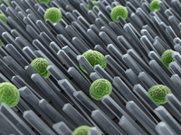
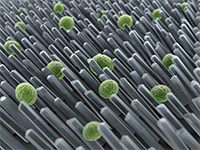
Microvesicles (exosomes) are released from all cells and can be isolated from biofluids, including serum, plasma, urine, and CSF. These exosomes carry RNA (mRNA, microRNA, and other small RNAs) and protein from the donor cell that can be easily isolated even after long-term storage in freezers. The use of exosomal RNA has sparked a tremendous interest in the field of biomarkers, where access to the genetic status of disease in a non-invasive way is highly desirable. To develop a clinical test using microvesicle RNA, isolation of exosomes in a robust way that is easy, scalable, reproducible and allows for high-throughput processing in a clinical setting is critical. The RNA needs to be of high quality with markers that allow quality assessment of clinical samples. The use of exosomes is a great platform that enables longitudinal monitoring of a variety of mutations and transcript levels in tumors using qPCR, NGS and digital PCR. |
| 10:00 | Coffee Break and Networking in the Exhibit Hall | 11:00 | Plasma Vesicles Content as a New Sample Type for Clinical Diagnosis and Prognosis of Myocardial Infarction and Stroke
Dominique PV de Kleijn, Professor Experimental Vascular Surgery, Professor Netherlands Heart Institute, University Medical Center Utrecht, The Netherlands, Netherlands
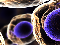
Extracellular vesicles, including microvesicles, microparticles and exosomes are abundant in plasma. All cells secrete extracellular vesicles with release being very rapid after innate immunity activation of inflammatory cells. These vesicles are important in cell-to-cell communication in a variety of processes including coagulation, antigen presentation and tissue damage. Plasma extracellular vesicles can be easily isolated from frozen plasma or serum and contain protein, miRNA and RNA depending on source and stimulus. Isolating these vesicles from the blood and studying their content may provide rapid information on the pathological status of tissues. We showed that plasma vesicles content can be used for the diagnosis of Acute Coronary Syndrome in an emergency department cohort of 471 patient with chest pain. Next to this, we showed that vesicle content in a cohort of 1060 patients can be used for the prognosis of secondary cardiovascular events on top of existing risk factors and are expecting to have a CE certified kit available this year.
We anticipate that plasma extracellular microvesicles in the near future will emerge as an important source for diagnostic and prognostic markers for cardiovascular disease and will help us in understanding the underlying biology.
| 11:45 | Patients’ Blood miRNA Profiling as a New Tool for Prognosis in Multiple Myeloma
Flavia Pichiorri, Assistant Professor, The Ohio State University, United States of America
Multiple Myeloma (MM) is an incurable cancer where prognosis is determined by the combination of the International Staging System (ISS) and fluorescent in-situ-hybridization (FISH) analysis of MM plasma cells (MM-PCs). However, the outcome predictions based on the current criteria are not precise and patients with similar characteristics have different outcome, thereby supporting the need for novel and more accurate prognosticators. microRNAs are short- non-coding-RNAs whose expression levels change in the transition from normal PCs to MM-PCs. miRNAs are master regulators of gene expression and have been found altered in all cancers. Their identification in body fluids suggests their possible use as novel biomarkers in MM patients. A comprehensive profile of circulating miRNAs for disease prognostication has not been reported. We profiled more than 654 different miRNAs using nCounter technology (nanoString, Seattle, USA) on samples from 54 newly diagnosed MM patients enrolled on the randomized phase 3 study of Velcade Melphalan Prednisone Thalidomide versus Velcade Melphalan Prednisone (NCT#01063179). Stem loop real-time PCR (qRT-PCR) was performed on an additional 234 MM patients enrolled in the same trial. We found that 3 miRNAs [miR-16 (P=0.0019), -19b (P=0.043) and miR-25(P=0.0012)] were associated with overall survival (OS) of myeloma patients. By incorporating these indices into the ISS score, we generated a novel miRNA-integrated score that is highly predictive of survival duration (P<0.00001) and substantially improved the currently used prognostic scores. These observation are perfectly aligned with our previously published data in which these miRNAs, are tightly regulated in MM cells and are shown to regulate their survival. We can then conclude that circulating microRNAs can be considered a new class of non-invasive prognosticators in MM.
| 12:15 | Lunch and Networking in Exhibit Hall | 12:45 |  Technology Spotlight: Technology Spotlight:
New High-throughput Biomarker Discovery Platform for Exosome Isolation and poly(A)+ mRNA Analysis
Masato Mitsuhashi, Chief Scientific Officer, Hitachi Chemical Research Center, Inc.
We have developed a unique 96-well filterplate which captures exosomes and microvesicles (EMV) from various biological fluids, such as plasma, urine, saliva, cell culture media, etc. An additional adaptor will accommodate large volumes of culture media and urine (~10-30 mL). After centrifugation for only 5 min at 2,000 x g, EMV can be trapped on the membrane. For mRNA analysis, EMV is lysed on the filterplate, and the resultant lysates are transferred to a 96-well oligo(dT)-immobilized microplate for poly(A)+ mRNA isolation followed by cDNA synthesis and PCR. The results are comparable to those of standard ultracentrifugation method. In urine, glomeruli-, proximal tubules-, distal tubules-, and collecting ducts-specific mRNAs can be quantified, and are currently used for the analysis of kidney function in areas such as diabetic nephropathy and kidney transplantation. In plasma, various cell/tissue-specific mRNAs can be quantified, including hematopoietic cells in bone marrow, endothelial cells, neuronal cells in brain, skeletal muscle cells, fat cells, etc. The identification and quantification of tissue/cell specific mRNAs in EMV are applicable to various biomarker discovery projects.
| 13:15 | Poster Viewing and Networking Session in Exhibit Hall | |
Session Title: LDTs, CLIA, and Opportunities in the Biomarker Interrogation Space |
| | |
Session Chair: Hannah Mamuszka, Vice President of Business Development, Exosome Diagnostics |
| | 14:15 | Companion Diagnostics Development at the Siemens Clinical Laboratory
Sunil Pandit, Senior Manager, Siemens Healthcare Diagnostics, United States of America
Siemens Clinical Laboratory (SCL) is an accredited CLIA laboratory within Siemens Healthcare Diagnostics Inc., a global diagnostics company. SCL provides external access to laboratory developed tests (LDTs) and cutting-edge diagnostic technologies, pharmaceutical clinical trial testing services, and a unique menu of clinical tests. Utilizing LDT assays as prototypes for globally distributed In Vitro Diagnostic (IVD) products allows efficient development of new technologies and validated assays for companion diagnostics applications and clinical utility studies. In this presentation, the speaker will describe experience gained in developing companion diagnostics assays at Siemens. | 14:45 | Cancer Biomarker Discovery: The Case for Pharma OutSourcing from a Dx Perspective
Premal Shah, Director, Genomic Health Inc, United States of America
Cancer therapeutic development is hard. Really hard. The successes have been few and far between. Many companies are looking to identify and incorporate biomarkers as part of their development plan. However, while companies often fixate on simple, “kittable” markers, the more powerful markers of the future—driven by leveraging NGS capabilities—will likely be complex. Identifying these markers requires niche expertise that can be provided only by companies with deep experience in this area. It is for this reason, combined with the costs that make outsourcing cancer biomarker discovery a necessity for today’s pharma companies. | 15:15 | Coffee Break and Networking in the Exhibit Hall | 15:45 | Extracorporeal Elimination of Circulating Exosomes as a Therapeutic Adjunct to Address Infectious Disease and Cancer
James Joyce, Chairman & CEO, Aethlon Medical, Inc., United States of America
The Aethlon Hemopurifier® is a first-in-class device that targets the rapid elimination of infectious disease and cancer glycopathogens from circulation. In cancer, the Hemopurifier has been demonstrated to capture exosomes underlying lymphoma, melanoma, ovarian, and breast cancer. These microvesicular particles trigger apoptosis of immune cells and have been reported to facilitate tumor growth, metastasis, and the development of drug resistance.
In design, the Hemopurifier consists of the affinity lectin Galanthus nivalis agglutinin (GNA) immobilized in the outer-capillary space of plasma membrane technologies. The resulting mechanism provides selective target elimination as GNA binds high mannose signatures abundant on the surface of exosomes and viral glycoproteins. Studies of hepatitis c (HCV) infected patients receiving a three-treatment Hemopurifier protocol combined with interferon-based standard of care resulted in undetectable HCV in as little as seven days in hard-to-treat genotype-1 patients. The studies also documented the ability of the Hemopurifier to capture as many as 300 billion HCV copies during a single six- hour treatment. The FDA recently approved an IDE that allows for the initiation of US feasibility studies. | 16:15 |  | Keynote Presentation Noninvasive Cancer Personal Genomics
Charles Cantor, Chief Scientific Officer, Sequenom Inc, United States of America
In clinical situations where there is significant programmed cell death like cancer or pregnancy, DNA and RNA molecules appear in the circulation derived from cells that have died. In pregnancy, mature diagnostic tests are now available. In cancer, things are the situation is at a much more preliminary stage. To utilize DNA from the apoptotic cells it must differ in some way from the considerable background of host DNA. These differences can be copy numbers, sequence variations or epigenetic variations. In this talk I will contrast the situations in the two indications and chart possible future developments. |
| 17:15 | Exosome Biogenesis: Role of Exosomal Cargo in the Production and Secretion of Exosomes
Suraj Bhat, Professor, University Of California Los Angeles, United States of America
The molecular information content of exosomes is fundamental to their function. Exosomes, carry cell-type specific cargo targeted for extracellular destinations. While a constitutive synthesis of exosomes is conceivable as part of the cellular basis of tissue homeostasis we assume that the production of exosomes containing cell type-specific gene products may not be random but dictated by tissue physiology and cellular function; exosomes may be assembled and secreted in response to instructions received from the neighboring cells or from distant tissues. Thus an efficient way of coordinating the process of exosome production and secretion, which is temporally precise and functionally relevant, is to have the cell-type specific cargo a role in their biogenesis.
We have used the human retinal pigment epithelium (RPE) as the functioning paradigm. RPE straddles the blood/retina barrier and physically and physiologically supports retinal photoreceptor function. Working with aB-crystallin (aB), a small heat shock and cell-type specific exosomal protein associated with a number of neuro-degenerations including Alzheimer’s, Parkinson’s disease, Multiple sclerosis and AMD (age-related macular degeneration), we demonstrate that this protein is essential for exosome biogenesis in the human RPE cells in culture. aB is found only in some cell types (for example most epithelial cells contain aB while most fibroblastic cell lines do not). Its presence in exosomes, therefore, allows us to address the question if the exosomal cargo has a role in exosome biogenesis? Understanding cell-type specific control of exosome biogenesis may allow us engineer exosomal content biologically. We will discuss our recent data obtained from these investigations. | 17:45 | Reception and Roundtable Discussions in Exhibition Hall | 19:15 | Close of Day One of Summit. | 19:15 | Exosome Night: Dinner: Premium Beers, California Wines, and Dinner for All Registered Exosome Track Delegates. Enjoy an Awesome Evening and Network with your Fellow Delegates. Sponsored by Exosome Diagnostics, Inc. |
Friday, 13 September 2013 |
Session Title: Circulating Biomarkers |
| | |
Session Chair: Dr. Johan Skog, CSO, Exosome Diagnostics |
| | 08:45 |  | Keynote Presentation Microarrays for Biomarker Discovery
Bertrand Jordan, Emeritus Research Director, National Centre for Scientific Research, France
It is clear that in many cases diagnostic, prognosis or prediction cannot be achieved with the measurement of a single biological parameter – i.e. biomarkers need to be comprised of several (often many) measured entities. Microarrays, being inherently multiplex devices, are particularly well suited for the ascertainment of complex patterns correlated to clinical information.
DNA microarrays are an established approach for mutation detection and pathogen characterization (not really a biomarker study), but they have also been used for expression profiles providing diagnostic (Pathwork diagnostics “TOO”, tumor of origin determination), as well as prognostic and predictive information (Agendia “Mammaprint”, breast cancer). Initial enthusiasm about this approach was dampened by the realisation of serious statistical issues in initial data analysis as well as by the requirement for fresh-frozen samples, but these hurdles have now been largely removed.
Protein and peptide microarrays are a more recent entrance in this contest. Beyond “simple” determination of specific antigens or antibodies in patient serum, complex patterns are finding clinical use. In particular, the capability to measure protein activity (not just protein amount) and even to assess drug sensitivity directly in patient samples in a multiplex fashion is opening new areas of application.
Microarrays thus have a place in the field of biomarker discovery and ascertainment that will be resistant to competition from other approaches (e.g. NGS). |
| 09:30 | BEAMing and Droplet Digital PCR Analysis of Mutant IDH1 mRNA in Tumor Extracellular Vesicles
Leonora Balaj, Instructor in Neurosurgery, Mass General Hospital (MGH)/Harvard Medical School, United States of America
Development of biofluid-based molecular diagnostic tests for cancer is an important step towards tumor characterization and real-time monitoring in a minimally invasive fashion. Extracellular vesicles (EVs) are released from tumor cells into body fluids and can provide a powerful platform for tumor biomarkers since they carry tumor proteins and nucleic acids. BEAMing (beads, emulsion, amplification, magnetics) PCR is substantially more sensitive than many other assays for detecting single nucleotide mutations when there is a significant background of wild-type sequences. Picodroplet digital PCR developed by RainDance Technologies is another highly sensitive technology that can be used to quantify rare mutations in a high wild type background. Here, we describe a novel approach that uses BEAMing droplet RT-PCR, as well picodroplet digital RT-PCR, to interrogate mRNA sequences contained within EVs from serum and cerebrospinal fluid (EV-BEAMing). Using both assays, we were able to reliably detect and quantify mutant and wild-type IDH1 transcripts in cerebrospinal fluid of patients with gliomas. EV-BEAMing and picodroplet dPCR are valuable new tools for cancer diagnostics which can be applied to a variety of biofluids and neoplasms. | 10:00 | Development of EXO50: Exosome Isolation and RNA Extraction Kit for Plasma
Hannah Mamuszka, Vice President, Exosome Diagnostics Inc, United States of America
Until recently, exosome isolation and nucleic acid extraction from biofluids has required the use of ultracentrifugation, which is time consuming, inefficient, produces unreliable results, and is not relevant for use in development of a diagnostic test. With the development of the EXO50 kit for use with serum and plasma, we have demonstrated a kit that achieves comparable or better results to ultracentrifugation, but in a shorter time, and with more reliable, reproducible, robust results. In validating the EXO50 kit, we compared expression levels of specific mRNAs as well as microRNA to ultracentrifugation and precipitation based methods. Interestingly, the kit clearly segregates microRNAs that are found on the inside of the exosome as compared to microRNAs that are Ago2-protein bound something that ultracentrifugation and precipitation methods often do not allow for. The EXO50 kit is scalable and linear, requiring as little as 200uL and as much as 4mL in the current version, allowing for competent analysis of a single mutation or biomarker discovery using array or sequencing of thousands of genes. The fast, easy, and robust protocol of the EXO50 kit will allow for plasma based IVD development. | 10:30 | Coffee Break and Networking in the Exhibit Hall | 11:15 |  | Keynote Presentation Use of Nextgen to Study Circulating Biomarkers
Mostafa Ronaghi, Senior Vice President And Chief Technology Officer, Illumina Inc, United States of America
A paradigm shift has happened with drastic cost reduction and improvements in the workflow and accuracy of Nextgen sequencing. In general, Nextgen is replacing several classical clinical tests by improved accuracy, enhanced safety for patient, and lower cost of test. In this talk, recent progress in Nextgen sequencing will be discussed. Furthermore, several examples will be provided on how Nextgen is being implemented to study circulating biomarkers in biological fluids. |
| 12:15 | Lunch and Networking in Exhibit Hall | 13:15 | Poster Viewing and Networking Session in Exhibit Hall | |
Session Title: CTCs and Other Classes of Circulating Biomarkers |
| | |
Session Chair: Dr. Leonora Balaj, MGH/Harvard Medical School |
| | 14:15 | NanoVelcro-Embedded MicroChips for Detection and Isolation of Circulating Tumor Cells
Hsian-Rong Tseng, Professor, Crump Institute for Molecular Imaging, California NanoSystems Institute, University of California-Los Angeles, United States of America
This presentation will introduce a new type of cell-affinity assay that is capable of detecting circulating tumor cells (CTCs) in blood samples collected from metastatic cancer patients. Similar to most of the existing approaches, anti-EpCAM was grafted onto the surfaces to distinguish CTCs from the surrounding hematologic cells. The uniqueness of our technology is the use of nanostructured surfaces, which facilitates local topographical interactions between CTCs and substrates at the very first cell/substrate contacting time point. We demonstrated the ability of these nanostructured substrates to capture CTCs in whole blood samples with significantly improved efficiency and selectivity.
Abstract. Our team at UCLA has demonstrated a highly efficient, inexpensive CTC assay capable of detecting and isolating CTCs in blood samples collected from metastatic cancer patients. First, we pioneered a unique concept of “NanoVelcro” cell-affinity substrates, by which capture agent (antibodies or aptamers)-coated nanostructured surfaces were utilized to immobilize CTCs in a stationary device setting. Second, by integrating the NanoVelcro substrate with an overlaid microfluidic component that can generate vertical flows, further improved CTC capture efficiency (>85%) has been achieved as a result of the enhanced collisions between CTCs and the substrate. Side-by-side analytical validation using both artificial and patient CTC samples suggested that the sensitivity of our CTC Assay outperforms that of CellSearch. In order to further exploit CTC’s diagnostic value, we combine the use of NanoVelcro Chip with the Laser MicroDissection (LMD) technique to enable highly efficient and specific isolation of viable/preservative-free CTCs from patient blood samples. Ultimately, our goal is to carry out sequential molecular and functional analyses of the single CTCs harvested by our NanoVelcro-embedded microchips. | 14:45 | Challenges of Implementing and Validating CTC Assays in Oncology Clinical Trials
Iman Jilani, Associate Director, Pfizer Inc, United States of America
This presentation will discuss the challenges of incorporating CTC assays in Oncology clinical trials. Content will include choosing a platform , method development, validation, and an example of an implementation into an ADC clinical trial. | 15:15 | Coffee Break and Networking in the Exhibit Hall | 15:45 | Tumor Cell Heterogeneity: Achieving Single Cell Resolution
Robert Proulx, President & GM, US Operations, Silicon Biosystems, United States of America
Advances in technologies for sorting cells and improvements in sensitivity for genomic analytical methods have driven an interest in studying the phenotypic and genotypic variation of disease at the single cell level. In this presentation we illustrate how single cell analysis can be achieved and applied in oncology to drive personalized medicine and cancer treatment decisions. | 16:15 | Pathology is Phenotyping. What Do Pathologists Do? How Do they Do It?
Dean Troyer, CEO, Provia Biologics, United States of America
Microscopic examination remains the gold standard for classification and staging of cancer and other diseases. Any diagnostic test that relies on tissue must fit into this work flow. Histology is the processing of tissue biopsies for microscopic evaluation. It is crucial to understand how a diagnostic test fits into the landscape of pathology and clinical medicine. This presentation will include a practical overview of diagnostic pathology as it relates to existing diagnostic tools such as immunohistochemistry and fluorescence in situ hybridization. Newer diagnostics must find their place in this workflow. Some rules of the road about diagnostic markers/tests: they should change clinical practice; tissue houses the most concentrated and unaltered forms of tumor markers; pricing is relevant; pathologists are cost effective – your marker should pick up where they leave off; multiplexing is preferable; they must fit into the existing work flow of histopathology. | 16:45 | Biomarker IP after Prometheus: Patenting the Unpatentable
Gregory Scott, Patent Attorney, Klarquist Sparkman, LLP, United States of America
Karri Bradley, Partner, Klarquist Sparkman, LLP, United States of America
In this tag-team presentation, two IP lawyers will discuss the latest trends in this evolving field:
The Supreme Court decision in Mayo Collaborative Services v. Prometheus Laboratories, Inc. shifted the biomarker IP landscape by raising the bar to obtain meaningful patent protection for diagnostic methods involving biomarker detection. We will review recent developments in patent law concerning diagnostic method claims, and the approaches we are currently using to successfully address the Prometheus standard.
| 17:15 | Biomarker Development to Improve Trials for Type 1 Diabetes
Matthias Herrath, Professor, The La Jolla Institute For Allergy and Immunology, United States of America
Immunotherapy of type 1 diabetes and other autoimmune disorders faces significant hurdles, because therapeutic efficacy needs to be balanced with acceptable side effects. Since development of disease also exhibits heterogeneity depending on age, environmental and genetic factors, it would be desirable to rationally develop a suitable and practical set of biomarkers, that should be able to segregate responders from non-responders and ideally predict efficacy of immune-based intervention on an individual basis. We have developed panel of biomarkers that are specifically based on novel insight into the pathogenesis of T1D achieved as part of the nPOD (national pancreatic organ donor) consortium. This strategy, its implication and realization in future clinical trials and potential impact on individualized medicine will be discussed. | 17:45 | Close of Conference. |
|


 Add to Calendar ▼2013-09-12 00:00:002013-09-13 00:00:00Europe/LondonExosomes and Circulating Biomarkers Summit 2013Exosomes and Circulating Biomarkers Summit 2013 in San Diego, CASan Diego, CASELECTBIOenquiries@selectbiosciences.com