Wednesday, 24 October 201808:00 | Conference Registration, Materials Pick-Up, Morning Coffee and Breakfast Pastries | |
Session Title: Conference Opening Session Framing the Key Topics and Opportunities in Liquid Biopsy |
| | 09:00 |  | Conference Chair Conference Chairman Welcome and Opening Remarks
Dominique PV de Kleijn, Professor Experimental Vascular Surgery, Professor Netherlands Heart Institute, University Medical Center Utrecht, The Netherlands, Netherlands
|
| 09:15 |  | Keynote Presentation Mt-SEA Pipeline: High Throughput Flow Cytometric Analysis of Exosomes in Clinical Biofluids
Jennifer Jones, NIH Stadtman Investigator, Head of Transnational Nanobiology, Laboratory of Pathology, Center for Cancer Research, National Cancer Institute, United States of America
Because Extracellular Vesicles (EVs) carry surface receptors that are characteristic of their cells of origin, EVs have tremendous potential as non-invasive biomarkers for diagnosis, risk-stratification, treatment selection, and treatment monitoring. We developed a first-in-class pipeline to characterize EV heterogeneity and provide high-sensitivity quantification of informative EVs in biofluids before, during, and after treatment. By combining multiplex assays with high-resolution, single EV flow cytometric methods together into a Mutiplex-to-Single EV Analysis (Mt-SEA) pipeline, we are able to characterize a broad range of relevant EV subsets, while also accurately measuring the concentration of specific EV populations. Detection of tumor-associated EVs and detection of EV repertoire changes during treatment paves the way to future evaluation of EVs as as biomarkers for use in personalized, adaptive therapies. |
| 09:45 | 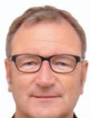 | Keynote Presentation Vesicles and Cellular Communication within the Bone Environment
Hans van Leeuwen, Professor and Dean, Erasmus University Medical Center, Netherlands
Bone cells are not only important in bone metabolism and building and maintaining healthy bone but are also involved in numerous other processes. This presentation will focus on two of these. One is the interplay with the cells in the bone marrow in particular the hematopoietic stem cells that give rise to the cells of our immune system. The other is the role in cancer metastasis. I will discuss the presence of extracellular vesicles in the bone environment and how these are involved in the interplay between bone cells, the osteoblasts, and hematopoietic stem cells and prostate cancer cells. In addition, I will address our studies on unraveling the content of these osteoblast as well as prostate cancer cells-derived extracellular vesicles. Hereby I will introduce extracellular vesicles in the bone environment and a role of bone cells beyond bone metabolism. The understanding of these extracellular vesicles in the either physiological or pathophysiological condition may provide novel diagnostic and therapeutic tools. |
| 10:15 | Morning Break and Networking in the Exhibit Hall | 10:45 |  | Keynote Presentation Liquid Biopsy: The New Diagnostic Concept in Oncology
Klaus Pantel, Chairman, Department of Tumor Biology, University Medical Center Hamburg-Eppendorf, Germany
The analysis of circulating tumor cells (CTCs) in blood may provide clinically relevant information as “liquid biopsy” (Alix-Panabieres & Pantel, Nature Rev Cancer 2014) and provide new insights into tumor biology (Uhr & Pantel, PNAS, 2011, Lu et al., Cancer Cell, 2011; LeBleu et al., Nat. Cell Biol., 2014; Werner et al., Cancer Discovery, 2015). Besides CTCs the molecular analysis of ctDNA provides important complementary information as “liquid biopsy” (Pantel & Alix-Panabieres, Cancer Res., 2013; Pantel et al., Nature Med. 2013; Heitzer et al., Genome Med. 2013; Schwarzenbach et al., Nature Rev. Clin. Oncol., 2014, Joosse & Pantel, Cancer Cell 2015; Bardelli & Pantel, Cancer Cell 2017). Moreover, circulating microRNAs, extracellular vesicles and tumor-educated platelets are also interesting new members of the “Liquid Biopsy family” with potential clinical relevance in the future. In particular exosomes have gained great interest because they act as biomarkers with important functional properties for tumor progression. E.g., the integrin composition of exosomes can determine the organ site of metastases (Hoshino, Pantel, Lyden et al., Nature, 2015) and microRNAs in exosomes can impact the biology of the recipient cells (Anfossi, Bababayan, Pantel, Calin, Nature. Rev. Clin. Oncol. 2018). Detection of these miRNAs can contribute to early detection of cancer (Meng et al., Oncotarget, 2016). Liquid biopsy analyses with validated platforms provides reliable information on prognosis and may serve to identify therapeutic targets or mechanisms of resistance on metastatic cells. Metastatic cells might have unique characteristics that can differ from the bulk of cancer cells in the primary tumor currently used for stratification of patients to systemic therapy. Moreover, monitoring of blood samples before, during and after systemic therapy (e.g., chemotherapy, hormonal therapy, antibody therapy) might provide unique information for the future clinical management of the individual cancer patient and might serve as surrogate marker for response to therapy. In conclusion, liquid biopsies can be used to improve the management of individual cancer patients and contribute to the vision of personalized medicine (Alix-Panabieres & Pantel, Cancer Discovery, 2016; Bardelli & Pantel, Cancer Cell 2017). Validation of liquid biopsy assays is essential and is currently being performed by the EU/IMI consortium CANCER-ID (www.cancer-id.eu). |
| 11:15 | 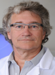 | Keynote Presentation Circulating DNA as a Next Generation Diagnostic Tool
Alain Thierry, Director of Research, Institut de Recherche en Cancérologie de Montpellier-INSERM, France
Circulating tumor DNA (ctDNA) holds promises in tumor burden monitoring or malignancies early detection. Based on crucial observation on the structure and origin of ctDNA we developed a multiparametric method for specifically analyze ctDNA. This technology enables the first clinical validation of the analysis of circulating DNA in oncology, in a prospective blinded multicentric study for the detection of KRAS and BRAF mutations from colorectal patient plasma samples. Aims of our research are now focusing on various aspects of the potential of ctDNA: (i), detecting the emergence of the mutations following targeted therapy; (ii), developing the Intplex test for the multimarker quantitative analysis of ctDNA; (iii), studying the follow up of CRC patients; (iv), the prognostic power of ctDNA and (v) the screening power of ctDNA. |
| 11:45 |  | Keynote Presentation Functional Analyses of Circulating Tumor Cells in Cancer Patients
Catherine Alix-Panabières, Director of the Laboratory of Rare Human Circulating Tumor Cells, University Medical Centre of Montpellier, France
Circulating tumor cells (CTCs) in blood are promising new biomarkers potentially useful for prognostic prediction and monitoring of therapies in patients with solid tumors including colon cancer. Moreover, CTC research opens a new avenue for understanding the biology of metastasis in cancer patients. However, an in-depth investigation of CTCs is hampered by the very low number of these cells, especially in the blood of colorectal cancer patients. Thus, the establishment of cell cultures and permanent cell lines from CTCs has become the most challenging task over the past year. We described, for the first time, the establishment of cell cultures and a permanent cell line from CTCs of one colon cancer patient (Cayrefourcq et al. Cancer Res. 2015). The cell line designated CTC-MCC-41 is in culture for more than three years and has been characterized at the genome, transcriptome, proteome and secretome levels. This thorough analysis showed that CTC-MCC-41 cells resemble characteristics of the original tumor cells in the colon cancer patient and display a stable phenotype characterized by an intermediate epithelial/mesenchymal phenotype, stem-cell like properties and an osteomimetic signature indicating a bone marrow origin. Functional studies showed that CTC-MCC-41 cells induced rapidly in vitro endothelial cell tube formation and in vivo tumors after xenografting in immunodeficient mice. More recently, we defined the molecular portrait of these metastasis-competent CTCs (Alix-Panabières et al. Clin Chem. 2017). These results highlight that CTC-MCC-41 line display a very specific transcription program completely different than those of the primary and metastatic colon cancer cell lines. Interestingly, among the 1,624 transcripts exclusively up-regulated in CTC-MCC-41 samples, key genes related to energy metabolism, DNA repair and stemness genes were observed. Such data may supply insights for the discovery of new biomarkers to identify the most aggressive CTC sub-populations and for the development of new drugs to inhibit metastasis-initiator CTCs in colon cancer. Moreover, the development of new immunotherapeutic strategies is of utmost importance. Antibodies against proteins that block the immune response of T-cells such as PDL1 have been approved for treatment of cancer patients after showing remarkable long-term remissions in subsets of patients. It is now important to develop predictive biomarkers to identify patients with the highest benefit from these therapies. In 2015, we could show for the first time that PD-L1 is heterogeneously expressed on CTCs from metastatic breast cancer patients (Mazel et al. Mol Oncol 2015). Further functional analysis of this interesting subset of CTCs might reveal special immunosuppressive properties. |
| 12:15 | Networking Lunch in the Exhibit Hall -- Meet the Exhibitors and View Posters | |
Session Title: Circulating Biomarkers for Cancer |
| | 13:30 |  mRNA Profiles of Circulating Tumor Cells and Extracellular Vesicles in Matched Samples From Metastatic Breast Cancer Patients mRNA Profiles of Circulating Tumor Cells and Extracellular Vesicles in Matched Samples From Metastatic Breast Cancer Patients
Corinna Keup, Researcher, Universitätsklinikum Essen, Klinik für Frauenheilkunde und Geburtshilfe
The analysis of RNA enclosed in circulating tumor cells (CTCs) or extracellular vesicles (EVs) may be sensitive enough to detect disease progression earlier than contemporary visual staging methods. A prediction of the ideal therapy strategy via characterization of CTCs or EVs would be even more beneficial. We, for the first time, compared RNA profiles of CTCs and EVs in metastatic breast cancer (MBC) patients to get insights about their feasibility for therapy stratification.
CTCs were isolated by positive immunomagnetic selection targeting EpCAM, EGFR and HER2 with the AdnaTest EMT-2/StemCell SelectTM, QIAGEN GmbH and EVs were isolated by affinity-based binding to a spin column with the exoRNeasy Serum/Plasma protocol, QIAGEN GmbH. mRNA was purified, reverse transcribed and pre-amplified cDNA was analysed for 18 genes by multimarker qPCR assays (AdnaTest TNBC Panel prototype, QIAGEN GmbH).
| 14:00 |  | Keynote Presentation Early Detection of Cancer by Serum Exosomes
Takahiro Ochiya, Professor, Department of Molecular and Cellular Medicine, Tokyo Medical University, Japan
Recent studies have revealed a novel biomolecule, the exosome, which supports cancer metastasis and many other cancer–associated events. Exosomes are a subset of extracellular vesicles (EVs), which are lipid bilayer vesicles secreted from cells. Exosomes have a diameter of 50–150 nm, and they transfer miRNA, mRNA, DNA, and proteins to other cells, thereby mediating intercellular communications. Application of these EVs for biomarker analysis, biofunctions and cell-free regeneration medicine are actively studied, however, common methods for EVs isolation is not always accurately conducted. Especially, EVs represent an important source of potential biomarkers in many diseases that could provide a non-invasive method of early diagnosis or prognosis. Here we will discuss on our current progress on EVs detection as ExoScreen method (Yoshioka et al., Nat Commun, 2014) and usage for early detection of colorectal and pancreatic cancer with high sensitivity and specificity. |
| 14:30 | Liquid Biopsy: Screening for Single Nucleotide Variants and Indels From Cell-Free Tumoral DNA
Yves Rozenholc, Professor, University of Paris Descartes, France
Analyzing circulating free DNA from blood sample of patient known to have a cancer may help to build a precise picture of the tumoral mutations driving the carcinogenesis process, which can be used as input of a personalized treatment and offers the opportunity to realize a precise follow-up of patient under treatment. However, detecting single-nucleotide variations and insertions/deletions in circulating tumor DNA is challenging because of their low allele frequency. Based on quantification of error rate at each base position [position error rate (PER)], I will present a screening tool called PlasmaMutationDetector, which allows to identify mutations as the result of a statistical test comparing minor-allele frequency to measured PER at each base position. The method is based on a non-optimized commercial kit and allows actually the screening of about 20000 mutations (SNV and INDEL) potentially occurring along several oncogenes. It shows excellent sensibility and specificity and allows detection when minimal mutated allele frequencies is as low as 0.3% for single-nucleotide variations and 0.1% for insertions/deletions. During this presentation, to help adaptation of this method to other oncogenes and/or other NGS techniques, I will present the main technical steps, which allow to deal with multiplicity, hotspot and PER estimation. | 15:00 | Afternoon Coffee Break and Networking in the Exhibit Hall | 15:30 |  | Keynote Presentation ISET: A New Approach for the Follow-Up and Early Diagnosis of Invasive Cancers
Patrizia Paterlini-Brechot, Professor of Oncology/Molecular Biology, University Paris Descartes, France
Circulating Tumor Cells (CTC) and Circulating Tumor Microemboli (CTM) are Circulating Rare Cells which herald tumor invasion and are expected to provide an opportunity to improve the management of cancer patients and help to detect the most aggressive invasive cancers before other diagnostic approaches like imaging. An important issue in the CTC field is how to obtain highly sensitive and unbiased collection of these fragile and heterogeneous cells for their diagnosis and molecular study when they are extremely rare, particularly at the beginning of the invasion process. We report on the ISET® (Isolation by SizE of Tumor/Trophoblastic Cells) open system for marker-independent isolation of tumor cells from blood. ISET® allows to reliably diagnose and count CTC with unparalleled sensitivity and specificity. Independent published data from different research teams have demonstrated the superior clinical sensitivity and specificity of ISET®. They have also demonstrated the prognostic relevance of CTC detection by ISET® in patients with melanomas, as well as lung, colorectal, liver, pancreatic, head and neck, and ovarian cancers. Furthermore, the utility of theranostic characterization of CTC by ISET® has been demonstrated for non-small-cell lung cancers, castration-resistant prostate cancers, colorectal cancers, hepatocellular carcinomas and melanomas. |
| 16:00 |  Efficient, Automated Purification of ccfDNA From Plasma Efficient, Automated Purification of ccfDNA From Plasma
Douglas Horejsh, Senior Research Scientist, Promega Corporation
While circulating cell-free DNA (ccfDNA) is one of the most promising sources of DNA-based information for oncology research available today, scientists continue to look for better ways to actively target the precious small fragment DNA necessary for liquid biopsy applications.
The use of ccfDNA as a sample type is not without its challenges. The low concentration and highly fragmented nature of ccfDNA, coupled with the low frequency of biomarkers of interest, present many challenges to the adoption of ccfDNA monitoring. Variations inherent to the sample type, as well as sample handling, can affect the amount of contaminating high molecular weight gDNA. Presence of high levels of contaminating gDNA may result in difficulty detecting low-level mutations present in the minor component ccfDNA
We have developed novel nucleic acid purification chemistries that improve upon current manual and automated methods for the purification of DNA from plasma and demonstrate their use in downstream assays. In addition, we seek to provide tools to assess DNA quantity to understand the composition of the nucleic acid obtained. In those cases where samples have been contaminated by gDNA, we have created applications where we can enrich for the ccfDNA component of a sample, thereby eliminating larger material and effectively decreasing background without affecting levels of the 170bp fragment.
| 16:30 | Diagnostic Leukapheresis (DLA) to Improve CTC-Based Liquid Biopsies
Nikolas Stoecklein, Professor, Experimental Surgical Oncology, Heinrich Heine University Düsseldorf, Germany
The direct access to systemically spread cancer cells is the true potential of CTC-based liquid biopsies. However, the major obstacle to implement CTC-based liquid biopsies into clinical routine applications is the extreme low concentration of CTCs and the minimal amount of investigated blood in standard CTC-tests. To tackle this problem, we introduced Diagnostic Leukapheresis (DLA), which resulted in an increase in CTC detection frequency and an escalation of the median CTC numbers. We hope that DLA, or similar approaches derived thereof, will advance the clinical utility of CTC-based liquid biopsies. | 17:00 |  | Keynote Presentation The Isolation and Analysis of Rare Cells, Exosomes, and Circulating Nucleic Acids For Liquid Biopsy
Daniel Chiu, A. Bruce Montgomery Professor of Chemistry, University of Washington, United States of America
This presentation will describe a rare-cell isolation instrument we call eDAR (ensemble decision aliquot ranking) for the automated enrichment and analysis of rare cells, a nanofluidic technology for the high-sensitivity and high-purity isolation of exosomes, and a digital-nucleic-acid detection platform based on our SD (self digitization) chip. I will outline the workings of these techniques, describe their performance, and discuss the clinical questions we are addressing to demonstrate the utility of liquid biopsy in precision medicine. |
| 17:30 | 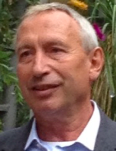 | Keynote Presentation Circulating Tumor Cells to Guide Treatment of Cancer Patients
Leon Terstappen, Chair Medical CellBiophysics, MIRA Research Institute for Biomedical Technology and Technical Medicine, University of Twente, Netherlands
The presence of Circulating Tumor Cells (CTCs) enumerated with the CellSearch system is associated with relatively poor survival and their reduction after the first weeks of therapy indicates a response to therapy. Present research focus is on the ability to extract treatment relevant information from the proteins, RNA and DNA in these CTC. |
| 18:00 | Networking Reception with Beer and Wine. | 19:00 | Close of Day 1 of the Conference. |
Thursday, 25 October 201807:00 | Morning Coffee, Breakfast Pastries and Networking | 07:30 |  | Keynote Presentation Breakfast Lecture: Multiplex Real-Time PCR Assays that Assess the Abundance of Rare Somatic Mutations Associated with Cancer
Fred Kramer, Professor of Microbiology, Biochemistry & Molecular Genetics, New Jersey Medical School Rutgers University, United States of America
“SuperSelective” PCR primers, due to their unique design, exponentially amplify rare mutant DNA fragments present in a sample, while ignoring abundant related wild-type DNA fragments, even if the only difference between the mutant and the wild-type is a single-nucleotide polymorphism. Different target mutations whose presence suggests the same therapeutic action can be detected with molecular beacons bearing the same colored fluorophore. Differently colored molecular beacons are simultaneously present in these assays to determine which therapeutic action should be taken and which therapeutic action should be discontinued. These multiplex, homogeneous assays are sensitive, rapid, inexpensive, utilize existing PCR instruments, and can be carried out frequently during the course of treatment utilizing non-invasive liquid biopsy samples. |
| |
Session Title: Deploying Exosomes, CTCs, and Circulating Nucleic Acids in Cancer, Cardiovascular Disease and CNS Diseases |
| | 08:30 | Exploiting Extracellular Nucleic Acids From Body Fluids For Precision Medicine Purposes
Jo Vandesompele, Professor, Ghent University and CSO, Biogazelle, Belgium
In contrast to general belief, a substantial part of the human transcriptome is abundantly present in the blood and other body fluids as extracellular mRNAs, lncRNAs and microRNAs, ready to exploited. I will discuss various workflows for RNA sequencing of body fluid derived RNA, including probe-based target capture as a sensitive RNA sequencing workflow to study thousands of mRNA and lncRNA genes in cell-free RNA from patients’ plasma and urine. Apart from RNA abundance profiling, this type of data can also be used to detect structural RNA variants, such as somatic mutations, fusion genes and RNA editing events, all known to play an important role in disease, including cancer. The resulting RNA profiles can be deconvoluted to enumerate the cells, tissues and organs that contribute to the extracellular RNA. Human body fluid RNA sequencing enables liquid biopsy guided precision oncology, such as therapy stratification, treatment response monitoring and early detection of relapse. I will also discuss the pre-analytical jungle of RNA targeted liquid biopsies and need for standardization, as part of the ongoing exRNAQC study. | 09:00 | | Keynote Presentation Vesicles For Ischemic Heart Disease
Dominique PV de Kleijn, Professor Experimental Vascular Surgery, Professor Netherlands Heart Institute, University Medical Center Utrecht, The Netherlands, Netherlands
Cardiovascular Disease (CVD) is with the cardiovascular events of Ischemic Heart Disease and Stroke, the number 1 and 2 cause of death in the world and expect to increase especially in Asia. Ischemic heart disease (IHD) comprises 3 entities: stable coronary artery disease (SCAD), unstable angina (UA) and myocardial infarction (MI). Because IHD is associated with an increased risk of adverse clinical events such as heart failure and death, early recognition of IHD is of utmost importance. However, to diagnosis IHD is challenging, as many patients present with atypical symptoms. It is known that women have a different symptom sensation than men. Troponins are the main diagnostic tool for detection of MI. Blood biomarkers for SCAD (typically causing stable angina) and UA, however, are not available. These diagnoses frequently require hospital visits/admissions for time-consuming and costly (non)invasive tests. We use plasma extracellular vesicle protein content of vesicles from plasma subfractions as an accurate source for early diagnosis of SCAD and UA. Diagnosis of ischemic heart disease as well as normalization and characterization of extracellular vesicles in plasma subfractions will be discussed. |
| 09:30 | Accuracy, Reproducibility and Consequences of Bias in Quantitative Small RNA-seq For Circulating Cell-free RNA Profiling
Maria Giraldez, Researcher, University of Michigan, United States of America
Small RNA-seq is increasingly being used for profiling of small RNAs, including cell-free RNA such as microRNAs present in plasma and other biofluids. In practice, small RNAseq can involve many different methods for library preparation, with distinct protocols and technical variations that could cause substantial differences in results, yet these have not been fully and systematically studied. We report here the results of a study using common references (synthetic RNA pools of defined composition, as well as plasma-derived RNA) to evaluate the accuracy, reproducibility and bias of small RNA-seq library preparation for five distinct protocols and across nine different laboratories. We observed protocol-specific and sequence-specific bias, which was ameliorated using adapters with randomized end-nucleotides for ligation. Despite this technical bias, relative quantification using small RNA-seq was remarkably accurate and reproducible, even across multiple laboratories using different methods. These results provide a foundation for reproducible, cross-laboratory small RNAseq studies of cell-free, circulating RNA in a variety of clinical contexts. | 10:00 |  MicroRNA Based Liquid Biopsy: New Extraction, Preservation and Detection Technologies to Generate miRNA Signatures for Cancer Screening MicroRNA Based Liquid Biopsy: New Extraction, Preservation and Detection Technologies to Generate miRNA Signatures for Cancer Screening
Pablo Castan, Research Director, Arcis Biotechnology
The development of minimally-invasive tests, such as liquid biopsy, for early cancer detection in its preclinical phase is crucial to improve the outcome of oncologic patients. MicroRNAs (miRNAs) are small, non-coding and tissue-specific-RNAs, which have been reported as extracellular messengers derived from the cross-talk between tumours and their surrounding microenvironment. The development of easy (de-skilled and cost-effective) extraction and preservation solutions teamed with fast and reliable detection could thus represent the ideal format for early detection of defined types of cancer. In this presentation, a new methodological workflow for the prospective validation of a circulating miRNA test using custom made microfluidic cards and microsensing technology in plasma is discussed and compared to state of the art solutions currently used for liquid biopsy.
| 10:30 | Interpreting Extracellular Vesicles as Snapshots of Cells
Esther Nolte-‘t Hoen, Assistant Professor, Department of Biochemistry & Cell Biology, Faculty of Veterinary Medicine, Utrecht University, Netherlands
Our research focuses on the role of EV in communication between cells at steady state and during immune activation or microbial infection. Using high-resolution flow cytometric analysis of single EV, we previously showed that EV populations are highly heterogeneous. Our recent work on EV released by immune cells and virus-infected cells indicates that both spatial and temporal elements drive EV heterogeneity. This has implications for interpreting EV as snapshots of cells. Recognition and in-depth analysis of EV heterogeneity can facilitate the search for refined EV-based biomarkers. Moreover it can help to dissect differences in the formation and function of distinct EV populations. Examples will be shown on how environmental triggers can lead to changes in the type of released EV and their small non-coding RNA content. Additionally, it will be explained how advanced flow cytometry-based technologies can be applied to study EV release dynamics and to isolate EV subpopulations for downstream functional analysis. | 11:00 | How Clinical Biobanks Support Precision Medicine: From Standardized Preprocessing of Liquid Biopsies to Treatment Guidance
Jens Habermann, Head, Section of Translational Surgical Oncology and Biobanking & Scientific Director, Surgical Center for Translational Oncology, Department of Surgery & Institute of Human Genetics, University of Lübeck and University Hospital Schleswig-Holstein, Campus Lübeck, Germany
CTCs and Exosomes harbor an enormous potential for precision medicine. However, clinical implementation requires standardization. This is in contrast to current praxis, which is still hampered by major inter-study heterogeneity. This talk will address (i) current challenges and solutions for standardized CTC / Exosome assessment and (ii) how clinical biobanks can support CTC / Exosome research for precision medicine, e.g. in colorectal cancer. | 11:30 | Boosting Clinical Validation of ctDNA Diagnostics
Gerrit Meijer, Professor of Oncologic Pathology, Netherlands Cancer Institute, Netherlands
| 12:00 | Networking Lunch in the Exhibit Hall and Poster Viewing | 13:00 | Molecular and Functional Characterization of CTCs in Non-Small Cell Lung Cancer (NSCLC)
Francoise Farace, Head of Rare Circulating Cells Translational Laboratory, INSERM Gustave Roussy Institute, France
The presentation will focus on the liquid biopsy potential of CTCs to analyze resistance mutations to tyrosine kinase inhibitors in ALK-rearranged and EGFR-mutant patients. Our work on NSCLC CDX (CTC-derived xenograft) will be presented and discussed. | 13:30 |  | Keynote Presentation Exosomal Integrins: Biomarkers for Liquid Biopsy
Lucia Languino, Professor of Cancer Biology, Thomas Jefferson University, United States of America
Cancer Extracellular Vesicles (EVs) found in blood are heterogeneous and show unique protein compositions. Our analysis shows that a prostate cancer EV subset, designated exosomes, carries a unique repertoire of integrins, which are surface receptors for extracellular matrix. Using iodixanol gradients, immunoblotting and size measurements we show that integrins are differentially expressed in cancer exosomes. Our recent studies demonstrate that exosomes from prostate cancer cells contribute to horizontal propagation of unique integrin-associated invasive phenotypes. We also show that exosomal integrins are transferred to cells in the tumor microenvironment and mediate response to therapy. This presentation will discuss the role of exosomal integrins in cancer progression and therapy response, and their potential use as clinical biomarkers for non-invasive diagnosis of cancer. |
| 14:00 | Single-Cell Transcriptome Analysis For Circulating Tumor Cells based on Microcavity Array
Tomoko Yoshino, Professor, Tokyo University of Agriculture and Technology, Japan
Circulating tumor cells (CTCs) are one of the powerful biomarkers for cancer diagnosis and potential targets for drug discovery. Genetic analysis of CTCs could provide detailed information of cancer patient. However, the recovery of such extremely rare cells from the peripheral blood and the achievement of genetic analysis without loss of CTCs presents a technical challenge. In this presentation, I will introduce several techniques towards single-cell transcriptome analysis for CTCs. Filtration using metal microfilters (microcavity array: MCA) has been used to develop precise CTC recovery technique. Furthermore, recovered cells on the MCA is easily manipulated by encapsulation by photopolymerizable hydrogel using multiple light projection system, resulting in simply manipulate multiple CTCs at a single cell level. The cells were applied RNA-seq analysis, and we investigated single cell gene expression analysis to reveal the cancer-specific transcripts. We believe our system have a potential to apply high-throughput single-CTC analysis and contribute for development of non-invasive cancer diagnosis method. | 14:30 | Liquid Biopsy in Colon Cancer
Pierre Laurent, Professor, Universite Paris Descartes, France
RAS mutations are currently sought for in tumor samples, which takes a median of almost 3 weeks in western European countries. This creates problems in clinical situations that require urgent treatment and for inclusion in therapeutic trials that need RAS status for randomization. Analysis of circulating tumor DNA might help to shorten the time required to determine RAS mutational status prior to anti-EGFR antibody therapy for metastatic colorectal cancer. Here we compared plasma versus tissue RAS analysis in a large prospective multicenter cohort. | 15:00 | Droplet-Based Digital PCR Procedures and Optimized NGS to Track Circulating Tumor DNA in Liquid Biopsies: Application to Cancer Patient Follow-Up
Valérie Taly, CNRS Research Director, Professor and Group leader Translational Research and Microfluidics, Université Paris Cité, France
Tracking of circulating cell-free nucleic acids in body effluents has been greatly facilitated by recent technological developments including droplet-based digital PCR and optimized NGS. In particular, droplet based digital PCR has allowed to reach unprecedented sensitivity and accuracy for rare sequences detection. Complementarity of these technologies will be presented through several retrospective and prospective studies. Moreover, we will present their application for the detection of circulating tumor DNA and its potential use for cancer patient treatment management. | 15:30 | Afternoon Coffee Break | 16:00 | Approaches For Validation of Liquid Biopsy Testing of Circulating Tumour DNA
Alison Devonshire, Science Leader, LGC Limited, United Kingdom
The typically low concentration of ctDNA in cancer patients’ samples makes validation of analytical sensitivity a critical element in the assessment of a diagnostic test. The potential for real-time monitoring of tumor dynamics through liquid biopsy testing also necessitates a systematic evaluation of quantitative accuracy. This presentation will discuss approaches to validate the performance of targeted methods for cancer mutations, including pre-analytical controls, and the development of reference materials and reference measurement procedures, which also support longer-term comparability and standardization of patient testing. | 16:30 |  | Keynote Presentation Circulating Biomarkers in the Management of Solid Tumors
Minetta Liu, Associate Professor and Chair, Oncology Research, Mayo Clinic, United States of America
The real-time, serial detection of tumor specific molecular alterations will facilitate improved clinical outcomes through individualized treatment approaches. The advent of technologies that allow for the isolation and characterization of circulating tumor cells (CTCs) and cell free nucleic acids (cfNA) in the peripheral blood has led to great interest in the “liquid biopsy”. These assays offer a less invasive, potentially more cost effective tool to assess prognosis and response to treatment, and they may facilitate the early diagnosis of recurrence or primary malignancy itself. The challenges of validating these molecular biomarkers and the need to provide solutions to promote rapid translation into clinical practice will be discussed. |
| 17:00 | Successful Implantation: How Small Molecule Dynamics Play a Big Role in Cell-Cell Communication
Yael Heifetz, Principal Investigator, The Hebrew University of Jerusalem, Israel
Embryo-maternal communication is crucial for achieving successful pregnancy and for the future health of the progeny. A relay of molecular signals between mother and gametes is critical as soon as after copulation, during gamete transport in the female reproductive tract, gamete maturation, and fertilization. The next step, the process of embryo implantation, is a major obstacle for successful maintenance of pregnancy to term. For implantation to succeed, a synchronized network of molecular interactions between mother and embryo must occur. Although key molecules have been identified, we still lack comprehensive understanding of the early dynamics of implantation which set-up the stage for a successful pregnancy. To examine the early sequence of events that initiate the embryo-maternal cross-talk we used in-vitro (human endometrial and trophoblast cell lines) and in-vivo (domestic pig Susscrofa domestica) models, and various molecular and imaging approaches to monitor the spatial and temporal dynamics of intercellular interactions. This talk will focus on extracellular vesicles (EVs), which have been suggested to act as essential mediators of embryo-maternal communication. Studying the dynamics of the cross-talk, and specifically signals that promote successful implantation at an early stage, will shed more light on the implantation process and possibly guide new strategies in diagnosing and preventing implantation failure. | 17:30 | Panel Discussion: Emerging Themes and Trends in Liquid Biopsy Development
| 18:30 | Close of Day 2 of the Conference. |
Friday, 26 October 201807:30 | Morning Coffee, Breakfast Pastries and Networking | |
Session Title: Exosomes, Microvesicles and Extracellular Vesicles (EVs) -- Research, Diagnostic and Therapeutic Opportunities as Well as Synergy with Other Circulating Biomarker Classes |
| | 08:00 | Improving the Detection of Circulating Tumour DNA in Plasma by Selecting Short DNA Fragments
Florent Mouliere, Researcher, Nitzan Rosenfeld Group, University of Cambridge, United Kingdom
The sensitivity for detecting the presence of genomic changes in circulating tumour DNA (ctDNA) is limited by its low concentration in plasma. Here I will present new approaches tailoring sequencing to the biological properties of ctDNA for improving the detection of rare mutant molecules. In particular, we studied the feasibility for enrichment of ctDNA by physical and in-silico size selection in plasma samples collected from multiple cancer types. Size selection of short DNA fragments enriches for ctDNA, unlocking un-targeted genome-wide sequencing for liquid biopsy. | 08:30 | EV-Associated Biomarker Discovery in Liquid Biopsies
An Hendrix, Professor, Ghent University, Belgium
An Hendrix will discuss the impact of pre-analytical factors and isolation methods on the identification of extracellular vesicle-associated biomarkers in liquid biopsies. She will present the development and implementation of reference materials for extracellular vesicles and how this can help to compare and normalize results in research and clinical setting. | 09:00 | Technology Spotlight Presentation: Imaging Flow Cytometry Enhances the Detection and Quantification of Rare Events Such as Circulating Tumor Cells and Minimal Residual Disease
Peter Rhein, Instrument Application Scientist, Merck KGaA, Germany
Using the ImageStream®, we can combine flow cytometric and microscopic assays such as immunophenotyping and fluorescence in situ hybridization (FISH), which contributes to significantly improve the detection and functional analysis biological rare events. | 09:30 |  Biomarker detection using Particle Metrix ZetaView® and Nanoparticle Tracking Analysis (NTA) Biomarker detection using Particle Metrix ZetaView® and Nanoparticle Tracking Analysis (NTA)
Sascha Raschke, Technical and Scientific Support, Particle Metrix GmbH
By using the ZetaView® NTA instrument Extracellular Vesicles (EVs) and other biological nanoparticles in a heterogeneous sample can be individually analyzed in terms of their size, number and concentration. Furthermore, they can be phenotypically characterized using fluorescence-labeled antibodies or intercalating membrane dyes. Two different fluorescence channels of the ZetaView® machine allow analysis of two different fluorescent labels in rapid succession with just one sample, eliminating the time and effort required to produce a second fluorescently labeled sample.
| 10:00 | Coffee Break in the Exhibit Hall and Poster Viewing | 10:30 |  | Keynote Presentation Exploiting the Unique Protein Distribution in Different Plasma EV Types For Biomarker Discovery
Sai Kiang Lim, Research Director, Institute of Medical Biology, A*STAR, Singapore
Intercellular communication via secreted lipid membrane vesicles or extracellular vesicles (EVs) is an emerging research area. Many cell types are known to secrete EVs and these EVs are heterogenous in size, density, composition and biogenesis pathway. EVs in the size range of 100-200 nm are among the most intensely studied EVs, and are frequently referred to as exosomes which are EVs derived from endosomes. Recently, we demonstrated by pulse chase experiments that Mesenchymal Stem Cell (MSC) exosomes can be identified by their high affinity for cholera toxin B chain (CTB) which binds GM1 gangliosides. MSC was also observed to secrete at least two other EV types that can be distinguished by their affinity for lipid-binding proteins namely, AnnexinV (AV) that binds phosphatidylserine and Shiga toxin B chain (STB) that binds globotriasosylceramide. These three EV types have different protein and RNA cargo. Several other cell types were also observed to produce CTB-, AV- and ST-binding EVs (CTB-EVs, AV-EVs and ST-EVs). Evaluation to date suggest that these different EV types are unique, and carry different protein and RNA cargos. We have since demonstrated that plasma also have CTB-EVs, AV-EVs and ST-EVs. Hence plasma biomarkers can now be analyzed in four permutations: plasma, plasma CTB-EVs plasma AV-EVs or plasma ST-EVs. The distribution of some plasma proteins in these four permutations is different among individuals, and could be associated with diseases. As such, plasma CTB-EV and AV-EV could be sources of novel biomarkers or novel permutations of known biomarkers. Here I will describe using this approach to discover three biomarkers in plasma, plasma CTB-EV and plasma AV-EV of 28-32 weeks pregnant women that could predict development of PE at an average of seven weeks later. This study was done using a prospective biobank of 843 low risk patients with a 2.1% PE risk. At 100% sensitivity, the three biomarkers have a combined AUC of 0.96, 78.6% specificity and a PPV of 9.9%. I will also present the discovery of ovarian cancer biomarker candidates using similar approaches. |
| 11:00 |  | Keynote Presentation Cancer-derived Extracellular Vesicles and Tumor Microenvironment
Giovanni Camussi, Professor, University of Torino, Italy
This presentation will cover the role of extracellular vesicles in cell-to-cell communication. It will also address the correlation between the molecular composition of extracellular vesicles and their biological activity. The ability of tumor and cancer stem cell derived extracellular vesicles to induce epigenetic changes in tumor micro-environmental cells, to inhibit the differentiation process of DCs and to favor the formation of a pre-metastatic niche will also be covered.
|
| 11:30 |  Multilevel Analysis of Individual EVs, Direct From Sample With Minimal Purification Multilevel Analysis of Individual EVs, Direct From Sample With Minimal Purification
Andrew Malloy, VP of Sales and Marketing, NanoView Biosciences
Current analytical tools have detection limitations when used for exosome characterization due to their small size. Specifically, exosomes have fewer surface epitopes available and low-signal relative to background levels, especially in complex samples. Current workflows require multiple measurement technologies that result in the inability to link phenotypic and physical characterization on individual EVs across a biologically relevant size range. In addition, sample purification often results in loss of sample and technique dependent biases.
NanoView Biosciences has developed an imaging technique that allows individual EVs as small as 40nm to be characterized direct from sample with no purification in most cases. Physical characteristics such as size and EV count can be presented alongside phenotypic information at the single EV level. Low volumes of sample can be probed and a panel of up to 10 surface markers can be interrogated in parallel, in a single measurement. Lastly, through the addition of fluorescent labeling, three additional markers can be probed and colocalized across the existing panel of 10 markers. These capabilities make the tool ideally for basic EV research, biomarker discovery and the development of EV based liquid biopsies and therapeutics.
| 12:00 | Exosome-derived Epigenetic Biomarkers For Saliva Diagnostics
Christa Noehammer, Senior Scientist, Austrian Institute of Technology, Austria
The aim of our research activities at AIT, the Austrian Institute of Technology, is to define reliable biomarkers suitable for early and non-invasive disease diagnosis and prognosis. To this end we have been establishing and optimizing a whole range of multiplex capability technologies (e.g. microarrays, quantitative PCR, Luminex bead technology) to meet the special demands and challenges of diagnostic biomarker discovery - and validation in body fluids. Using this specific technology expertise we e.g. successfully discovered autoantibody- as well as DNA methylation -based diagnostic marker panels for the big 4 cancer entities (breast, colon, prostate, lung) in serum or plasma. Based on these success stories and the evident advantages of saliva as a diagnostic matrix our recent special interest is to go for saliva diagnostics and to evaluate saliva for its suitability for circulating biomarker-based diagnostics. Along these lines we will report on the evaluation of different commercially available strategies for isolation of exosomes from human serum and saliva. We will further present data from genome-wide microRNA – as well as DNA-methylation profiling in salivary - and serum-derived exosomes which will also include our experiences when comparing the performance of high-density microarrays and next generation sequencing for miRNA profiling. Last but not least we will report on first salivary and plasma exosome-derived epigenetic biomarkers for type 2 diabetes diagnosis. | 12:30 | Lunch and Networking in the Exhibit Hall | 13:15 |  Luncheon Tech Spotlight: Seeing is Believing - Meet the Nanoimager Luncheon Tech Spotlight: Seeing is Believing - Meet the Nanoimager
Katarzyna Ginda-Mäkelä, Application Specialist, Oxford Nanoimaging (ONI)
ONI crafts desktop-compatible microscopes with miniaturized Nobel Prize winning technology capable of detecting individual molecules using fluorescence. This exquisite sensitivity is used to create images and videos of biological samples in an unprecedented fashion, opening a whole new world of discovery, leading to a better understanding of the fate and genesis of exosomes and a potential development of novel therapeutic tools. Our team is a blend of life science and microscopy experts, software and mechanical engineers as well as data scientists.
| 13:30 |  | Keynote Presentation Stem Cell-Derived Extracellular Vesicles (EVs) as Therapeutics For Extending Healthspan
Paul Robbins, Professor, Department of Biochemistry, Molecular Biology, and Biophysics, and the Institute on the Biology of Aging and Metabolism, University of Minnesota Medical School, United States of America
A universal characteristic of aging is loss of tissue regenerative potential due to stem cell dysfunction, contributing to the dramatic increase in the risk of morbidity and mortality. We have used both naturally aged mice and mouse models of accelerated aging to demonstrate that transplantation of young, functional adult stem cells results in an extension of healthspan. In addition, we have demonstrated that stem cell-derived EVs are a key factor for conferring the observed extension of healthspan by young stem cells. Progress towards developing clinically-relevant approaches using stem cell-derived extracellular vesicles to age-related pathologies will be presented. In addition, progress towards using circulating EVs as biomarkers of biological age will be presented. |
| 14:00 |  | Keynote Presentation Engineered Extracellular Vesicles for CRISPR/Cas9 Gene Editing
Niek Dekker, Principal Scientist, Discovery Sciences, AstraZeneca, Sweden
Extracellular vesicles (EVs) represent an exciting opportunity as biological delivery vehicles for therapeutic cargo with excellent safety, low intrinsic immunogenicity, cell-specific tropism and biological delivery efficiency. There are multiple approaches for the introduction of protein and RNA cargo into EVs, including physical, chemical and cell engineering. Application of CRISPR/Cas9 requires the functional delivery of the ribonucleoprotein (RNP) complex of Cas9 enzyme and guide RNA. Engineering of the production cell line can be used to introduce cargo into the EVs, and examples for the introduction of fluorescent and luminescent proteins will be disclosed. Uptake in recipient cells can be monitored using high content imaging to provide insight in kinetics and localization. Functional delivery can be analyzed using several assays for the gene editing event (eg Cel1 and TIDE). The use of EVs for delivery of CRISPR/Cas9 has a range of exciting applications for ex vivo manipulation of primary cells and in vivo editing of genes in diseased tissue. |
| 14:30 | Challenges in the Detection, Isolation and Quantification of Extracellular Vesicles and Exosomes
Alain Brisson, Professor Emeritus, University of Bordeaux, France
The emerging field of extracellular vesicle (EV) and exosome research faces a notorious lack of reliable characterization methods. This is mainly due to the small size of EV/exosomes (mostly from 30 to 500 nm) and the heterogeneity of their suspensions. This presentation will illustrate the advantages of novel methods allowing to image, phenotype, quantify and isolate EVs in complex fluids. | 15:00 | Production of Functionally Bioactive EVs from an Immortalized Human Neural Stem Cell (hNSC) Line
Randolph Corteling, Head of Research, ReNeuron Ltd., United Kingdom
To ensure the scale required for clinical research and development, producer cell immortalization and clonal isolation is a practical strategy to produce consistent, functionally bioactive exosomes for use as therapeutic agents. | 15:30 | Afternoon Coffee Break | 16:00 | Circulating Microvesicles – Biomarkers and Effectors and Cardiovascular Health and Disease
Felix Jansen, Leader of the Microvesicle Research Group, Department for Cardiology, Pulmology and Angiology, University Hospital Bonn, Germany
My talk will summarize the diagnostic potential and current obstacles in using circulating MVs as biomarkers in CVD. Furthermore, I will highlight the regulatory role of MV in the progression of atherosclerosis. | 16:30 | Liquid Biopsy for Breast Cancer Biomarkers Discovery
Clotilde Costa, Translational Medical Oncology Group, Joint Unit Roche-Chus, Oncomet, Universitary Cinical Hospital of Santiago de Compostela, Spain
Metastasis is responsible for 90% of cancer related deaths, thus, biomarkers discovery to predict and monitor tumor progression is an urgent need in oncology. Liquid Biopsy analysis permit to obtain tumoral information in real time, in a non-invasive way and in a systemic manner. Thus, molecular characterization of circulating material of metastatic breast cancer patients in different time points of the disease (before and after treatment) had led us to identify biomarkers with interest for clinical oncology translation. | 17:00 | Pre-Clinical Test Requirements For EVs Drugs
Marcin Jurga, R&D Director, The Cell Factory bvba, Netherlands
Extracellular vesicles (EVs) are promising drug candidates and drug delivery tools in treatment of multiple diseases. Currently, one of the most popular source of EVs are mesenchymal stromal cells (MSCs) with a broad spectrum of anti-inflammatory and immunosuppressive activity. Translation of the EVs drug candidates into clinic requires well-established production system and effective quality control procedures. The presentation will give an overview of the clinical-grade production process of the EVs drugs and will describe the quality control and activity assays developed and validated for clinical translation of the EVs drugs. | 17:30 | Relevance of EV Heterogeneity For Their Application in Liquid Biopsy
Irina Nazarenko, Exosome and Tumor Biology Group Leader, University of Freiburg, Germany
The need to develop better approaches allowing comprehensive molecular characterization of a progressing tumor has led to the development of the “Liquid Biopsy” concept, which is based on the application of body fluids as a biomarker source. In view on the complexity of blood and availability of components of different origins, including circulating tumor cells, free-circulating nucleic acids and Extracellular Vesicles (EVs), a question of a specific impact of each of these different compartments on the outcome of liquid biopsy-based diagnostic may arise. We address this question comparing amounts of mutated oncogenes in different EV subtypes and in the free-circulating fractions in blood of cancer patients. Our results show that different tumor types at different stages may contain varying portions of potential biomarkers in the free circulating fraction and in different EV subtypes, indicating a potential relevance of a comparative analysis of different blood compartments prior the choice of the most appropriate source and technique for the liquid biopsy-based diagnostic. | 18:00 | Glioblastoma Extracellular Vesicles As a Tool To Investigate The Hijacking of Multiple Microglial Functions To Promote Tumor Growth
Niek Maas, Clinical Researcher, University Medical Center Utrecht, Netherlands
This presentation will focus on the role of extracellular vesicles (EVs) in the microenvironment of glioblastoma malignant brain tumors. We will present data on EV associated modification of microglia and macrophages in vivo. | 18:30 | Close of Conference. |
|


 Add to Calendar ▼2018-10-24 00:00:002018-10-26 00:00:00Europe/LondonExosomes and Liquid Biopsies Europe 2018Exosomes and Liquid Biopsies Europe 2018 in Rotterdam, The NetherlandsRotterdam, The NetherlandsSELECTBIOenquiries@selectbiosciences.com