Monday, 9 September 201908:00 | Conference Registration, Materials Pick-Up, Morning Coffee and Tea | 08:30 | 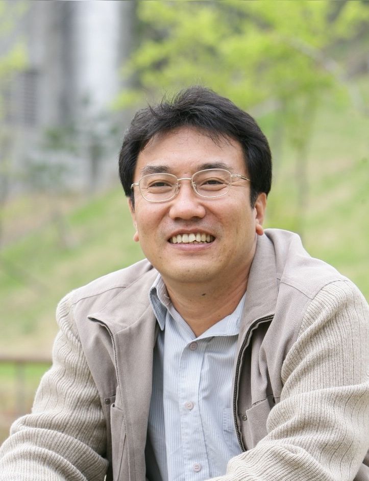 | Conference Chair Chairman's Welcome and Introduction to the Conference
Yong Song Gho, Professor, Pohang University of Science And Technology (POSTECH), Korea South
|
| 08:45 |  | Conference Chair Chairperson's Welcome and Introduction to the Conference
Chulhee Choi, Professor and Chair, BioMedical Imaging Center, Korea Advanced Institute of Science and Technology (KAIST), President, ILIAS Biologics Incorporated, Korea South
|
| |
Session Title: Conference Opening Session -- Emerging Trends and Themes in the Extracellular Vesicles Field, circa 2019 |
| | 09:00 |  | Keynote Presentation Emerging Applications of Extracellular Vesicles in Medicine
Jan Lötvall, Professor, University of Gothenburg; Founding President of ISEV; Editor-in-Chief, Journal of Extracellular Vesicles, Sweden
Extracellular vesicles (EVs) are released by all cells in the human body, and can reflect the status and function of the releasing cell. Further, EVs can shuttle cargo molecules from one cell to another, including proteins and RNA. The human body is exceptionally rich in EVs, and it has over the last ten years been acknowledge that the phenotype of these vesicles change in disease, and that EVs indeed can be utilized for diagnostic purposes, for example in cancer. Further, the functionality of EVs can in multiple ways be harnessed for the development of future biological medications. This presentation will discuss the diversity of EVs isolated from human tissues, and will also touch upon the opportunities of developing EVs as powerful therapeutics. |
| 09:30 |  | Keynote Presentation Plasma Extracellular Vesicles as a Biomarker Source for Cardiovascular Disease
Dominique PV de Kleijn, Professor Experimental Vascular Surgery, Professor Netherlands Heart Institute, University Medical Center Utrecht, The Netherlands, Netherlands
Cardiovascular Disease (CVD) is with cardiovascular events of Ischemic Heart Disease (IHD) and Stroke, the number 1 and 2 cause of death in the world and expect to increase especially in Asia. We use protein signatures measured in 3 different subsets of plasma extracellular vesicle (EV) as an accurate source for early diagnosis and prediction of cardiovascular events like IHD and Stroke.
The diagnosis IHD is challenging, as many patients present with atypical symptoms. It is known that women have a different symptom sensation than men. Troponins are the main diagnostic tool for detection of MI. Blood biomarkers for stable angina (SA) and unstable angina (UA), however, are not available. These diagnoses frequently require hospital visits/admissions for time-consuming and costly (non)invasive tests. Using a simplified method that is suitable for automation and microfluidics to isolate the plasma EV subfractions in 25 ul plasma, we now validated our data in a large case control study (150 cases vs 300 controls) that showed that EV subfractions can diagnose accurately SA in a drop of blood. Next to this, using another signature in plasma EV subfractions for patient that undergo a carotid endatherectomy (CEA) we could accurately determine which patient will get a second cardiovascular event (myocardial infarction, stroke or death) or not within 3 years after CEA.
Plasma EV subfractions are a very valuable biomarker source for the diagnosis and prognosis of cardiovascular disease. |
| 10:00 |  | Keynote Presentation Immunomodulatory MSC Exosome and Small Extracellular Vesicles
Sai Kiang Lim, Research Director, Institute of Medical Biology, A*STAR, Singapore
Mesenchymal stromal cell (MSC) are the most clinically tested cell type for regenerative medicine. Despite much setbacks, MSC is now gaining traction as a safe. viable regenerative medicine with recent market approval for use in GVHD and Crohn’s disease. However, it is increasingly clear that small extracellular vesicles (sEVs) of 50 to 200 nm diameter such as exosomes mediate the therapeutic potency of MSCs. Since the first demonstration that MSC secretes cardioprotective sEVs in a mouse and pig models of myocardial ischemia/reperfusion injury, these sEVs were widely reported to therapeutically efficacious against at least 30 other indications. A common observation in many MSC sEV-mediated efficacies is a significant immunomodulatory effect. This talk will describe our studies on the immunomodulatory potential of MSC sEVs. |
| 10:30 | Morning Coffee and Tea Break and Networking in the Exhibit Hall | 11:15 |  | Keynote Presentation Microfluidics Technologies for Cancer Diagnosis - From Cell Capture to Single Cell Analysis
Chwee Teck Lim, NUS Society Chair Professor, Department of Biomedical Engineering, Institute for Health Innovation & Technology (iHealthtech), Mechanobiology Institute, National University of Singapore, Singapore
The presence of Circulating Tumor Cells (CTCs) in bloodstream of patients is an important intermediate step in cancer metastasis and can provide valuable insights into disease detection and personalized treatment. As compared to obtaining a tissue biopsy which is invasive and painful, ‘‘liquid biopsy’’ for CTCs detection can be easily performed via a routine blood draw. The presence and number of CTCs in peripheral blood has been associated with the severity of the disease and have potential uses for early detection, diagnosis, prognosis and treatment monitoring. However, the presence of the large number of blood cells complicates as well as makes detection of rare CTCs in blood of cancer patients extremely challenging. Here, we address these issues and demonstrate that physical biomarkers such as the unique size and deformability of CTCs can be effectively used for their detection and separation from blood via microfluidics. We do this by leveraging on the many inherent advantages of microfluidics such as high sensitivity and spatial resolution, short processing time and low device cost. We developed a suite of microfluidic biochips that exploit the principles of cell size/deformability based separation as well as inertial focusing to perform high throughput continuous detection and separation of diseased cells. These simple, efficient and cost effective microfluidic platforms will be invaluable in extracting viable CTCs for many downstream clinical applications including single cell analysis as well as personalized treatment. |
| 11:45 |  | Keynote Presentation Innovative Microfluidic Isolation of Circulating Biomarkers: cfDNA and Exosomes
Sehyun Shin, Professor & Director, Nano-Biofluignostic Engineering Research Center, Korea University and Anam/Guro Hospital of Korea University, Korea South
Liquid biopsy, non-invasively analyzing biomarkers circulating in various body fluids, has been emphasized as a promising technology to accelerate the realization of personalized treatment of cancer. Among the circulating biomarkers, cell-free DNA (cfDNA) and exosome have been implicated as important biomarkers in cancer management. Efficient techniques for isolation of such biomarkers from blood are prerequisites for precision liquid biopsy. Also, a sample-to-answer system for such biomarker extraction is highly required for clinical application. Thus, in the present study, two of technologies (PIBEX and PLL clustering) to isolate main circulating biomarkers (cfDNA and exosome) of liquid biopsy are presented. First, the PIBEX system is a centrifugation-free cfDNA extraction microfluidic chip capable of extracting cfDNA from plasma samples through microfluidic circuits within 15 min under vacuum pressure using an immiscible solvent. Second, the PLL clustering method is the microfluidic chip capable of isolating exosomes from plasma samples within an hour under vacuum pressure using a cationic polymer solution. These chips effectively eliminates the repetitive centrifugation processes and dramatically shortened the sample preparation time. The proposed microfluidic platform could facilitate the development of a sample-to-answer system for use in liquid biopsy of cancers. |
| 12:15 |  | Keynote Presentation Identification of Different Subpopulations of Circulating Tumor Cells in the Blood of Localized and Metastatic Cancer Patients Using Microfluidics
Steve Soper, Foundation Distinguished Professor, Director, Center of BioModular Multi-Scale System for Precision Medicine, The University of Kansas, United States of America
Liquid biopsies are becoming an attractive source of biomarkers that can be used to manage a variety of cancer-related diseases. One of the principle biomarkers for oncology-related diseases found in blood is circulating tumor cells (CTCs). The challenge associated with using CTCs as biomarkers for many cancer-related diseases has been the modest clinical sensitivity demonstrated using the FDA-approved platform. CTCs expressing invasive phenotypes down-regulate epithelial antigens, such as the epithelial cell adhesion molecule – EpCAM, which is typically used for the affinity selection of CTCs. As such, CTCs may have a continuum of phenotypes and thus, a single selection marker may not address all cells comprising the tumor microenvironment. We have developed a CTC selection strategy that employs two polymeric microfluidic chips modified with antibodies and connected in series with each selecting a distinct CTC subpopulation from a single blood sample. In addition to the common marker used for CTC positive selection (EpCAM), Fibroblast Activation Protein alpha (FAPa) expressing CTCs can also be selected. Using the dual selection strategy, both CTC types were detected from patients with clinical sensitivity that showed significant improvement compared to selection in which only EpCAM was used. Approximately 90% of the selected CTCs were found not to co-express both antigens. Due to the high purity (>80%) and clinical yields (>95% recovery) of the dual selection strategy, molecular analysis of both EpCAM and FAP-alpha CTCs could be carried out including next generation sequencing, and droplet digital PCR. Our results suggest FAP-alpha and EpCAM CTCs can be used in concert to guide therapeutic decisions for cancer patients. In this presentation, I will discuss the microfluidic chips we use for the selection of CTCs, the clinical results secured using these chips, and the molecular profiling of both CTC subpopulations. |
| 12:45 | Networking Lunch, Meet with Exhibitors and View Posters | |
Session Title: Biomarker and Diagnostic Potential of Extracellular Vesicles (EVs) |
| | 14:00 |  Multiparameter Phenotyping and Characterization of EVs - Direct from Complex Biofluids With No Purification Multiparameter Phenotyping and Characterization of EVs - Direct from Complex Biofluids With No Purification
Andrew Malloy, VP of Sales and Marketing, NanoView Biosciences
When studying EVs in a diagnostic capacity, understanding the nature and role of EV sub-populations is of paramount importance. Existing technologies either struggle to measure EVs due to their small size and/or are unable to link proteomic information to physical characterization at the single EV level. NanoView provides a multiparameter measurement of single EVs at the smallest sizes. The technology is able link proteomic information of surface and luminal proteins to physical characterization, without the need for any sample purification. Up to 4 surface and luminal proteins can be colocalized on single EVs
| 14:30 |  | Keynote Presentation Circulating EV-miRNAs For a Novel Diagnosis and Early Detection of Cancer
Takahiro Ochiya, Professor, Tokyo Medical University, Japan
Currently, it was discovered that extracellular miRNAs circulate in the blood of both healthy and diseased patients. Most of the circulating miRNAs are included in protein, lipid or lipoprotein complexes, such as RNA-binding proteins, apoptotic bodies, extracellular vesicles (EVs) named as exosomes, and are, therefore, highly stable. The existence of circulating miRNAs and exosomes in the blood of cancer patients has raised the possibility that disease-specific miRNAs and exosomes may serve as a novel diagnostic marker for early detection of cancer and monitoring for cancer development. Here we present our current progress on our project for miRNA-based novel cancer diagnosis in Japan. |
| 15:00 |  | Keynote Presentation Lab-on-a-Disc for Liquid Biopsy
Yoon-Kyoung Cho, Professor, Biomedical Engineering, Ulsan National Institute of Science & Technology; Group leader, IBS; FRSC, Fellow of Royal Society of Chemistry, Korea South
The lab-on-a-disc systems to isolate and detect liquid biopsy markers such as circulating tumor cells (CTCs) and cell-free DNA(cfDNA) or Extracellular vesicles (EVs) are developed and tested with clinical samples such as whole blood or urine from cancer patients. First, we will introduce the fluid-assisted separation technology (FAST), which enables ultrafast, uniform, clog-free, and highly efficient isolation of CTCs from whole blood with pressure drop much less than in conventional filtration. In addition, we demonstrate a fully automated cfDNA enrichment device integrating the plasma separation, sample lysis, DNA binding and elution on a single device. We used the lab-on-a-disc to isolate cfDNA from patients with non-small cell lung cancer and successfully detected epidermal growth factor receptor gene mutations (L858R, T790M) during targeted drug therapy. Next, we will present a rapid, label-free, and highly sensitive method for EVs isolation and quantification using a lab-on-a-disc integrated with nanofilters (Exodisc). Urinary EVs from prostate cancer patients could be automatically enriched within 30 min using a tabletop-sized centrifugal microfluidic system followed by molecular analysis or on-chip ELISA. As a proof of concept study, we analyzed androgen receptor splice variant 7 (AR-V7) which is associated with castration-resistant prostate cancer (CRPC) and resistance to anti-androgen therapy in the RNA isolated from the urine-derived extracellular vesicles (EVs) without the need for blood withdrawal. Further, the device also facilitates temporal monitoring of tumor progression within live mouse xenograft models over a period of 13 weeks whilst using minimal volume of weekly collected blood samples. Taken together, Exodisc is a powerful tool to provide a rapid, sensitive, and point-of-care-type method for extracting intact EVs from urine or small volumes of blood samples for disease diagnosis and monitoring. We believe that this revolutionary method can contribute to accelerate the acceptance of liquid biopsy-based cancer diagnostics as a standard practice in clinical settings. |
| 15:30 |  Multifluorescence NTA - Next Generation EV Characterization with Particle Metrix ZetaView Multifluorescence NTA - Next Generation EV Characterization with Particle Metrix ZetaView
Sven Rudolf Kreutel, Chief Executive Officer, Particle Metrix GmbH and CEO, Particle Metrix Inc., USA
Nanoparticle Tracking Analysis (NTA) has emerged to a vital and fast characterization technology for exosomes, microvesicles or viruses. In combination with fluorescence detection (F-NTA), NTA enables the user to perform biomarker detection on the single particle level, thus enhancing real EV concentration measurements. Classic NTA instruments however are equipped with one laser, requiring phenotyping in sequence. Here we report the multi-fluorescence detection of four independent biomarkers (CD63, CD81, CD9 and CD41) in one NTA sample with the new Particle Metrix ZetaView QUATT for the first time.
| 16:00 | Afternoon Coffee and Tea Break and Networking | 16:30 |  | Keynote Presentation An Integrated Microfluidic Device for Selective Isolation and Detection of Cancer-specific Exosomes
Hyo-Il Jung, Professor, Yonsei University, Korea South
Intercellular communications can be accomplished by means of extracellular vesicles (EVs) responsible for transferring biological cargo such as lipids, proteins, functional messenger RNAs, microRNAs, and double-stranded DNA. One of the EVs, exosome, is the size of 30-150 nm membrane vesicles released from both normal and cancer cells. Therefore the isolation and detection of exosomes are of great importance in the liquid biopsy-based cancer diagnosis and monitoring of treatment response as the functionality of the exosomes reflects the cancer progression and metastasis. There have been a myriad of researches on the microfluidic devices for the probing tool and some of them demonstrated a breakthrough to replace the time-consuming and tedious steps of ultracentrifugation in purifying exosomes. Here I will describe the microfluidic devices for the isolation and detection of cancer-specific exosomes and then discuss the critical concerns and future directions in the field of exosome work. |
| 17:00 |  | Keynote Presentation Protein Content of Extracellular Vesicles as Biomarker in Cancer
Andreas Möller, Associate Professor, Group Leader and Head, Tumour Microenvironment Laboratory, QIMR Berghofer Medical Research Institute, Australia
Despite significant therapeutic advances, cancer remains a leading cause of death worldwide. A significant clinical problem is the generally late discovery of a cancer and the uncertainty of choosing the most effective therapy for the individual patient. Several novel biomarkers are proposed, ranging from genetic and genomic evaluations of the cancer or the cancer material in circulation to assessing nucleic acids, proteins or lipids. Accurate cancer biomarkers, in particular non-invasive liquid biomarkers based on blood samples or other body fluids, will allow clinicians to identify cancer patients early, triage them to the most appropriate intervention and follow the response of the cancer in real time over the course of the therapy.
In this presentation, I will share data on a novel biomarker for Non-Small-Cell Lung Cancer (NSCLC). We have developed a blood-based multi-protein signature capable of accurately prognosticating clinical outcome in NSCLC patient cohorts. This signature is contained in small circulating nano-vesicles termed exosomes. Overall, this work describes a novel prognostic biomarker signature to identify early stage NSCLC patients at risk of developing metastatic NSCLC, thereby enabling implementation of personalized adjuvant treatment decisions. |
| 17:30 |  | Keynote Presentation Extracellular Vesicles (EVs)-derived exRNA as Liquid biopsy Using NGS
Hidetoshi Tahara, Deputy Executive Director, Industry-Academia Collaboration; Professor, Department of Cellular and Molecular Biology; Head, The Research Center for Drug development and Biomarker Discovery, Hiroshima University, Japan
MicroRNAs are small non-coding genes that regulate numerous pathways through targeting the number of mRNAs. Dysregulation of miRNAs including genomic alteration, DNA methylation, transcriptional regulation and abnormal miRNA biogenesis are reported in most cancers. miRNA also found in body fluids including blood, salvia and urea, as important sources for early detection of cancer. Therefore, circulating microRNA may serve as novel diagnostic tools to detect early cancer as well as other diseases. We examined the expression of microRNA using serum/plasma samples obtained from pancreatic cancer patients, head and neck cancer patients hepatic carcinoma cell patients compared with healthy controls. To identify significant biomarker from body fluids, proper normalization strategy is of critical importance. We used microRNA array, qRT-PCR and next generation sequencing (NGS) platform to analyze microRNA, and found that appropriate normalized microRNAs in each platform. We identified a number of biomarker candidates such as mature miRNAs, isomiR, tRNA-derived fragments (tRFs), and other ncRNA, are known to regulate expression of genes involved in cell metabolism and are released into body fluid from various cells with extracellular vesicles. To test the possibilities that these candidate small RNAs are secreted from cancer cells, we purified EVs from serum in health volunteer and cancer patients, and analyzed small RNAs using NGS, and found that some of candidate small RNAs can be found in EVs. Interestingly, these small RNAs are also found in culture media in breast cancer cell lines. Based on our alogism, we identified significant biomarker secreted from cancer cells for early detection of cancer. |
| 18:00 | Networking Reception with Beer and Wine -- Meet the Exhibitors and Network with Colleagues | 19:00 | Close of Day 1 of the Conference | 19:30 | Professor Steve Soper Training Course on Microfluidics and Liquid Biopsy [Separate Registration Required to Attend this Training Course] |
Tuesday, 10 September 201908:00 | Morning Coffee, Tea and Networking in the Exhibit Hall | |
Session Title: Extracellular Vesicle-based Delivery and Therapeutics Development |
| | 09:00 | | Keynote Presentation Extracellular Vesicles and Mimetic Technologies for Theranostics
Yong Song Gho, Professor, Pohang University of Science And Technology (POSTECH), Korea South
Communication between cells and environment is an essential process in living organisms. The secretion of extracellular vesicles is a universal cellular process occurring from simple organisms to complex multicellular organisms, including humans. Throughout evolution, both prokaryotic and eukaryotic cells have adapted to manipulate extracellular vesicles for intercellular communication via outer membrane vesicles in the case of Gram-negative bacteria and ectosomes (also known as microvesicles) or exosomes in eukaryotic cells. Recent progress in this area has revealed that extracellular vesicles play multiple roles in intercellular and interspecies communication, suggesting that extracellular vesicles are NanoCosmos, i.e., extracellular organelles that play diverse roles in intercellular communication (http://evpedia.info). This presentation will briefly introduce the state-of-art extracellular vesicle research and focuses on our recent progress in novel extracellular vesicle-mimetic technologies for targeted drug delivery, theranostics, and epigenetic reprogramming as well as for adjuvant-free, non-toxic vaccine delivery system against bacterial infection. Future research directions on our extracellular vesicle isolation technology ‘ExoLutE’ and bacterial extracellular vesicles-based cancer immunotherapy for basic researches and clinical application will be briefly introduced. |
| 09:30 | | Keynote Presentation Exosome-based Delivery of Therapeutic Proteins: Principles and Applications
Chulhee Choi, Professor and Chair, BioMedical Imaging Center, Korea Advanced Institute of Science and Technology (KAIST), President, ILIAS Biologics Incorporated, Korea South
Our group has recently developed an opto-genetically engineered exosome system, named ‘exosomes for protein loading via optically reversible protein–protein interaction” (EXPLOR) that can deliver soluble proteins into the cytosol of target cells via controlled, reversible protein–protein interactions (PPI). By integrating a reversible PPI module controlled by specific wavelength of light with the endogenous process of exosome biogenesis, cargo proteins of our interest can be loaded into the exosomes. Engineered exosomes were shown to significantly increase intracellular levels of cargo proteins and their function in the recipient cells in both a time- and dose-dependent manner. In this presentation, I will introduce the basic principles of EXPLOR technology and follow-up studies for therapeutic applications focusing on novel anti-inflammatory exosomes in various disease models. |
| 10:00 | New Oral Absorption Pathway of Nanoparticle
You Han Bae, Distinguished Professor, Department of Pharmaceutics and Pharmaceutical Chemistry, University of Utah, United States of America
All nanomedicine assumes injection for administration. Biologics show extremely poor oral bioavailability due primarily to limited solubility, low permeability and digestive enzymes. Clinical trials of various small-sized peptides (not larger than insulin) with permeation enhancers and enzyme inhibitors have documented only limited oral bioavailability. If protective nanoparticles loaded with such active ingredients were absorbed from the gastrointestinal tract, it would overcome most of issues relevant to poor bioavailability. Various approaches for nanoparticle oral delivery, based on receptor-mediated endocytosis and M-cell uptake, have been explored, the uptake rates have, however, been limited and their clinical translation yet remains to be proven.
We are exploring paradigm-shifting approaches for intestinal absorption of intact nanoparticles by combining fat digestion processes and enterohepatic recycling of bile acids.
Nanoparticles of which surfaces are modified with bile acids are transported, after oral administration, to the ileum, bind apical sodium-dependent bile acid transporter (ASBT), and are absorbed by unknown endocytic pathways. The nanoparticles then seem to share the transport pathway of chylomicron. This presentation will demonstrate solid evidences supporting a new nanoparticle uptake pathway, report a platform nanotechnology for oral drug delivery, and summarize high oral bioavailability of selected small molecules, peptides/proteins and nucleic acids at a reasonable dose range to meet corresponding therapeutic windows in rodent models. | 10:30 | Morning Coffee and Tea Break and Networking in the Exhibit Hall | 11:00 | Innovative Therapeutics Development Using BioDrone® Platform Technology
ShinGyu Bae, CEO, MDimune Inc., Korea South
MDimune Inc. was founded in 2015 to bring hope to many suffering patients by enabling more efficient drug delivery. Our innovative therapeutics platform called the “BioDrone® Technology” leverages the cell-derived vesicles (CDVs) obtained from various human cells to achieve target-specific drug delivery. Using BioDrone® Technology platform, we are striving to move forward the drug development endeavors against diverse challenges in human health.
Exosomes have emerged as one of the most promising therapeutics in the last decade and are actively pursued to address diverse disease areas including cancer and regenerative medicine. Despite their therapeutic potentials, a small quantity of naturally released exosomes and subsequent purification steps have hindered broader application of exosomes. The BioDrone® Technology recently developed by MDimune drastically improved the yield by producing CDVs using a proprietary extrusion method. Using this innovative technology, researchers can also deliver CDVs loaded with drugs to the specific target (e.g., lesion) more efficiently, which mimics a “drone” that has revolutionized the logistics industry. This technique is a new concept of drug delivery platform technology that can significantly reduce the adverse effects as well as maximize the therapeutic efficacy with a small amount of drug. Recently, the research and development of stem cell-based regenerative medicine has continually expanded, and the efficacy of stem cell-derived exosomes has gained broad attraction. We demonstrated the regenerative potentials of BioDroneTM Technology in multiple disease models. CDVs derived from stem cells enhanced the survival of the alveoli in chronic obstructive pulmonary disease (COPD) animal model. CDVs also alleviated the pain and promoted cartilage regeneration in the osteoarthritis (OA) animal model. Furthermore, CDVs have regenerative potential for damaged neurons as demonstrated in vitro in the neurodegenerative disease models such as Alzheimer's disease and Parkinson's disease. Currently, MDimune is striving to expand partnerships with biotech, pharmaceutical companies and hospitals to develop innovative therapies based on BioDrone® Technology. | 11:30 | Bacterial Extracellular Vesicles as Next-Generation Cancer Immunotherapy
Tae Ryong Lee, Chief Scientific Officer, Rosetta Exosome, Korea South
Coley’s toxin is a mixture of bacterial species developed as a treatment for cancer by Dr. William Coley in the late 19th century and there have been many attempts to develop modern versions of Coley's toxins since then. These attempts were not so successful until the recent development of ‘cancer immunotherapy’ especially due to the toxic side effects. Extracellular vesicles (EVs) are considered to represent the characteristics of their origin, so we assumed that bacterial EVs can be key components of Coley’s toxin with reduced side effects. Therefore, we investigated bacterial EVs as a therapeutic agent for treating cancer. Several different cancers were grafted subcutaneously to mice and these mice were treated with EVs obtained from several Gram-negative and Gram-positive bacterial species including toxicity-reduced EVs from the msbB-deleted or LTA-mutant bacterial strains. Treatments of all EVs significantly reduced the tumor volumes in all tested cancers. In vivo IVIS imaging analyses showed that EVs were highly enriched in tumor tissue rather than spreading out the other organs. Cytokine analyses have revealed that IL12p40, IFN-? and CXCL10 cytokines increased in blood and tumor tissue upon EVs treatment. These results suggest that bacterial EVs are promising immunotherapeutic agents to treat various cancers and these could bring a new insight in the development of novel cancer immunotherapy. | 12:00 | The Effects of ASC-Exosome on Dermatitis
Sun Jae Kwon, Director, ExoCoBio Exosome Institute, ExoCoBio Inc., Korea South
Exosome from human adipose tissue-derived mesenchymal stem cells (ASC-EXOSOME) was isolated from the serum-free conditioned media by sequential filtration and characterized as recommended by the International Society for Extracellular Vesicles (ISEV). Previously, we revealed that the ASC-EXOSOME alleviates the atopic dermatitis in an inflammatory murine model through down-regulation of multiple cytokine mRNA expression. As a subsequent study, we further analyzed the effects of ASC-EXOSOME on an alternative murine model of atopic dermatitis. The effects of ASC-EXOSOME on the skin barrier function will be presented and discussed. | 12:30 |  Nano-Flow Cytometry: A Versatile Platform For Comprehensive EV Analysis Nano-Flow Cytometry: A Versatile Platform For Comprehensive EV Analysis
Jinliang Zhuang, Application Director, NanoFCM Inc.
Studying extracellular vesicles (EVs) is the key to unveil their biological functions and explore their clinical applications. However, the investigation of EVs, particularly, in a single particle level, remains significant challenges due to their extreme small size, large population heterogeneity, and low quantities of carriers. Herein, we demonstrate that our NanoFCM Analyzer is an ideal platform to address the mentioned problems. The extremely high resolution of NanoFCM Analyzer with regard to size and fluorescence enables customers to accomplish complete EV analysis for heterogeneous samples in a simple, highly efficient, and accurate fashion.
| 13:00 | Networking Lunch in the Exhibit Hall and Poster Viewing | 14:00 | Inhibition of Exosome Secretion in Cancer Cells by an Antibiotic
Moon-Chang Baek, Director, Exosome Convergence Research Center (ECRC), Kyungpook National University School of Medicine, Korea South
Inhibitors of the secretion of cancer exosomes, which promote cancer progression and metastasis, may not only accelerate exosome biology research but also offer therapeutic benefits for cancer patients. We identify sulfisoxazole (SFX) as an inhibitor of small extracellular vesicles (sEV) secretion from breast cancer cells through interference with endothelin receptor A (ETA). SFX, an FDA-approved oral antibiotic, showed significant anti-tumor and anti-metastatic effects in mouse models of breast cancer xenografts, reduced the expression of proteins involved in biogenesis and secretion of sEV, and triggered co-localization of multivesicular endosomes with lysosomes for degradation. The important role of ETA, as a newly-identified target of SFX, is demonstrated by gain- and loss-of-function studies of ETA protein through a direct binding assay, pharmacological and genetic approaches. These findings may provide a foundation for sEV-targeted cancer therapies and the mechanistic studies on sEV biology. | 14:30 | Microfluidic Chips Using Hydraulic Electric Analogy and Movable Layers for cfDNA Extraction and Amplification
Sung-Jin Kim, Associate Professor, Department of Mechanical Engineering, Konkuk University, Korea South
We present two microfluidic chips that drastically reduce the use of dynamic external controllers for cfDNA extraction and amplification. The first chip generates pulsatile pressures to significantly reduce filter clogging without hemolysis, and consists of an oscillator, a plasma-extraction pump, and filter units. The oscillator autonomously converts constant gravity pressure to pulsatile pressure, and the pump uses the pulsatile pressure to extract plasma through the filter. In this way, we achieve plasma extraction with 100% purity and 90% plasma recovery at 15% hematocrit and without any significant filter clogging by red blood cells. This enables us to implement successful on-chip amplification of a nucleic acid (miDNA21) in plasma filtered from blood. The second chip has open chambers in vertically movable top layers and rotationally movable bottom layers to exploit surface tension in biochemical solutions, and implements control over fluid motion. This chip structure enables us to successfully manipulate solutions and magnetic beads without using on-chip valves and pumps that increase operational and structural complexity. The chip performs automated DNA extraction for cfDNA analysis. | 15:00 | In situ Detection of Exosomal Biomarkers for Cancer Diagnosis
Won Jong Rhee, Associate professor, Division of Bioengineering, Incheon National University, Korea South
Exosomes are small extracellular vesicles that contain biomolecules produced from their originating cells and known to travel through the circulatory system. They are biomarker reservoirs that provide disease information with a high accuracy, especially when specific markers, including miRNAs and proteins, are combined. Since the tumor is extremely heterogeneous, a single biomarker cannot reflect the exact symptoms of the disease or its stage. These make exosomal biomarkers from the body fluids attractive biomarkers that can lead to a paradigm shift in the diagnosis of disease including cancer. However, currently available exosomal miRNA and protein detection methods are time consuming, expensive, and laborious. Meanwhile, simultaneous detection of an exosomal miRNA and protein in a single reaction is even more challenging. Thus, the development of an efficient method for detecting multiple miRNAs and proteins in a single exosomal reaction is highly needed. Herein, to increase the value of using exosomes over other circulating biomarkers for cancer liquid biopsy, a method for simultaneous multiplexed in situ detection of exosomal miRNAs and proteins was developed. Exosomal miRNAs and surface proteins were simultaneously detected in captured exosomes with a high specificity, using nano-sized molecular beacons and fluorescent dye-conjugated antibodies. The method allowed the quantitative analysis of various disease-specific miRNAs and surface proteins in cancer cell line-derived exosomes in a single exosomal reaction. Overall, simultaneous multiplexed in situ detection of exosomal miRNAs and surface proteins can be developed as a simple, cost-effective, non-invasive liquid biopsy method for diagnosing cancer. | 15:30 | Afternoon Coffee and Tea Break, and Networking | 16:00 | Role of Extracellular Vesicles as Communication Signals between Feto-Maternal Tissues
Ramkumar Menon, Distinguished John D. Stobo, MD, Endowed Chair and Professor, The University of Texas Medical Branch (UTMB), United States of America
During pregnancy, feto-maternal communication can be mediated through extracellular vesicles. Exosomes carry cellular signals and traffic between fetal and maternal tissues to produce functional changes in recipient cells. Exosomes may function as a biomarker indicative of the physiologic status of their tissue of origin. These properties of exosomes during pregnancy are not well studied. To test exosome trafficking and function, we utilized a transgenic mouse model containing membrane-targeted, red fluorescent protein tdTomato (mT) and enhanced green fluorescent protein (mG) Cyclic recombinase (Cre)-reporter construct expressed only in fetal tissues. This model allows fetal tissues and their exosomes to express mT under normal conditions or mG expression if fetal tissues are exposed to Cre that will excise mT. As maternal tissue remains negative for this construct, mT/mG expression and their switching can be utilized to determine fetal specific cell and exosome trafficking. mT/mG homozygous males were mated with wild type (WT) females to have all fetal tissues express the mT/mG allele. Red florescence due to mT expression of the mT/mG allele in fetal tissues (placenta, fetal membranes) was confirmed by confocal microscopy on embryonic day (E) 16. Localization of fetal exosomes in maternal uterine tissues were performed by immunostaining for exosome marker CD81 and mT expression followed by confocal microscopy. Fetal exosomes (mT+) in maternal plasma were immunoprecipitated using anti-mT and followed by confirmation by flow cytometry. To further illustrate the fidelity of fetal exosomes in maternal samples, exosomes bioengineered to contain Cre (1.0x10¹⁰ exosomes) were injected intraperitoneally (IP) on E13. On E16, fetal (placenta and fetal membranes) tissues were imaged to show mT to mG transition.mG expressing exomes were localized in maternal tissues (confocal microscopy) and plasma (flow cytometry). Mating between male with the mT/mG construct and null female resulted in fetal tissues and their exosomes expressing mT+.Total fetal exosomes in maternal plasma was about 35%. mT+ exosomes were isolated from maternal plasma and immunostaining localized mT+ exosomes in maternal uterine tissues. Maternal IP injection of Cre-enriched exosomes crossed placenta, excised mT from the mT/mG construct in the fetal tissues and caused mG expression in fetal cells. Further, mG+ exosomes released from fetal cells were isolated from maternal blood. Additionally, we show trafficking of mT+ fetal macrophage to maternal uterine tissues. We report feto-maternal and maternal-fetal trafficking of exosomes indicative of paracrine signaling during pregnancy. Exosomes from the maternal side can produce functional changes in fetal tissues. Trafficking of exosomes suggests their potential role in pregnancy as biomarkers of fetal functions and usefulness as a carrier of drugs and other cargo to the fetal side during pregnancy. Isolation and characterization of fetal exosomes can advance fetal research without performing invasive procedures. | 16:30 | Cancer Diagnosis Using Deep-Learning Analysis of Exosome Raman Signal
Yeonho Choi, CEO, EXoPERT, Korea South
We investigated the similarity of cancer by detecting SERS (Surface Enhanced Raman spectroscopy) from exosomes. By acquiring comprehensive exosomal signals, it is practicable to diagnose cancer without specific bio-markers. However, these signals are complex to analyze with ease. It is feasible to diagnose cancer by evaluating the similarity of the exosome by analyzing it through deep-learning. | 17:00 | Fluid Shear Stress Regulates the Landscape of microRNAs in Endothelial Cell-Derived Exosomes and Modulates the Function of Endothelial Cells
Kihwan Kwon, Professor, Ewha Womans University College of Medicine, Korea South
Endothelial cells (ECs) are constantly exposed to fluid shear stress (FSS) by blood flow that modulates endothelial function and vascular pathophysiology. Exosomes are potent mediators of intercellular communication, and their contents reflect cellular stress. We explored the differential regulation of miRNA profiles of EC-exosomes by atheroprone and atheroprotective flow and conducted a network analysis to identify the main biological processes modulated by exosomal miRNAs.
Exosomes were collected from culture media of human umbilical vein endothelial cells exposed to laminar shear stress (LSS; 20 dyne/cm2) and low-oscillatory shear stress (OSS; ±5 dyne/cm2). The differential expression of exosomal miRNA profiles was analyzed by miRNA sequencing and validated by qRT-PCR. A total of 26 miRNAs were selected for Gene Ontology (GO) analysis and the main biological processes regulated by EC-exosomes were involved in endothelial activation such as angiogenesis, cell migration, and vascular inflammation. OSS-exosomes promoted tube formation, cell migration, monocyte adhesion, and apoptosis. Shear stress-induced EC-exosomes had the same effects on endothelial mechanotransduction signaling as direct stimulation by FSS. In vivo studies using partial carotid artery ligation models showed that treatment with LSS-exosomes reduced the expression of pro-inflammatory genes, whereas treatment with OSS-exosomes had the opposite effect. In miRNA-target network and protein expression analyses, OSS-exosomes upregulated the expression of proteins that promoted angiogenesis, cell migration, and inflammatory responses, whereas they downregulated the expression of proteins that prevent apoptosis and inflammatory responses. In conclusions, understanding the landscape of EC-exosomal miRNAs regulated by differential FSS establishes their biological functions on a system level such as angiogenesis, cell migration, and vascular inflammation. | 17:30 | Close of Conference |
|


 Add to Calendar ▼2019-09-09 00:00:002019-09-10 00:00:00Europe/LondonCirculating Biomarkers, Exosomes and Liquid Biopsy Asia 2019Circulating Biomarkers, Exosomes and Liquid Biopsy Asia 2019 in Seoul, KoreaSeoul, KoreaSELECTBIOenquiries@selectbiosciences.com