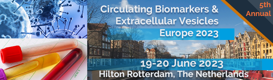
Co-Located Conference AgendasCirculating Biomarkers and Extracellular Vesicles Europe 2023 | Lab-on-a-Chip and Microfluidics Europe 2023 | Organoids and Spheroids Europe 2023 | Point-of-Care, Biosensors and Rapid Diagnostics Europe 2023 | 

Monday, 19 June 2023 |
For Logistics and Programming Details for Day 1-Morning of the Conference Please Consult the Agenda for Lab-on-a-Chip and Microfluidics Track |
| | |
Session Title: Circulating Biomarkers and Extracellular Vesicles, Emerging Themes and Current Status 2023 |
| | |
Session Chairperson: Professor Dr. Dominique de Kleijn, University Medical Center Utrecht |
| | |
Venue: Conrad Room -- Hilton Rotterdam |
| | 14:00 |  | Conference Chair Plasma Extracellular Vesicles for Cardiovascular Disease
Dominique PV de Kleijn, Professor Experimental Vascular Surgery, Professor Netherlands Heart Institute, University Medical Center Utrecht, The Netherlands, Netherlands
Cardiovascular Disease (CVD) is with the cardiovascular events of Ischemic Heart Disease and Stroke, the number 1 and 2 cause of death in the world and expect to increase especially in Asia. We use plasma extracellular vesicle (EV) protein content of vesicles from plasma subfractions on plasma of stroke and periferal artery disease(PAD) patients, patients after carotid atherectomy (CEA) and patients suspected for chronic coronary syndrome (CCS). Using 25 ul of plasma, we developed an automated 96-well based protocol using sequential precipitation. Using samples of the AtheroExpress, the largest ongoing CEA biobank we try to early detect the risk of a second Major Adverse Cardiovascular Event (MACE: myocardial infarction, stroke or cardiovascular death) in PAD and CEA patients. Identification of such high risk patients is very important for possible (expensive) add-on pharmaceutical therapy or the decision to operate or not. Sequential procipitation for EV isolation is also used for the diagnosis of CCS. |
| 14:30 |  | Keynote Presentation Versatile and Multiplexed Extracellular Vesicle and Single Extracellular Vesicle Analysis
David Juncker, Professor and Chair, McGill University, Canada
Extracellular vesicles (EVs) have emerged as a fundamental cell signaling and cargo system. EV proteomic analysis face challenges such difficulty to detect internal EV proteins, difficult to normalize concentration, co-expression, and lack of reproducibility. Single EV analysis, while highly desirable, still suffers from imprecision owing to lack of a protein markers universally expressed in EVs, low fluorescence of single molecules, and low scattering signals and the ensuing difficulty to detect small EVs. Here our efforts to address these challenges will be presented including multiplexed (i) inner and outer protein profiling in EVs; (ii) co-enrichment analysis which builds on co-expression analysis and quantifies protein co-enrichment, either negative or positive, and is illustrated with >100 protein pairs in EVs from metastatic, organotropic breast cancer cell lines. Next, (iii) a single EV analysis platform for label-free interference scattering (iSCAT) on a common (fluorescence) microscope will be introduced, and sizing of EVs from ~35 nm to ~200 nm in diameter shown, outperforming existing methods such as nanoflow cytometry; (iv) the co-expression and co-enrichment profiles of ~20 proteins on single EVs will be demonstrated by combining iSCAT, fluorescence imaging, and microfluidic exchange of DNA-barcoded antibodies. These new EV analysis methods will be useful to detect, profile and analyse EVs in health and disease, and deepen our understanding of their function. |
| 15:00 |  | Keynote Presentation Chip-Based Flow-Cytometry: From CTCs to Extracellular Vesicles
Andrew J deMello, Professor of Biochemical Engineering & Institute Chair, ETH Zürich, Switzerland
Imaging flow cytometry (IFC) marries the advantages of optical microscopy and flow cytometry to enable the high-throughput imaging of cells within flowing environments. I will describe how we have leveraged the capabilities of simple microfluidic systems to form novel platforms able to manipulate, process and assay cells in a controlled and high-throughput manner. Using inertial and viscoelastic effects, these systems can operate at throughputs exceeding 400,000 cells/s, extract fluorescence, brightfield, and darkfield images and are capable of multi-parametric quantification and sub-cellular localization of structures down to 500 nm. Additionally, I will describe more recent activities in which similar viscoelastic microfluidic platforms are used to measure the mechanical properties of cells at high-throughput and also isolate and analyse CTCs and extracellular vesicles in a rapid and efficient manner. |
| 15:30 | Mid-Afternoon Coffee and Tea Break and Networking in the Exhiibt Hall | 16:00 |  | Keynote Presentation Extracellular Vesicles: From Technology Towards Biomedical Applications
An Hendrix, Professor, Ghent University, Belgium
Extracellular vesicles (EVs) are membrane-enclosed communicative particles released in body fluids that carry cell-type-specific biomolecular patterns. Knowledge on their origin, fate and function in the human body is required to accelerate clinical applications but hampered by a plethora of technological pitfalls (PMID:33568799). We created the EV-TRACK knowledgebase to stimulate transparency and steer reproducibility (PMID:28245209). We designed reference EVs, that are trackable and distinguishable from sample EVs, to support instrument calibration and data normalization (PMID:31337761; 33452501). We established reproducible protocols to separate EVs from other particles (PMID:31776460). This supporting ecosystem has steered us towards the pioneering discovery of systemic bacterial EVs in non-septicemic patients, including cancer patients (PMID:30518529; 35033427). |
| 16:30 |  | Keynote Presentation Title to be Confirmed.
Jennifer Jones, NIH Stadtman Investigator, Head of Transnational Nanobiology, Laboratory of Pathology, Center for Cancer Research, National Cancer Institute, United States of America
|
| 17:00 |  Improvements in Fluorescence Nanoparticle Tracking Analysis Improvements in Fluorescence Nanoparticle Tracking Analysis
Sven Rudolf Kreutel, Chief Executive Officer, Particle Metrix GmbH and CEO, Particle Metrix Inc., USA
| 17:30 |  | Keynote Presentation Screening Tests Using Micro- and Nanofluidics for Early Disease Detection at the Point-of-Care
Steve Soper, Foundation Distinguished Professor, Director, Center of BioModular Multi-Scale System for Precision Medicine, The University of Kansas, United States of America
We are developing screening tests consisting of novel hardware, biomarkers, and assays to service a number of diseases, including the early detection of cancer and viral infections. The commonality in these tests is that they consist of microfluidic devices made from plastics via injection molding. Thus, our tests can be mass produced at low-cost that facilitates bench-to-bedside transition and point-of-care testing (PoCT) for large scale screening. The assays are based on the use of liquid biopsy markers as the input, which can be secured in a non- to minimally-invasive manner appropriate for screening. Recently we have focused on developing plastic nanofluidic devices, which provides unique opportunities for single-entity analyses. In this presentation, we will talk about the evolution of our fabrication efforts of plastic-based microfluidic and nanofluidic devices as well as their surface modification to make devices biocompatible. Then, we will discuss two applications of these devices and the assays for selection of rare liquid biopsy targets from clinical samples to serve as screening tests: (1) Analysis of extracellular vesicles (EVs) for the early detection of ovarian cancer; and (2) detection of viral particles from saliva samples, in particular SARS-CoV-2. Ovarian cancer is the 5th most deadly cancer for women in the US and has a 46.2% 5-y survival rate. Unfortunately, ~85% of cases are diagnosed at a late stage of disease, which demands new strategies for early detection. The screening test we are developing consists of a microfluidic chip for EV selection, which consists of a high density array of pillars surface-decorated with antibodies to efficiently select EVs followed by the label-free enumeration to determine elevated levels of EVs in the plasma of patients suspected of having ovarian cancer. Unique surface proteins were discovered for selection of ovarian cancer EV selection specifically for the early detection of disease. The infectious disease diagnostic accepts saliva samples and searches for viral particles using aptamers and counts the number of viral particles. For both disease examples, the selected particles were counted using a label-free approach; nano-Coulter Counter chip (nCC) consisting of in-plane nanopores. Both steps of the screening test described here were carried out using a microfluidic and nanofluidic chip. The chips were integrated to a control board for automating sample processing with results in <20 min. |
| 18:30 | Networking Reception with Dutch Beer and French Wine Tasting Sponsored by Kloé: Engage and Network with Colleagues, Engage with Exhibitors and View Posters | 19:30 | Close of Day 1 Conference Programming |
Tuesday, 20 June 202308:00 | Morning Coffee, Tea and Networking in the Exhibit Hall | |
Session Title: Circulating Biomarkers and Extracellular Vesicles -- Technologies Driving Applications |
| | |
Session Chairperson: Professor Dr. Dominique de Kleijn, University Medical Center Utrecht |
| | |
Venue: Conrad Room -- Hilton Rotterdam |
| | 08:30 | Global Inter-Laboratory Comparison Study to Standardize Measurements of Extracellular Vesicle Concentrations
Edwin van der Pol, Assistant Professor, Amsterdam University Medical Center, Netherlands
Introduction: Concentrations of extracellular vesicles (EVs) in body fluids are upcoming biomarker for health and disease. The concentration of cell-type specific EVs can be measured with state-of-the-art flow cytometers. However, flow cytometers have different detection limits and therefore measure different EV concentrations in the same sample. Consequently, clinical research studies reporting EV concentrations lack reproducibility and are typically single-center studies, which precludes future clinical implementation. To overcome these problems, the European Union invested 1.8 million euro into the “METVES II” consortium, which aimed for developing reference materials and methods to calibrate flow cytometers. The developed infrastructure was tested in a global inter-laboratory comparison study including 39 flow cytometers from 24 laboratories.
Methods: Concentrations of platelet-derived (CD61-APC) and erythrocyte-derived (CD235a-PE) EVs were measured in stabilized and pre-labeled human plasma EV test samples. The flow rates were calibrated using metrologically traceable silica beads, fluorescence intensities were calibrated using beads with a known number of fluorescent molecules, and light scattering intensities were calibrated using polystyrene beads and Mie theory. EV concentrations were compared between flow cytometers within calibrated fluorescence and size ranges.
Results: Preliminary results from 25 flow cytometers show that calibration leads to reproducible EV concentrations. For the platelet EV concentration, the coefficient of variation of measured EV concentrations decreased from 75% without calibration to 25% after calibration.
Conclusions: This is the first inter-laboratory comparison study demonstrating that full flow cytometer calibration improves the comparability of EV concentration measurements between flow cytometers, thereby paving the road to multi-center clinical research studies on EVs. | 09:00 | Red Blood Cells Protein Profile Is Modified in Breast Cancer Patients
Clotilde Costa, Translational Medical Oncology Group, Joint Unit Roche-Chus, Oncomet, Universitary Cinical Hospital of Santiago de Compostela, Spain
Metastasis is the primary cause of death for most breast cancer (BC) patients who succumb to the disease. During the hematogenous dissemination, circulating tumor cells interact with different blood components. Thus, there are microenvironmental and systemic processes contributing to cancer regulation. We have recently published that red blood cells (RBCs) that accompany circulating tumor cells have prognostic value in metastatic BC patients. RBC alterations are related to several diseases. Although the principal known role is gas transport, it has been recently assigned additional functions as regulatory cells on circulation. Hence, to explore their potential contribution to tumor progression, we characterized the proteomic composition of RBCs from 53 BC patients from stages I to III and IV, compared with 33 cancer-free controls. In this work, we observed that RBCs from BC patients showed a different proteomic profile compared to cancer-free controls and between different tumor stages. The differential proteins were mainly related to extracellular components, proteasome, and metabolism. Embryonic hemoglobins, not expected in adults’ RBCs, were detected in BC patients. Besides, lysosome-associated membrane glycoprotein 2 emerge as a new RBCs marker with diagnostic and prognostic potential for metastatic BC patients. Seemingly, RBCs are acquiring modifications in their proteomic composition that probably represents the systemic cancer disease, conditioned by the tumor microenvironment. | 09:30 |  | Keynote Presentation Nanotechnologies for Isolating and Characterizing Extracellular Nanocarriers of Biomarkers
Hsueh-Chia Chang, Bayer Professor of Chemical and Biomolecular Engineering, University of Notre Dame, Interim Chief Technology Officer, Aopia Biosciences, United States of America
We review a suite of nanotechnologies from our lab for high-throughput and scalable purification, enrichment and characterization of extracellular vesicles. The technologies are designed for medical diagnostics and biomarker discovery with physiological samples and for large-volume manufacturing of therapeutic exosomes. The relevant nanocarriers are exosomes, lipoproteins or protein-RNA complexes that carry potential protein and nucleic acid biomarkers for cancer, cardiovascular, neurodegenerative and even mental diseases. The technologies include size-based ultrafiltration membranes with conic nanopores to reduce protein fouling, bipolar membranes that can split water and actuate on-chip pH gradient to allow rapid and continuous isoelectric separation of nanocarriers by charge, superparamagnetic traps of immuno-nanobeads for rapid affinity and activity assay of specific nanocarriers, electrokinetic nanoporous microsensors that can profile surface proteins of nanocarriers, solid-state nanopore sensor for profiling microRNA cargoes etc. |
| 10:00 | Mitochondria-Containing Extracellular Vesicles From Macrophages Support Active Resolution of Inflammatory Pain
Niels Eijkelkamp, Associate Professor, University Medical Center Utrecht, Netherlands
Pain normally serves as a warning sign of inflammation and damage, that disappears when inflammation and damage resolves. However, in a substantial number of patients with inflammatory diseases such as rheumatoid arthritis pain persists even after cessation of inflammation. In this presentation I will discuss some of our recent findings that the immune system is actively involved in the resolution of inflammatory pain and that the road towards new pain therapeutics may hold promise for extracellular vesicles as vehicles of mitochondrial transfer. | 10:30 | Cytiva BioSciences Technology Spotlight Presentation | 11:00 |  NanoFCM Brings Flow Cytometry Capabilities to the Nanoscale! NanoFCM Brings Flow Cytometry Capabilities to the Nanoscale!
Natalia Gebara, Application Scientist, NanoFCM Co., Ltd.
Conventional flow cytometers often struggle to meet the sensitivity requirements for the analysis of nanoscale particles, such as exosomes, nanomedicine, and viruses. To meet this challenge, NanoFCM has developed the NanoAnalyzer, a dedicated nano-flow cytometry platform, which offers a flexible and high-throughput solution for sub-micron analysis. By using the NanoAnalyzer, single-particle characterization can be achieved which simultaneously measures the side scatter (40 -1000nm) and fluorescent properties of particles. The size detection of the NanoAnalyzer favorably compares to electron microscopy and covers the entire size range of EVs, offering a detailed analysis of size, concentration, and biochemical properties by direct correlation of the physical and phenotypic data. It is by combination of all these properties that the NanoAnalyzer is an ideal next-generation technique/ instrument for the analysis of EVs.
| 11:30 | Modeling the Feto-Maternal Communication by Extracellular Vesicles using Intrauterine Tissue Microphysiologic System
Ramkumar Menon, Distinguished John D. Stobo, MD, Endowed Chair and Professor, The University of Texas Medical Branch (UTMB), United States of America
The fetal inflammatory response in response to intrauterine inflammation is a major determinant of adverse pregnancy outcomes, specifically preterm birth (PTB). Inflammation causes intrauterine tissue (fetal membrane/amniochorion) senescence and generate damage-associated molecular pattern markers (e.g., High mobility group box 1 protein [HMGB1]). HMGB1 is released via extracellular vesicles. We tested the hypothesis that exosomal HMGB1 is one of the fetal signals capable of increasing Feto-Maternal interface (FMi) inflammation, predisposing to PTB. To test this hypothesis, exosomes from amnion epithelial cells (AECs) from the intrauterine fetal membranes grown under normal conditions were engineered to contain HMGB1 by (eHMGB1). eHMGB1 was characterized, and its propagation through FMi was tested using a four-chamber microfluidic organ-on-a-chip device (FMi-OOC) that contained four distinct cell types (amnion epithelium, amnion mesenchyme, chorion trophoblast and decidual cells) connected through microchannels. eHMGB1 propagated through the fetal cells to the maternal decidua and increased inflammation associated with PTB. To physiologically validate this finding, eHMGB1(containing 10 ng) was intra-amniotically injected into CD-1 mice on embryonic day 17 which resulted in PTB. In vivo kinetics was determined by injecting carboxyfluorescein succinimidyl ester labeled eHMGB1. We report that eHMGB1 trafficking in mice causing PTB was associated with increased FMi inflammation. Our study determined that fetal exosome-mediated paracrine signaling can generate inflammation and induce parturition. | 12:00 | A Digital Nanotechnology For Lung Cancer Screening and Long COVID
Alain Wuethrich, Group Leader / Researcher, The University of Queensland, Australia
Profiling circulating biomarkers has potential to deliver minimally invasive approaches for lung cancer screening and to better understand emerging diseases such as long COVID. Nanotechnology and microfluidics are well positioned to deliver innovative diagnostic systems that enable highly sensitive and multiplex analysis of circulating biomarkers. This presentation will describe a digital nanotechnology based on plasmonic barcodes and a nanostructured array for counting of single extracellular vehicles (EVs) and cytokines. We show the capability of phenotyping EVs in plasma of patients with benign and malignant lung nodules as a potential non-invasive approach for lung cancer screening. By digitally profiling trace-level cytokines at concentrations multiple orders of magnitude lower than detectable by conventional assays, we will further describe how the digital nanotechnology provided a new window into long COVID and could serve as diagnostic for this emerging disease. | 12:30 |  | Keynote Presentation Plasma Extracellular Vesicle-Associated microRNAs for On-Treatment Response Monitoring and Prediction
D. Michiel Pegtel, Associate Professor, Amsterdam University Medical Center, Netherlands
Response monitoring and outcome prediction is essential in the clinical management of hematological malignancies. Extracellular Vesicle associated microRNAs (EV-miRNAs) are considered promising liquid biopsy-based biomarkers for hematological malignancies. We performed small RNA sequencing of plasma samples collected during therapy and applied machine learning to build signatures for response prediction in Multiple Myeloma (MM) and patients with high grade B-cell lymphoma (HGBL). We collected plasma samples from multiple clinical trials and obtained 'real-world' samples. In HGBL, response was assessed by an end-of-treatment (EOT) PET/CT while in MM we defined response based in part on M protein levels. We isolated plasma EVs with size exclusion chromatography as confirmed with transmission electron microscopy (TEM), tunable resistive pulse sensing (TRPS), and western blotting. Library preparation was done according to our IsoSeek method. We applied machine learning to build models with EV-miRNAs for early response prediction (HGBL) and monitoring (MM). We could generate robust signatures for HGBL that can predict EOT response after one cycle of R-CHOP. If validated in independent cohorts, this novel approach could potentially, in combination with other modalities, guide early risk-adapted treatment strategies. In addition, we show that EV-miRNAs distinguish MM patients with active disease from those in remission. Together our data suggests plasma EV-miRNA sequencing is a versatile platform technology for minimally invasive response evaluation and prediction. |
| 13:00 | Networking Luncheon in the Exhibit Hall -- Network with the Exhibitors and View Posters | 13:30 | Biotechnology in Space: New Opportunities for Superior R&D -- Panel Discussion from 13:30 to 14:30 in the Rotterdam Room |
|


 Add to Calendar ▼2023-06-19 00:00:002023-06-20 00:00:00Europe/LondonCirculating Biomarkers and Extracellular Vesicles Europe 2023Circulating Biomarkers and Extracellular Vesicles Europe 2023 in Rotterdam, The NetherlandsRotterdam, The NetherlandsSELECTBIOenquiries@selectbiosciences.com