Impedance Spectroscopy Tools for Quantifying Transport Phenomena in Nanochannels and Tissues
Shaurya Prakash, Assistant Professor, Ohio State University
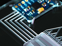
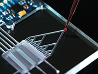
Over the past century, electrical or electrochemical impedance spectroscopy (EIS) has been used by chemists and biologists to study reaction rate kinetics, corrosion phenomena, battery aging, and tissue characteristics to name a few applications. EIS measures a current or voltage response of a system to an alternating voltage or current signal and records the response as complex impedance. The key idea is that the input is a small amplitude signal and therefore permits use of small-signal theory and linearization to analyze system response through data containing both magnitude and phase information. In this talk, I will present the development of EIS as a new tool for probing and quantifying microscale and nanoscale transport phenomena by using two representative case studies. First, we implemented a standard 4-electrode measurement system with two voltage sense or reference electrodes located close to the entrance and exit of nanocapillary array membranes (NCAMs) with remaining two current (also called counter) injection or measurement electrodes located away from the NCAMs in a permeation cell. The impedance data was fit to our variable topology model, to determine nanocapillary entrance (and exit) radius of 2.19 ± 0.01 nm and a center radius of 4.05 ± 0.01 nm in contrast to the manufacturer report of the nominal radius of 5 nm, demonstrating a new approach to quantify nanocapillary geometry in situ. Second, EIS was used to generate images of morphologically distinct regions in excised human liver tissue to differentiate, based on differences in electrical conductivity and permittivity, the tumor and non-tumor regions. The impedance data can be reduced to equivalent tissue structure images showing the ability to use EIS as an imaging technique, with direct comparisons to visual representation by digital photography and clinical validation by histopathology images.
|
|


 Add to Calendar ▼2014-09-18 00:00:002014-09-19 00:00:00Europe/LondonLab-on-a-Chip, Microfluidics and Microarray World CongressLab-on-a-Chip, Microfluidics and Microarray World Congress in San Diego, California, USASan Diego, California, USASELECTBIOenquiries@selectbiosciences.com