Exosomes and Viral Infections: Struggles of the Host Cell with the Intracellular Pathogen
Fatah Kashanchi, Professor, George Mason University
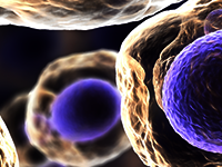
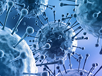
In order to perform normal functions, cells release various types of vesicles, such as exosomes and microvesicles (MVs) derived from endosomes and the plasma membrane. These extracellular vesicles (EVs) play a significant role in intercellular communication by serving as a carrier for the transfer of membrane and cytosolic proteins, lipids, and RNA between cells. In recent years, using state of the art technologies such as RNA seq, RPMA, and single cell omics, we have found that virally infected cells including HIV, HTLV, Rift Valley Fever, VEEV, Zika and Ebola virus secret exosomes that contain biomarker of infections. These markers include viral RNA, viral proteins and a range of unique cellular proteins that are released from infected cells. EVs were characterized using electron microscopy, nanoparticle tracking analysis (NTA), and common structural and functional proteins, such as Rab GTPase, SNAREs, annexins, Alix, Tsg101, flotillin and tetraspanins. These exosomes can be found in bodily fluids including urine, saliva, CSF, and blood. Interestingly, these exosomes can be detected during most inhibitory treatments (i.e. IFN, cART) indicating that infected cells may be present and active in tissues reservoirs (i.e. CNS, Lymph nodes). They also contain the usual viral proteins and RNAs and may be part of an early overexpression by these viruses to favor virus gene expression, assembly, and budding. Therefore, exosomes from infected cells pose a superior predictor of host response biomarker of infection since they contain and protect the viral biomarkers in various bodily fluids. In recent years, the diverse biological cargo contained within certain uninfected stem cell EVs (i.e., iPSC or MSC) are used for repair purposes. In a series of recent studies using large scale purification methods via tangential flow filtration (TFF), we have isolated iPSCs and MSCs EVs which significantly enhance the processes of cellular migration and angiogenesis in healthy recipient cells. In models of induced cellular damage (i.e., irradiation or infections), these EVs had the ability to rescue viability in multiple 2D and 3D cultures. Interestingly, there seems to be a “dual” mode of action, where non-coding RNAs activate innate immune molecules followed by presence of cytokines for activation and repair. Collectively, these results from past few years demonstrate the potential of EVs as diagnostic markers of infection, and stem cell EVs as a “holistic” therapeutic approach to reverse or to reduce cellular damage.
|


 Add to Calendar ▼2020-02-17 00:00:002020-02-18 00:00:00Europe/LondonEV-based Diagnostics, Delivery and TherapeuticsEV-based Diagnostics, Delivery and Therapeutics in Coronado Island, CaliforniaCoronado Island, CaliforniaSELECTBIOenquiries@selectbiosciences.com