07:00 | Morning Coffee, Breakfast Pastries and Networking in the Exhibit Hall |
|
Session Title: From Technologies to Utility -- Applications of Microfluidics/LOAC in Life Sciences and Beyond |
| |
07:30 | Micro-encapsulation of Carbon Dioxide Capture Solvents through Microfluidic Processing
Congwang Ye, Research Staff, Lawrence Livermore National Laboratory, United States of America
Microfluidics have been developed for many applications such as life science research and lab-on-a-chip. At LLNL, our work focuses on using microfluidics to produce microcapsules for energy applications, particularly the capture of CO2. Recently, new designer solvents have been developed to reduce energy cost for carbon capture. However, they suffer from many drawbacks such as high viscosity, solid precipitates and corrosivity. Separating the capture solvent with a gas permeable shell provides a practical pathway to use these solvents. In our lab, double emulsion drops were generated within a micro capillary device and are composed of liquid capture solvents as the core and different polymer precursor solutions as the shell layer. After crosslinking the polymer shell, the resulting microcapsules are mechanically robust and can be handled in fixed-bed, moving-bed or fluidized-bed reactors for different applications. While the mass transfer of gas through the capsule shell is slightly lower when compared to neat liquid sorbents, the increase in surface area enhances the overall CO2 absorption rate by 3.5-10x based on experimental data. The absorbed CO2 can then be released by heat or diffusion for reuse applications or underground storage. |
08:00 | Step by Step Fabrication of Biomaterials based on Cell Adhesion Control
Kennedy Okeyo, Senior Lecturer, Institute for Frontier Life and Medical Sciences, Kyoto University, Japan
On-chip fabrication of biomaterials such as cell sheets based on self-assembly organization of cells into tissues continues to attract attention due to potential applications in regenerative medicine as well as in the development of in vitro models for drug screening. We recently developed a technique for step-by-step fabrication of cell sheets based on adhesion control. It involves minimization of cell-substrate interaction to initiate self-assembly of cells, not into spheroids, but into planar cell sheets which can be then overlaid or stacked into 3D tissue models. For cell adhesion minimization, we developed a mesh culture system where cells are seeded and grown on suspended ultra-thin (~2µm thick) mesh scaffolds consisting of considerably large apertures (exceeding 100 µm in size) and thin mesh lines (3-5 µm in width).
|
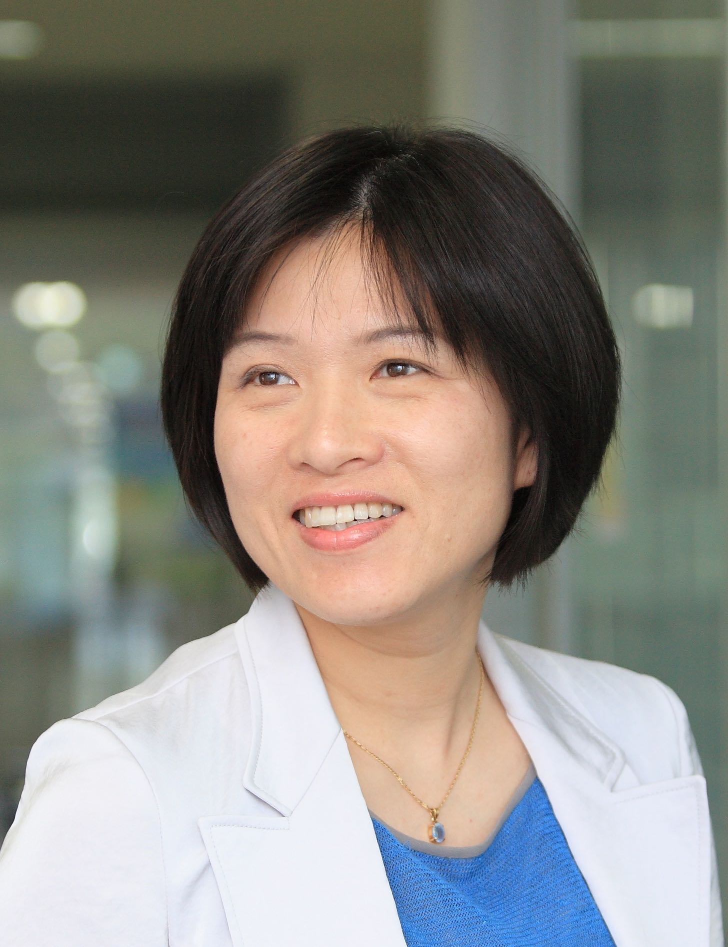
08:30 | 3D Microfluidic Mixer for Rapid Smartphone-based Diagnosis of Anemia
Mei He, Assistant Professor, University of Kansas and Chief Science Officer, Clara Biotech, United States of America
Clinical diagnosis requiring a central facility and site visits can be burdensome for patients in resource-limited or rural areas. Therefore, development of a low-cost microfluidic chip that utilizes smartphone data collection and transmission would beneficially enable disease self-management and point-of-care diagnosis. We demonstrated 3D simulation-guided printing for fabricating micro-, millifluidic mixers, which allows rapid study and testing of various geometries in lab-on-a-chip devices. Combined with smartphone, a 3D printed microfluidic mixer for auto-mixing of reagents via capillary force has been implemented, for measuring hemoglobin levels in finger-prick blood. Self-diagnosis of anemia has been successfully demonstrated using smartphone detection, showing consistent results with clinical measurements. Capable of 3D fabrication flexibility and smartphone compatibility, this work presents a novel diagnostic strategy for advancing personalized medicine and disease management. |
09:00 |  Technology Spotlight: Technology Spotlight:
High-Sensitivity, Cost-Effective Nanophotonic Biosensor Substrates
Arash Farhang, Optics Scientist, MOXTEK, Inc.
Numerous publications demonstrate that nanophotonic substrates offer the extraordinary surface sensitivity that enables many biosensing applications. Thus far colloidal-nanoparticle-based arrays and thin metallic films are the only such truly large scale substrates that have been available commercially. Moxtek has developed nanophotonic technologies and cost-effective volume production capabilities that enable and improve numerous biosensor applications. This technology greatly enhances fluorescence and label-free assay sensitivity through the utilization of surface plasmon, guided-mode, and gap-mode resonances. Such heightened sensitivity enables significantly-lower detection limits in molecular/chemical detection, microarrays, and other bioassay technologies, applications. These in turn open the doors to additional markets such as medical/point-of-care diagnostics, forensic testing, environmental monitoring, and food safety. Theoretical and measured data on the performance of these nanophotonic technologies, possible applications, and an overview of the available developmental and commercial manufacturing capabilities will be presented.
|
09:30 |  | Keynote Presentation Biological Imaging Using Microfluidics and Electrochemistry
Charles Henry, Professor and Chair, Colorado State University, United States of America
Chemical gradients drive many processes in biology, ranging from nerve signal transduction to ovulation to cancer metastasis. At present, microscopy is the primary tool used to understand these gradients by imaging both the gradients and the resulting cell motion. Microscopy has provided many important breakthroughs in our understanding of fundamental biology, but is limited due to the need to incorporate fluorescent molecules into biological systems through either labeling or genetic manipulations. To better understand some processes, there is a need to develop tools that can measure chemical gradient formation in biological systems that do not require fluorescent modification of the targets, can be multiplexed to measure more than one molecule, and are compatible with a variety of biological sample types, including in vitro cell cultures and ex vivo tissue slices. In response to this need, we have developed a high-density electrode array containing 8,192 individual electrodes to image release of electrochemically active metabolites like nitric oxide and norepinephrine from live tissue slices. The electrode array has a resolution of 30 µm and covers and area of 2 mm by 2 mm, easily large enough to image release of neurotransmitters across an ex vivo tissue slice from a mouse model. In this talk, electrochemical characterization of this system will be discussed first using simple chemical models. Then use of the system in combination with microfluidics, for imaging spatiotemporal resolution of neurotransmitter release at the organ scale will be presented. Use of microfluidics to deliver stimulants that induce changes in release profile will be shown. |
|
10:00 | Negotiating Your Way into an Assay: Specificity has Tolerances
David Wright, CEO, Wi, Inc., United States of America
|
10:30 | Coffee Break and Networking in the Exhibit Hall: Visit Exhibitors and Poster Viewing |
11:15 | Applications of Fully Integrated Active and Passive Flow Control in the Organ-on-Chip, Analytical and Diagnostics Field
Marko Blom, Chief Technical Officer, Micronit Microtechnologies, Netherlands
We will present various applications and examples in the organ-on-chip, analytical and diagnostics field relying on fully integrated active and passive, capillary driven flow control. Active valving fabrication strategies will be shown which do not require adhesives for the integration of materials relevant to the above mentioned fields.
The applications will range from accurate dosing, distribution and dispensing for cell culturing and Organ-On-Chip (OOC) applications to examples of sequential capillary flow in polymeric devices controlled by low-power electrical triggers realized using integrated electrodes. Specifically for the OOC field we have developed scalable Design-For-Manufacturability (DFM) concepts for such OOC applications, varying from resealable devices to fully integrated microtiter plate format culturing tools. The concepts are tested and validated in the field by end-users. Here, we demonstrate their use and applicability as versatile, flexible and enabling R&D tools for prototyping and (further) development of OOC applications. |
11:45 | Modeling and Simulation of Microfluidic Organ-on-Chip Devices
Matthew Hancock, Managing Engineer, Veryst Engineering, LLC, United States of America
Modeling and simulation are key components of the engineering development process, providing a rational, systematic method to engineer and optimize products and dramatically accelerate the development cycle over a pure intuition-driven, empirical testing approach. Modeling and simulation help to identify key parameters related to product performance (“what to try”) as well as insignificant parameters or conditions related to poor outcomes (“what not to try”). For microfluidic organ-on-chip devices, modeling and simulation can inform the design and integration of common components such as micropumps, manifolds, and channel networks. Modeling and simulation may also be used to estimate a range of processes occurring within the fluid bulk and near cells, including shear stresses, transport of nutrients and waste, chemical reactions, heat transfer, and surface tension & wetting effects. I will discuss how an array of modeling tools such as scaling arguments, analytical formulas, and finite element simulations may be leveraged to address these microfluidic organ-on-chip device development issues.
|
12:15 | Networking Lunch in the Exhibit Hall: Visit Exhibitors and Poster Viewing |
|
Session Title: Technology Advances in Microfluidics Drives Development of New Utility |
| |
13:30 |  | Keynote Presentation Human “Body-on-a-Chip” Devices as Tool to Improve Drug Development
Michael Shuler, Samuel B. Eckert Professor of Engineering, Cornell University, President Hesperos, Inc., United States of America
Alternatives or supplements to the use of animals in preclinical drug development that better mimic human response should reduce costs and increase the number of FDA approved drugs at the end of clinical trials. We have constructed micro-physiological (or “Body-on-a-Chip”) devices constructed from a combination of human tissue engineered constructs, micro-fabricated devices and physiologically based pharmacokinetic (PBPK) models. These human surrogates are constructed on a low cost, robust “pumpless” platform. In addition to measuring viability and metabolic responses, we can measure functional outputs such as electrical activity and force generation using integrated sensors (in collaboration with J. Hickman, University of Central Florida). We will focus our discussion on development of key organ modules and their integration with each other to form a model of the human body. |
|
14:00 |  | Keynote Presentation Microraft Array Platform for the Selection of Lymphocytes Based on Target-Cell Killing
Nancy Allbritton, Frank and Julie Jungers Dean of the College of Engineering and Professor of Bioengineering, University of Washington in Seattle, United States of America
Adoptive cellular therapy (ACT) is an emerging therapeutic in which cytotoxic T lymphocytes (CTLs) that recognize tumor cell epitopes are introduced into patients providing immunity against the cancer cells. For ACT to succeed, CTLs with high tumor-killing efficiency must be identified, isolated, and characterized. Current technologies do not enable simultaneous assay of cell behavior or killing followed by recovery of the most efficient killer cells. A microraft array technology that measures the ability of individual T cells to lyse a population of target cells followed by sorting of living cells into a multi-well plate for expansion and characterization was developed. The microraft array platform was combined with image processing and analysis algorithms to track and monitor killing assays over many hours. Automated cell collection was incorporated into the platform for facile cell collection from the array. As a proof of principle, human T cells directed against an influenza antigen were co-cultured with antigen presenting target cells on the microraft arrays. Target cell killing was measured by tracking the appearance of dead cells on each microraft over time. Microrafts with a single CTL demonstrating the greatest rate of target cell death were identified, cloned, and influenza-antigen reactivity confirmed. The platform is readily modified to measure the antigen-specific activity of individual cells within a bulk CTL culture or the cell heterogeneity within a population of gene-engineered T cells. |
|
14:30 | Free-Surface Microfluidics and SERS for High Performance Sample Capture and Analysis
Carl Meinhart, Professor, University of California-Santa Barbara, United States of America
Nearly all microfluidic devices to date consist of some type of fully-enclosed microfluidic channel. The concept of ‘free-surface’ microfluidics has been pioneered at UCSB during the past several years, where at least one surface of the microchannel is exposed to the surrounding air. Surface tension is a dominating force at the micron scale, which can be used to control effectively fluid motion. There are a number of distinct advantages to the free surface microfluidic architecture. For example, the free surface provides a highly effective mechanism for capturing certain low-density vapor molecules. This mechanism is a key component (in combination with surface-enhanced Raman spectroscopy, i.e. SERS) of a novel explosives vapor detection platform, which is capable of sub part-per-billion sensitivity with high specificity. |
15:00 | Bipolar Electrode Coupling of Nanoscale Electron Transfer Reactions to Remote Chromogenic and Luminogenic Reporter Reactions
Paul Bohn, Arthur J. Schmitt Professor of Chemical and Biomolecular Engineering and Professor of Chemistry and Biochemistry, University of Notre Dame, United States of America
The combination of fluorescence and absorption spectroscopy with electrochemistry presents new avenues for the study of redox reactions, with potential for enhanced throughput, sensitivity, and spatial resolution. Here we present a novel configuration for coupling high sensitivity voltammetric measurements implemented in nanoscale architectures - such as nanopore-confined recessed ring-disk electrode arrays - with remote electrochemically-triggered chromogenic and fluorigenic reporter reactions. Coupling is mediated by a mm-scale bipolar electrode which communicates the local solution potential in the analyte-measuring portion of the device to an opposing chromogenic or fluorigenic reporter reaction in a remote location. Oxidation (reduction) of reversible analytes at the disk working electrode is accompanied by reduction (oxidation) on the nanopore portion of the bipolar electrode and then monitored by the accompanying oxidation of the reporter to produce a change in color or luminescence. The remote end of the bipolar electrode is placed in a cell far from the nanopore ring-disk array so that highly efficient reporter measurements can be carried out conveniently against low intrinsic backgrounds. The combination of bipolar eletrodes with luminescence in the dihydroresorufin/resorufin system has been used to study in situ generation of H2O2 in electrokinetic flow and for analytical determinations down to pM limits of detection. Applications of chromogenic reporter reactions for point-of-care (POC) use will also be described. |
15:30 | Inertial Focusing in Triangular Microchannels for Flow Cytometry
Ian Papautsky, Professor, University of Cincinnati, United States of America
This work presents a microfluidic device that takes advantage of asymmetric velocity profile in a low aspect ratio triangular microchannel to focus cells and microbeads into a single position with high efficiency, and without the need for secondary flow, sheath flow or external forces. |
16:00 | Plasmonic-Enhanced Single-Molecule Detection
Steve Blair, Professor, University of Utah, United States of America
The next generation of molecular diagnostics tools are targeted to have single molecule sensitivity. Plasmonic-enhanced fluorescence can be a key enabling factor in achieving this goal. Large-scale arrays of plasmonic structures meet the requirements of enhanced signal-to-background in fluorescence detection, along with compatibility with existing instrumentation and surface chemistry. Fluorescence enhancement results from a combination of plasmonic mediated excitation and emission enhancement. Even though molecules are confined within a plasmonic structure, the spectral region of enhancement depends strongly on the metal. As such, have also been working with structures in Al, which is mass-production friendly and provides balanced enhancement throughout the visible spectrum, opening up a wider range of applications. However, new chemical passivation strategies need to be devised due to the native oxide of Al. Tuning of the relative enhancements can be accomplished by adjusting the shape of the plasmonic structures, opening up the UV spectral range where the native fluorescence of biomolecules can be accessed. |
16:30 | Cation Dependent Transport in Gated Nanofluidic Systems
Shaurya Prakash, Associate Professor, Department of Mechanical and Aerospace Engineering, The Ohio State University, United States of America
A nanofluidic device with an embedded and fluidically isolated gate electrode, analogous to solid state semiconductor field effect transistors was developed for exquisite ionic transport control. Specifically, the gated electric field allows for tunable control of both the magnitude and direction of the net ionic current through the nanochannels as a function of electrolyte concentration and gate electrode location permitting device operation as a multi-state switch. Additionally, we show that ion transport is cation dependent for negatively charged walls demonstrating that engineered surface charge can potentially lead to significant advances in separations and nanoscale selective ion transport. |
17:00 | Surface Functionalization Strategies for the Design of Thermoplastic Microfluidic Devices for New Analytical Diagnostics
Fanny d'Orlyé, Associate Professor, Chimie ParisTech, Unité de Technologies Chimiques et Biologiques pour la Santé, France
Cyclic olefin copolymer (COC) and fluoropolymer (Dyneon THV) are emerging materials attractive for the conception of microfluidic chips thanks to their UV-visible transparency and high resistance to aggressive solvents. We propose to develop new analytical microsystems using these polymers for trace quantitation in complex matrices. This involves the immobilization of selective ligands in a confined zone of the microchannel for target extraction and preconcentration. This integrated pretreatment step will be followed inside the microdevice by electrokinetic separation and on-line detection. This requires new surface treatments for chemically inert COC and Dyneon to modify the microfluidic system at two scales: (1) on the entire surface to control surface properties and fluid flows during electrokinetic separation, or (2) locally to immobilize selective ligands (aptamers) on restricted areas for target extraction. For global modification, a plasma-induced immobilization of brominated derivatives allowed further ligand immobilization through alkyne-azide “click” chemistry reaction. For local ligand immobilization, we developed an original electrochemical strategy on Dyneon THV microchannel. Local electrochemical carbonization followed by covalent linkage of ligands (aptamers) through “click” reaction leads to immobilization zones in the 50 micrometer range. |
17:30 | Design of a Lab-on-a-Chip for the Quantitation of S-Nitrosothiols: From their Separation to their Decomposition and Electrochemical Detection
Anne Varenne, Professor, Chimie ParisTech, Unité de Technologies Chimiques et Biologiques pour la Santé, France
Abdulghani Ismail, Researcher, Chimie ParisTech, Unité de Technologies Chimiques et Biologiques pour la Santé, France
NO is a diatomic free radical that has in biological fluids extremely short half-life (<1 s). The addition of NO to functional proteins is as important as phosphorylation in its consequences on cellular activities. In order to be transported and stocked in biological fluids, NO binds to peptides and proteins forming S-nitrosothiols (RSNOs). The variation of their proportion has been recognized in many diseases. RSNO are sensitive to decomposition by light, heat, and metal ions. RSNOs can exchange the NO between one other by transnitrosation reaction. Recently we studied the separation of different RSNOs and their transnitrosation reaction using capillary electrophoresis with capacitively coupled contactless conductivity detection. Also, we developed a decomposition process of RSNO using copper to quantify RSNO by electrochemical techniques (ultra-micro electrodes). We are currently down scaling these approaches within microchip devices. This opens the way for an integrated lab-on-a-chip for S-nitrosothiols quantitation.
|
18:00 | Close of Day 3 of the Conference. |


