08:00 | Conference Registration, Coffee, and Breakfast Pastries in the Exhibit Hall. Exhibit Hall Opens. |
|
Session Title: Emerging Technologies and Trends in Single Cell Analysis (SCA) |
| |
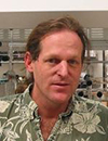
09:00 |  | Keynote Presentation Single Cell Barcode Chips for Quantitative Proteomics and Metabolomics Assays from Single Cancer Cells
James Heath, Elizabeth W. Gilloon Professor of Chemistry, California Institute of Technology (CalTech), United States of America
I will discuss microchip platforms for integrated and quantitative assays of functional proteins and metabolites from statistical numbers of single cells. I will further describe thermodynamics-derived approaches for using such platforms to understand cellular transitions associated with carcinogenesis. |
|
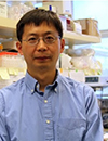
09:30 |  | Keynote Presentation Single-Cell Sequencing of the Human Brain
Kun Zhang, Professor, Department of Bioengineering, University of California San Diego, United States of America
|
|
10:00 | Integrated Lens-Free Imaging Technologies Enabling High-Throughput Label-Free Single-Cell Analysis
Richard Stahl, Senior Researcher, IMEC VZW, Belgium
Imagine an inexpensive, portable and label-free (blood) cell analyzer that can be used in emergency or resource-limited settings. Our goal is to use the versatility and scalability of semiconductor technologies to develop such a high-throughput single-cell analyzer using miniaturized lens-free microscopes integrated into a self-contained single-chip system. While our previously reported results confirm that such devices can reach sufficient performance, a number of challenges needs to be addressed in the imaging, micro-fluidics as well as photonic sub-systems. Here we give an overview of the system design, report the progress in development and integration of the individual hardware components, and showcase the system demonstrators with integrated photonics and micro-fluidics. We also report the advances on image reconstruction and cell classification algorithms and discuss the latest results from experiments on analyzing and sorting subtypes of white blood cells with our cell-sorting devices. |
10:30 | Coffee Break, View Posters, Visit Exhibitors and Networking |
11:00 | Tracking Development and Aging of Individual Bacteria with Microfluidic Devices
Stephen Jacobson, Professor, Indiana University, United States of America
Analysis of single cells provides powerful insight into biological processes that are often missed when a population of cells is studied as an ensemble. We are developing microfluidic-based approaches coupled with optical microscopy to track individual bacteria and to improve the temporal and spatial resolution of single-cell measurements. To streamline our experiments, we designed and tested a microfluidic “baby machine,” which automates the culture, synchronization, and analysis steps. This microfluidic device is used to produce synchronized populations of cells and monitor adhesion of individual bacteria to surfaces. To study development and aging in bacteria, we integrated nanochannel arrays into the microfluidic platform that physically trap bacteria. With these nanochannel arrays, we are able to study a number of bacteria strains and observe them over multiple generations. Consequently, we are able to determine rates of cell growth and division, monitor accumulation of cellular damage, and observe epigenetic effects.
|
11:30 |  | Keynote Presentation NGS Keynote: Opportunities and Challenges of Implementing Next Generation Sequencing Technologies for Delivery of Genomic Medicine
Nazneen Aziz, Executive Director, Kaiser Permanente Research Bank, United States of America
Many important advances in technologies for molecular analyses have fostered the rapid growth in molecular medicine. The use of these technologies in testing of DNA, RNA, proteins for screening, diagnosing and prognosis of patient’s disease, and also in the development of new biologics and small molecule drugs have given rise to a science that 30 years ago was practically non-existent. Each of these technologies has led to significant advances, but one in particular, can be undoubtedly labeled as ‘disruptive’. Next generation sequencing (NGS), introduced in 2005, has revolutionized the field by increasing the speed at which the genome can be sequenced at a exponentially lower cost. Within 5 years of its introduction and widespread use in research, NGS is transforming molecular medicine. NGS has a higher throughput and lower cost per base and therefore has been rapidly adopted into clinical testing. What is particularly intriguing about NGS’s rapid adoption into clinical testing is that it has a number of intricacies associated with its implementation that is unfamiliar to clinical laboratory. Dr. Aziz will address the opportunities, complexities and challenges of NGS clinical testing. |
|
12:00 | Networking Lunch, Visit Exhibitors and View Posters |
13:00 |  Technology Spotlight: Technology Spotlight:
Single-Cell Analysis: Guiding the Future of Early Detection, Prognosis, and Course of Treatment for Disease
Naveen Ramalingam, Scientist II, Fluidigm Corporation
A number of breakthrough technologies in single-cell analysis are paving the way for increasing the sensitivity & reproducibility of diagnostic tests, while simultaneously reducing the required labor and turnaround time for results. In this session, we will discuss some of these new breakthroughs and their impact on the community.
|
|
Session Title: Applications of Single Cell Analysis (SCA) into the Diagnostics Realm |
| |
13:30 | Carbon Nanopipettes (CNPs) for Automated Microinjection and the Study of Sub-cellular tRNA Dynamics
Haim Bau, Professor, University Of Pennsylvania, United States of America
We describe the use of carbon nanopipettes (CNPs) for microinjection into individual cells. The CNP consists of a pulled-quartz micropipette with a thin layer of carbon deposited along its entire interior surface. The tip of the CNP is etched to expose a carbon pipe that varies in diameter from tens to hundreds of nm. The bore of the CNP facilitates microinjection, while the carbon film enables electrical measurements. We utilize the carbon film for impedimetric detection of cytoplasmic and nuclear penetration to trigger injection. We use the CNPs in a custom-developed, automated injection system. As a demonstration of an application, we describe microinjection of fluorescently-labeled tRNA to monitor subcellular tRNA dynamics in real time. |
14:00 |  | Keynote Presentation NanoVelcro-Embedded Microchips for Detection and Characterization of Rare Cells in Blood: Validation Studies in Oncology and OB/GYN Clinics
Hsian-Rong Tseng, Professor, Crump Institute for Molecular Imaging, California NanoSystems Institute, University of California-Los Angeles, United States of America
Our research team at UCLA has demonstrated a highly efficient cell-affinity assay (known as NanoVelcro Microchips) capable of detecting and characterizing rare cells, e.g., circulating tumor cells (CTCs) and circulating fetal cells (CFCs) in blood samples collected from cancer patients and expectant mothers, respectively. In addition to conducting the enumeration of these rare cells, we have been exploring the use of NanoVelcro Microchips for isolating single CTCs and CFCs without contamination by white blood cells (WBCs) in the background. The individually isolated CTCs and CFCs can then be subjected to molecular analyses by FISH, RT-PCR, microarray and/or next-generation sequencing, enabling a wide range of applications in the fields of cancer and prenatal diagnosis. |
|
14:30 |  | Keynote Presentation Extending the Capabilities of Flow Cytometry by Advances in Sensitivity and Resolution
John Nolan, CEO, Cellarcus Biosciences, Inc., United States of America
Driven by the need to understand complex cell systems, high performance instruments and new reagents and assays are pushing the boundaries of the current generation of commercial systems. Multiparameter cell measurement using fluorescent probes can exceed 20 colors, while mass spec-based cytometry (mass cytometry) pushing beyond 50 parameters. Optical detection, which is desired for many applications, including single cell sorting, is being extended by spectral flow cytometry, where high spectral resolution enables the use of new analysis methods, optical probes, and multiplexing approaches. Higher sensitivity instruments and brighter reagents are allowing single particle analysis to extend to the nanoscale, to study complex systems of extracellular vesicles that mediate communication between cells and through the organism. This presentation will focus on these new instruments, reagents and assays, and the biomedical applications they are enabling in cardiovascular disease, cancer, and other areas. |
|
15:00 | Coffee Break, View Posters, Visit Exhibitors and Networking |
15:30 | Single Cell Pipette Assisted Cancer Cell Genome Analysis
Lidong Qin, Professor and CPRIT Scholar, Houston Methodist Research Institute, United States of America
There is increasing evidence that solid tumors are likely comprised of many subpopulations of cells with distinct genotypes and phenotypes, which is a phenomenon termed intratumor heterogeneity. Such heterogeneity becomes a major obstacle to effective cancer treatment and personalized medicine. Because of this inherent heterogeneity, data collected from cancer cell population-averaged assays likely hides valuable but rare events such as dramatic variations in gene expression at the single cell level. Therefore, understanding cellular heterogeneity from cancer biospecimens, especially in the study of phenotype-genotype correlation, will facilitate identification of new cell subsets, and assist in cancer prevention, diagnosis, and therapy. In this proposal, we focus on developing a high throughput approach for single-cell isolation based on cells’ capability in generating protrusions. To be able to retrieve the desired single adherent cell, a Protrusion Analysis Chip (PAC) is proposed, in which the single cell is captured by a single hook and physically isolated by a barrier. The PAC is rapid, operationally simple, highly efficient, and requires low-volume sample introduction. After adhesion and spreading, cell phenotype is identified microscopically and then the desired cell is retrieved for genotype analysis. The proposed technology developments and study plans may potentially strengthen the understanding of the relationship between phenotype and genotype at the single cell level. |
16:00 | Microfluidics for Cell Encapsulation
Richard Gray, Regional Director, Blacktrace, Inc., United Kingdom
Microfluidic methods have recently been used to encapsulate thousands of single cells, allowing massively parallel and rapid processing whilst requiring small samples and relatively simple implementation. Applications recently reported in major journals include high throughput generation of single cell RNA transcriptomes, profiling natively linked T-cell receptors, isolating monoclonal antibodies from blood samples, and directed evolution by library sorting. This presentation will review the devices and systems required and illustrate typical protocols, encouraging wider adoption by scientists.
|
16:30 | Fabrication of Polymer Nanofluidic Lab-on-Chip Devices for DNA Manipulation and Large Scale Sequencing
Jeroen A. van Kan, Professor, Centre for Ion Beam Applications, Physics Department, National University of Singapore, Singapore
A new 3D-nanolithographic technique which can produce smooth 3D-nanostructures, will be presented. This technique is used to produce masters for PDMS replication of nanofluidics LOC devices down to 60 nm. These LOC devices are extensively used for DNA single molecule analysis. Here I will give an update on the progress of the single molecule study, eg compaction studies and large scale sequencing. Finally thermal nano imprint lithography will be evaluated for the fabrication of PMMA nanofluidics LOC devices. |
17:00 | Single Cell Random Access Memory
Benjamin Yellen, Associate Professor, Biomedical Engineering, Mechanical Engineering and Materials Science, Duke University, United States of America
The ability to manipulate small fluid droplets, colloidal particles and single cells with the precision and parallelization of modern-day computer hardware has profound applications for biochemical detection, gene sequencing, chemical synthesis and highly parallel analysis of single cells. Drawing inspiration from general circuit theory and magnetic bubble technology, we have recently demonstrated a class of integrated circuits for executing sequential and parallel, timed operations on an ensemble of single particles and cells. The integrated circuits are constructed from lithographically defined, overlaid patterns of magnetic film and current lines. The magnetic patterns passively control particles similar to electrical conductors, diodes and capacitors. The current lines actively switch particles between different tracks similar to gated electrical transistors. When combined into arrays and driven by a rotating magnetic field clock, these integrated circuits have general multiplexing properties and enable the precise control of magnetizable objects. The presentation will focus on our recent progress on optimizing these integrated circuits to manipulate magnetically labeled CD4+ T cells, including recent data on single cell switches, cell velocities on chip, as well as the ability of non-fouling surface coatings to resist non-specific adhesion between the cells and chip surface.
|
17:30 |  | Keynote Presentation Isolation and Analysis of DNA/RNA Biomarkers from Hematological Cancer, Solid Tumors and TBI Patient Samples
Michael Heller, Professor, Dept Bioengineering, University of California-San Diego, United States of America
Rapid isolation and fluorescent detection of ccf-DNA/RNA biomarkers from CLL, solid tumor and TBI patient samples (20-100ul) is achieved in 10-15 minutes using an AC dielectrophoretic (DEP) microarray device. In the case of chronic lymphocytic leukemia (CLL), both PCR and DNA sequencing analysis for CLL derived ccf-DNA produces analytical results comparable to conventional “gold standard” procedures. Now, in order to determine the origin of ccf-DNA fragments, apoptotic ~180bp or necrotic >180bp, or both, PCR based amplicon size analysis and sequencing is being carried out on ccf-DNA isolated by DEP from CLL and solid tumor (lung, breast, colon) samples. For CLL, 540 bp mutation-containing PCR amplicons are readily produced even from low levels of ccf-DNA isolated by DEP from both blood and plasma samples suggesting a necrotic origin. Identification of the size range of ccf-DNA cancer biomarkers for their apoptotic or necrotic origins may very well have its own diagnostic value. Ultimately, DEP based sample to answer microarray systems will enable liquid biopsy for cancer and other molecular diagnostic point of care (POC) applications.
|
|
18:00 | Cocktail Reception in the Exhibit Hall: Visit the Exhibitors, View Posters and Network with Your Colleagues. Beers, Wines, and Appetizers Served |
20:00 | Close of Day 2 of the Conference |


