08:00 | Continental Breakfast Served in the Exhibit Hall |
|
Session Title: Technologies and Themes in the Organs-on-Chips Space + Themes in Organoids Research |
| |
|
Venue: Marriott Coronado Island Ballroom C |
| |
08:30 |  | Keynote Presentation The NIH Microphysiological Systems Program: In Vitro 3D Models for Safety and Efficacy Studies
Danilo Tagle, Director, Office of Special Initiatives, National Center for Advancing Translational Sciences at the NIH (NCATS), United States of America
Approximately 30% of drugs have failed in human clinical trials due to adverse reactions despite promising pre-clinical studies, and another 60% fail due to lack of efficacy. A number of these failures can be attributed to poor predictability of human response from animal and 2D in vitro mkodels currently being used in drug development. To address this challenges in drug development, the NIH Tissue Chips or Microphysiological Systems program is developing alternative innovative approaches for more predictive readouts of toxicity or efficacy of candidate drugs. Tissue chips are bioengineered 3D microfluidic platforms utilizing chip technology and human-derived cells and tissues that are intended to mimic tissue cytoarchitecture and functional units of human organs and systems. In addition to drug development, these microfabricated devices are useful for modeling human diseases, and for studies in precision medicine and environment exposures. Presenattion will elaborate in the development and utility of microphysiologicals sytems and in the partnerships with various stakeholders for its implementation. |
|
09:00 |  | Keynote Presentation High Throughput Vascularized Organoids For Drug Screening
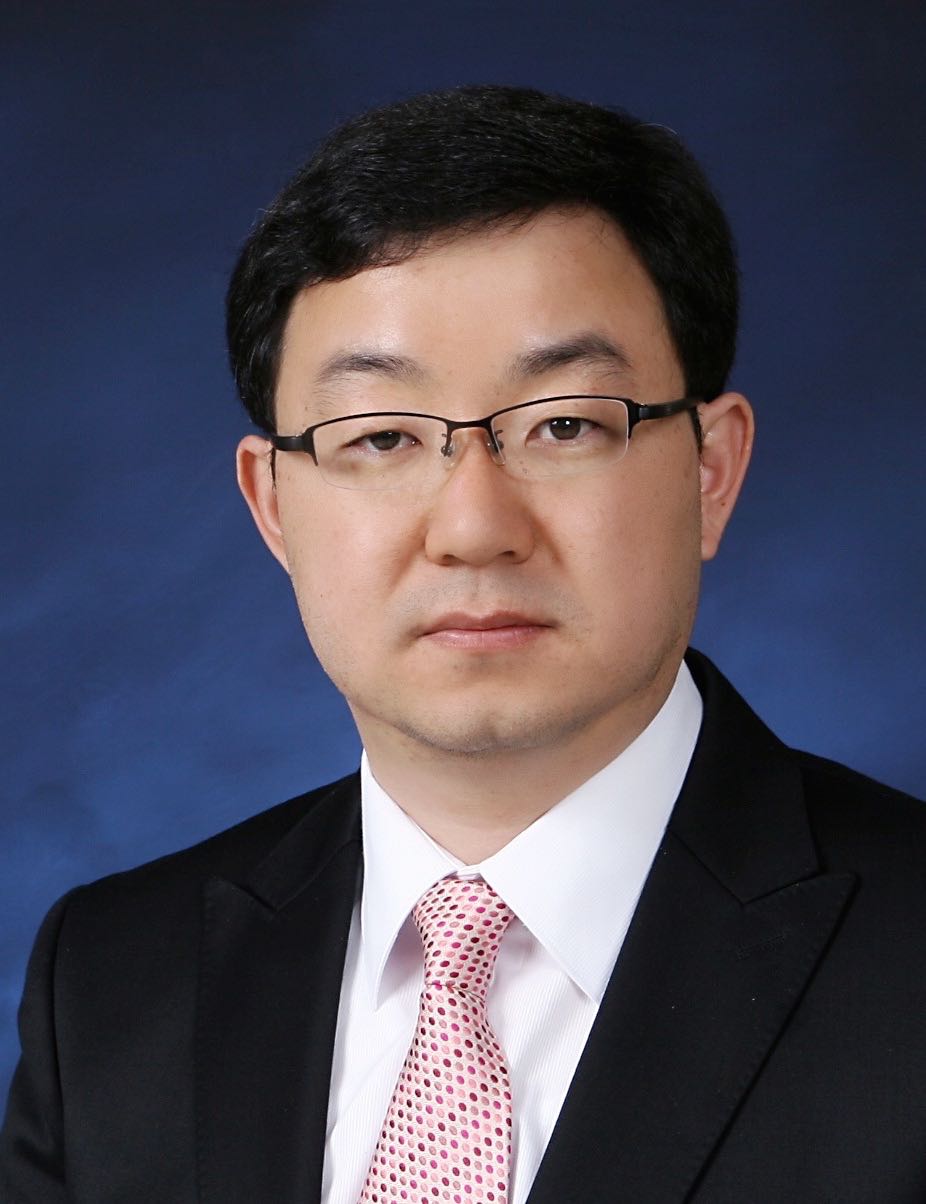
Noo Li Jeon, Professor, Seoul National University, Korea South
Recent developments in microfluidics based organ-on-a-chip approaches have progressed rapidly in last few years. This presentation will describe recent work in our laboratory where new patterning method based on open microfluidics combined with injection molded devices enable formation of array of vascularized tumor spheroids and organoids that can be used for drug screening. Utilizing a microfluidic design with spontaneous capillary flow (SCF) based patterning mechanism, we present a novel in-vitro platform, Sphero-IMPACT and T-IMPACT (Injection-Molded Plastic Array 3D Culture platform). Drugs and immune cells can delivered via the perfusable vessel networks and live time-lapse imaging can be used to follow dynamic changes in the tumor microenvironment. Recent results on vascularized iPSCs derived neurosphere will also be described. The IMPACT platform is a versatile, high throughput platform with potential applications in organoid based drug screening with the form factor and throughput of standard microtiter plates. |
|
09:30 |  | Keynote Presentation Body on a Chip: Will It Transform Drug Development?
Michael Shuler, Samuel B. Eckert Professor of Engineering, Cornell University, President Hesperos, Inc., United States of America
A physiologically representative, multi-organ microphysiological systems (MPS) based on human tissues (also known as “human-on-a-chip”) may be a transformative technology to improve the selection of drug candidates most likely to earn regulatory approval from clinical trials. Such microscale systems combine organized human tissues with the techniques of microfabrication based on PBPK (Physiologically Based Pharmacokinetic) models. I will describe such systems being constructed at Hesperos and at Cornell. They are “self-contained” in that they can operate independently and do not require external pumps as is the case with many other microphysiological systems. They are “low cost”, in part, because of the simplicity and reliability of operation. They maintain a ratio of fluid (blood surrogate) to cells that is more physiologic than in many other in vitro systems allowing the observation of the effects of not only drugs but their metabolites. While systems can be sampled to measure the concentrations of drugs, metabolites, or biomarkers, they also can be interrogated in situ for functional responses such as electrical activity, force generation, or integrity of barrier function. Operation up to 28 days has been achieved allowing observation of both acute and chronic responses with serum free media. We have worked with various combinations of internal organ modules (liver, fat, neuromuscular junction, skeletal muscle, cardiac, bone marrow, blood vessels and brain) and barrier tissues (eg skin, GI tract, blood brain barrier, lung, and kidney). We have achieved unidirectional flow in a pumpless system which is important for mimicking the response of vascular tissues and constructed blood brain mimics with human in vitro like characteristics. The use of microelectrode arrays to monitor electrically active tissues (cardiac and neuronal) and micro cantilevers (muscle) have been demonstrated. While most systems use 5 or fewer organ modules, we have demonstrated that a 13 “organ” compartment device can be constructed. Most importantly these technical advances allow prediction of both a drug’s potential efficacy and toxicity (side-effects) in pre-clinical studies and have been applied to circulating immune cells, cancer cells, and rare diseases such as Myasthenia gravis. |
|
10:00 |  | Keynote Presentation Using Microfluidics for Immune Cell Trafficking and Capture
Steven C. George, Edward Teller Distinguished Professor and Chair, Department of Biomedical Engineering, University of California-Davis, United States of America
Microfluidic technology has played a leading role to advance our understanding of fundamental biological processes including cell separation and isolation, next generation sequencing, and cell trafficking. Over the past five years our lab has applied the basic principles of microfluidics to control fluid shear and flow to create simple microphysiological systems to better understand: 1) how to capture and isolate rare immune cells from the peripheral circulation, and 2) the principles which guide and control immune cell (lymphocytes, monocytes, and neutrophils) trafficking in complex tissue microenvironments. For the former, we leverage the ability to coat surfaces with antigens that are recognized by rare populations of B lymphocytes in the peripheral circulation. We then control the shear force at the surface and can capture and isolate these rare cell populations. Understanding how these rare cell populations evolve over time following viral (e.g., SARS-CoV-2, influenza) infection is central to understanding immunity following infection or immunization. For the latter, we are pursuing three projects. The first involves neutrophil trafficking into the cardiac muscle during COVID19-induced “cytokine storm”, including counterstrategies that limit binding of neutrophils to the inflamed endothelium. The second involves modeling myeloid cell-directed immunosuppression in the tumor microenvironment, and how counterstrategies such as inhibiting STAT3 signaling, can enhance CAR-T cell trafficking and effector function. The third project describes small tumor cell-derived extracellular vesicle (sEV) transport (convection and binding to the extracellular matrix) and how the sEV can establish spatial gradients to guide monocyte migration in the tumor microenvironment. This talk will provide an overview of our major results from each of these projects. |
|
10:30 | Injectable and Drug Deliverable Nano-Matrix for Cartilage-on-a-Chip
Yupeng Chen, Associate Professor, University of Connecticut, United States of America
Janus base nano-matrix is a novel injectable solid scaffold that can be applied in the microchannels of tissue chips to achieve localized drug delivery and improve cartilage cells anchorage and functions. The injectable nano-matrix may be a suitable tissue engineering scaffold for cartilage-on-a-chip. |
11:00 | Tissue Organoids For Disease Modeling and Therapy Development
Shay Soker, Professor of Regenerative Medicine and Chief Science Program Officer, Wake Forest Institute for Regenerative Medicine, United States of America
Three dimensional tissue equivalents (organoids) replicate native human tissue structure and function and can be studied in vitro for several weeks to allow intensive investigations. Besides their advantages in drug toxicity testing and for development of new drugs, the human tissue organoid platform serves as a model system to explore human tissue development and disease. Our recent research was focused on the use of tissue organoids to study human development and congenital diseases as well as other common diseases such as tissue fibrosis and cancer. Once a disease model is established, patient-specific (personalized) therapies can be realized as pre-clinical testing platform. |
11:30 |  | Keynote Presentation Using Islet Spheroids to Model Beta Cell Targeted Interventions in Type 1 Diabetes
Matthias von Herrath, Vice President and Senior Medical Officer, Novo Nordisk, Professor, La Jolla Institute, United States of America
After investigating the human pathology of type 1 diabetes through organ donor pancreata (nPOD) it has become clear that beta cells become more recognizable by the immune system and that these beta cell Intrinsic events substantially contribute to their destruction. As immune reactivity to beta cells is even present to a low degree in healthy individuals, this ‘unmasking’ of the beta cell appears critical for T1D to develop. Upregulation of MHC is one of the key pathogenetic events. Thus, interventions should not only target the immune system, but should also be geared towards beta cells health and their recognizability. Here we have built, in collaboration with InSphero (Zurich, CH) a human islet spheroid immune attack model that replicates key events of human islet recognition and allows for the testing of drugs that can preserve beta cell function. |
|
12:00 | Pancreas Cancer Organoids for Research and Precision Medicine
Hervé Tiriac, Assistant Researcher, University of California-San Diego, United States of America
|
12:30 | Buffet Lunch and Networking in the Exhibit Hall with Exhibitors and Conference Sponsors |
13:00 |  | Keynote Presentation COVID-19 Therapeutics Safety ‘Clinical Trial’ with Cardiac Microphysiological Systems
Kevin Healy, Jan Fandrianto and Selfia Halim Distinguished Professorship in Engineering, University of California, Berkeley, United States of America
Three-dimensional (3D) microphysiological systems (MPSs) mimicking human organ function in vitro are an emerging alternative to conventional monolayer cell culture and animal models for myriad applications. Human induced pluripotent stem cells (hiPSCs) have the potential to capture the diversity of human genetics and provide an unlimited supply of cells. Combining hiPSCs with microfluidics technology in MPSs offers new perspectives for applications as diverse as drug development and organ preservation. This talk will address the use of our heart muscle MPS to assess the cardiac liabilities associated with repurposed therapeutics that treat SARS-CoV-2 infection. We propose that this MPS can help clinicians design their trials, rapidly project cardiac outcomes, and define new monitoring biomarkers to accelerate access of patients to safe COVID-19 therapeutics. |
|
|
Session Title: 3D-Bioprinting Technologies, Tools and Emerging Themes |
| |
|
Venue: Marriott Coronado Island Ballroom C |
| |
13:30 |  State-of-the-art Extrusion Bioprinting Technologies and Applications State-of-the-art Extrusion Bioprinting Technologies and Applications
Taci Pereira, Vice President and General Manager of Bioprinting, 3D Systems
In this talk, Taci Pereira, Vice President and General Manager of
Bioprinting at 3D Systems, will review key technologies from the Allevi
by 3D Systems bioprinting platform. These include hardware, software,
and material products for a wide range of research applications, such as
tissue engineering, regenerative medicine, disease modeling,
organs-on-chips, and bioink development.
|
14:00 |  Balancing Cost, Throughput and In Vivo Relevancy for In Vitro Liver Studies Balancing Cost, Throughput and In Vivo Relevancy for In Vitro Liver Studies
Peter Worthington, Director of Drug Discovery, Visikol
While we can generate ever more complex advanced cell culture models for use in our in vitro programs, not all research questions require this level of complexity. Through this presentation, Dr. Worthington will discuss how to balance cost, throughput and in vivo relevancy in the context of drug induced liver injury (DILI) and modeling liver disease (NASH, NAFLD). The presentation will review the use cases and applications for 2D cell culture models, 3D spheroid models and ex vivo precision cut tissue slice models.
|
14:30 |  Rapid Bioprinting for 3D Tissue Models Rapid Bioprinting for 3D Tissue Models
Shaochen Chen, Professor and Chair, NanoEngineering Department, University of California-San Diego
In this talk, I will present our laboratory’s recent research efforts in developing digital light processing (DLP) based rapid 3D bioprinting methods to create 3D tissue constructs using a variety of biomaterials and cells. These 3D printed scaffolds are functionalized with precise control of micro-architecture, mechanical (e.g. stiffness), chemical, and biological properties. Such functional scaffolds allow us to investigate cell-microenvironment interactions at nano- and micro-scales in response to integrated mechanical and chemical stimuli. From these fundamental studies we have been creating both in vitro and in vivo precision tissues for tissue regeneration, disease modeling, and drug discovery. Examples including 3D bioprinted liver and heart models will be discussed. I will also showcase 3D printed biomimetic scaffolds for treating spinal cord injury. Throughout the presentation, I will discuss engineer’s perspectives in terms of design innovation, biomaterials, mechanics, and scalable biomanufacturing.
|
15:00 |  Applications for Combining Extrusion and Light-based Bioprinting Techniques Applications for Combining Extrusion and Light-based Bioprinting Techniques
Nicole Diamantides, Bioprinting Field Application Scientist, CELLINK
Extrusion and light-based bioprinting are different 3D fabrication techniques that have been used for a wide variety of applications such as tissue engineering, 3D cell culture models for drug discovery, and creating models to study disease progression. Extrusion bioprinting is compatible with a wide variety of biomaterials and allows for the creation of multimaterial constructs that aid in mimicking the native tissue environment. Light-based printing, such as digital light processing, utilizes biomaterials that can be crosslinked upon exposure to light allowing for the creation of 3D structures with small features, such as channels which can act as vasculature. This talk will highlight how these two bioprinting techniques can be used in conjunction to create more complex and physiologically-accurate 3D models.
|
15:30 |  3D Printing is Allowing Users to Iterate a Wide Range of Microfluidic Devices Faster 3D Printing is Allowing Users to Iterate a Wide Range of Microfluidic Devices Faster
Hemdeep Patel, President, Co-Founder, CADworks3D
Over the last decade, 3D printing has changed the way designs are created, evaluated and iterated in all industries and in every facet of life. Since 2016, CADworks3D has been an active player in the world of microfluidics & bio-engineering by showcasing how 3D printers can radically improve the cycle of design, evaluation and iteration. The CADworks3D line of microfluidic 3D printers and 3D materials has users to test a wide range of devices from clear microfluidic encapsulated chips to master molds used for casting PDMS devices.
|
16:00 | Biochemical Compatibility of Stereolithographic Resins
Noah Malmstadt, Professor, Mork Family Dept. of Chemical Engineering & Materials Science, University of Southern California, United States of America
While 3D printing offers a promising route to rapidly prototyping and manufacturing microfluidic systems, it relies on materials that have yet to be fully tested in a biological context. Stereolithographic 3D printing, for instance, requires polymer precursors that can be cured via free radical chain reaction polymerization, as well as associated initiators and dyes. We have examined the ability of devices printed with a variety of common SLA resin formulations to support common reactions used in molecular biology laboratories, including PCR, translation, transcription, and reverse transcription. We found that all reactions are inhibited by all materials; some to the point where there are no products present at the limit of detection. This inhibition occurs not only when the material is incubated with the reaction, but if the reaction is performed in buffer that had previously been exposed to the material, suggesting that inhibitory molecules are leached from the cured resin and into buffer. In most cases, significant activity can be recovered by performing the reaction in the presence of BSA, which potentially acts to adsorb inhibitory molecules. |
|
15-Minute Oral Presentations of Selected Posters in the OOAC Track -- Venue: Coronado Ballroom C |
| |
16:30 | Generation of iPSC-derived 3D Neurospheres for Modeling Retroviral Infection and EV-mediated Repair
Heather Branscome, Senior Scientist, ATCC and Research Assistant, George Mason University, United States of America
We report the generation of iPSC-derived neurospheres and show that these cultures are permissive to HIV-1 infection. In addition, we examine the functional effects of stem cell extracellular vesicles (EVs) on HIV-1 infected neurospheres. Our data demonstrates the feasibility of iPSC-derived neurospheres for modeling HIV-1 infection and highlights the potential of stem cell EVs for rescuing cellular damage. |
16:45 | Establishing a Microfluidic in vitro Model of Remote Ischaemic Damage in Connected Neuronal Cultures
Jennifer Knight, Medical Student at the University of Oxford, University of Oxford, United Kingdom
This project consisted of designing a microfluidic device in which we fluidically isolated the somas, axons, and dendrites of stem-cell derived neurons. This allowed us to create a novel in-vitro model where we can study remote damage in ischaemic stroke. |
17:00 | Anaerobic Microbiota Cultivation by Gut on a Chip Developed from Highly Transparent Thermoplastic and Off-Stoichiometry Thiol-ene Polymer
Artis Galvanovskis, Student, Latvian Biomedical Research and Study Centre, Latvia
The gut microbiota and its products play a critical role in human health and in many physiological processes. An altered microbiota can lead to various diseases like autoimmune disorders, metabolic disorders and cancer. Therefore, there is an urgent need for in vitro test platforms for human microbiota, but the current research methods are limited. A promising, new method currently in microbiota research is the gut on-chip (GOC). However, the usual chips from polydimethylsiloxane are not suited for research of anaerobic microbiota due to their gas permeability and small molecular absorption. To tackle this problem, we developed GOC from highly transparent thermoplastic and Off-stoichiometry thiol-ene polymer, both materials have low gas permeability and low small molecule absorption. This approach allows us to create an oxygen gradient from an aerobic endothelial channel to the anaerobic channel representing the gut lumen. Therefore, this study aims to use alternative materials for GOC microfabrication and test this GOC for anaerobic microbiota cultivation. Additionally, we established anaerobic microbiota isolation from stool samples using GutAlive kit. Currently, we are optimizing the cultivation process of isolated microbiota from stool samples in anaerobic conditions in our GOC with CACO2 and HUVEC cells. |
17:15 |  | Keynote Presentation Closing Keynote Presentation: Developing a Neurovascular Microphysiological System to Model Metastasis to the Brain and Alzheimer’s Disease
Roger Kamm, Cecil and Ida Green Distinguished Professor of Biological and Mechanical Engineering, Massachusetts Institute of Technology (MIT), United States of America
System complexity is always an important consideration in the context of model design to recapitulate disease processes or for drug development, and few systems pose the array of potentially important cell types and system parameters as are found in the brain. Over the past several years, we have sought to create models for a variety of applications in neurological function and disease starting with the blood brain barrier and cancer metastasis to the brain, and progressing to various manifestations of Alzheimer’s disease including cerebral amyloid angiopathy, amyloid beta plaque formation, and amyloid clearance from the brain via the meningeal lymphatics. In this talk, I will present the various stages of model development in terms of the essential cell types, the challenges/barriers that arise when adding complexity, and the ways in which each stage of the model is assessed. The emphasis will be on models that draw upon natural self-organization and self-assembly of a diverse cell population into a model with the morphological and functional characteristics of the healthy and diseased brain. |
|
18:00 | Close of Conference |


