07:00 | Morning Coffee, Breakfast Pastries and Networking in the Exhibit Hall |
07:30 | Breakfast IP Briefing: The Intellectual Property Landscape of 3D-Bioprinting
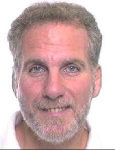
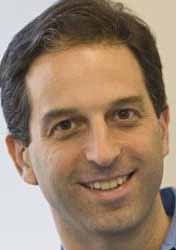
Deborah Sterling, Director, Sterne, Kessler, Goldstein & Fox PLLC, United States of America
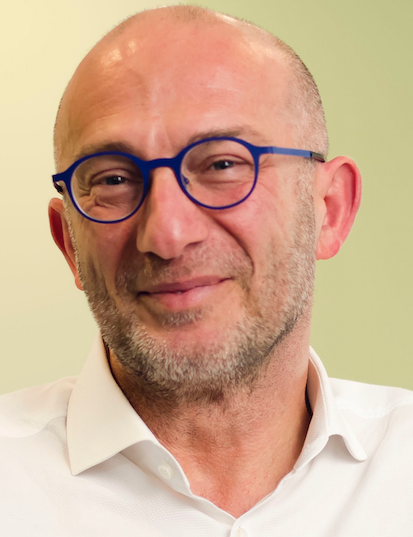
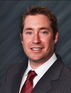
Stephanie Elmer, Associate, Sterne, Kessler, Goldstein & Fox PLLC, United States of America
A company seeking to make, use, or sell bioprinted organs and tissues must consider the patent landscape. This talk will focus on ways to protect bioprinted organs and tissues, patent filings directed to bioprinted organs and tissues, certain exceptions to patent infringement, as well as the possibility of future litigation. |
|
Session Title: Emerging Themes in Tissue Engineering |
| |
08:00 | Scaffold-Free Biofabrication with the ‘Regenova Bio 3D Printer’
Nicanor Moldovan, Associate Research Professor, Departments of Biomedical Engineering and Ophthalmology, Indiana University-Purdue University Indianapolis, United States of America
Bioprinting is one of the most advanced forms of biofabrication. ‘Scaffold-free’ bioprinting relies only on the cells’ ability to reconstitute complex bio-similar tissues, without the need of an artificial material as bioprinting support (or ‘scaffold’). I will illustrate this concept with the case of ‘Regenova Bio 3D Printer’, commercialized in Japan by Cyfuse Biomedical K.K., and in US by its subsidiary Amuza, Inc. This technology is now available in our recently-established 3D Bioprinting Core facility that serves the researchers at our University and in Midwest USA. |
08:30 | Cardiovascular Tissue Engineering using 3D Printing Technology
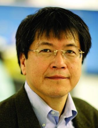
Narutoshi Hibino, Assistant Professor, Cardiac Surgery, Johns Hopkins Hospital, United States of America
Cardiovascular disease is one of the leading causes of death worldwide despite the variety of medical, mechanical, and surgical strategies. We have developed novel 3D printing technologies that could change the practice of cardiovascular disease treatment, including patient specific 3D printed tissue engineered vascular graft and bio 3D printed cardiac tissue. I will discuss insights of these new 3D printing technology as well as challenges we need to overcome for future clinical application and commercialization. |
09:00 | Recent Advances in Micro-Liver Tissue Engineering
Berk Usta, Instructor in Surgery, Massachusetts General Hospital – Harvard Medical School – Shriners Hospitals for Children, United States of America
The liver performs many key functions such as serving as the metabolic hub of the body. Accordingly, the liver is the focal point of many investigations aimed at understanding an organism’s toxicological response to endogenous and exogenous challenges. In this talk, I will review the recent advances in micro-tissue engineering by other groups and then focus on our own work at the Center for Engineering in Medicine at the Massachusetts General Hospital. Main subject areas will be our work on producing viable micro human/rat liver models, alternative biomaterials for micro-tissue engineering and finally our recent efforts towards recapitulation of physiological zonation of the liver. |
09:30 | Stimuli-Responsive Biomaterials for 3D Printing
Rachael Oldinski, Assistant Professor, Department of Mechanical Engineering, University of Vermont, United States of America
The development of stimuli-responsive, or ‘smart’ biomaterials is critical to the manipulative manufacturing of custom tissue engineering scaffolds and implants. This talk will focus on the modification of a seaweed derivative, alginate, for the design of sustainable materials for tissue engineering construct development using 3D printing technology. |
10:00 |  | Keynote Presentation A Modular Prefab Approach to Building Macrotissues
Jeffrey Morgan, Professor of Medical Science and Engineering, Brown University, United States of America
This presentation will focus on the use of micro-molds to direct the self-assembly of cells into large modular microtissues with complex geometries and a new instrument (BioP3) that picks, places and perfuses these modules as they are stacked and undergo fusion into a single macrotissue. |
|
10:30 | Coffee Break and Networking in the Exhibit Hall: Visit Exhibitors and Poster Viewing |
11:00 | Tissue Engineered Microenvironments For Studying Human Tumor Metastasis
Jungwoo Lee, Assistant Professor, University of Massachusetts-Amherst, United States of America
Disseminated tumor cells (DTCs) undergo varying periods of dormancy in ectopic tissue sites before developing overt metastatic tumors. Accumulating evidence suggests that metastatic dormancy and recurrence of DTCs is regulated by intrinsic genetic instability and close interaction with the surrounding microenvironment. Yet, the mechanism by which DTCs enter and escape dormancy remains largely uncertain due to the lack of model systems that can capture the initial activity of extremely rare DTCs. Here, we introduce a bioengineered approach to capture the critical events regulated by the extrinsic tissue microenvironments on DTCs with high experimental control and fidelity. We first developed human soluble factor enriched and vascularized microenvironments by subdermally implanting human bone marrow stromal cell preseeded scaffolds into immunodeficient NSG mice. Humanized tissue analogues recruited circulating human tumor cells released from physiologically relevant orthotopic xenograft tumors. Tail-vein delivery of human peripheral blood mononuclear cells further increased cellular complexity. Established human stromal-tumor-immune niches were serially transplanted into naïve NSG mice for continuous monitoring of metastatic tumor development. Our approach successfully recapitulated the heterogeneous phenotypes, dormant and aggressive, of DTCs and demonstrated human stromal and immune cell derived niche regulation. |
11:30 | Beyond Tissue Printing: Liquid Like Solids Support 3D Cell Science and Engineering
Thomas Angelini, Professor, Department of Mechanical and Aerospace Engineering, University of Florida, United States of America
Throughout the broad areas of tissue engineering, cellular therapeutics, and personalized medicine, there is an immediate need for research platforms with the facility and versatility of cell culture that rival or surpass the effectiveness of animal models, allowing biochemical, environmental, and mechanical parameters to be tuned precisely for each tissue, disease, patient and test condition. In this presentation I will describe a recently developed three-dimensional microenvironment for cell culture, as well as a 3D printing technique for creating micro-tissues within this 3D growth medium. The method leverages the fluid-solid jamming transition within liquid-like solids, which allows robotically controlled nozzles to create cellular structures, deposit molecules, and exchange fluids directly to suspended cell populations. This system enables the large scale, rapid generation of micro-tissues of reproducible size and geometry with access to nutrients or exogenously added bioactive molecules through unrestricted diffusion. This research is the focus of an ongoing collaboration between multiple laboratories from the college of engineering and the college of medicine at the University of Florida. The ultimate goal is to discover the chemical and physical environmental conditions necessary to create in vitro replicas of in vivo tissue groups for high-throughput drug screening, and implantation for tissue therapy or repair. |
12:00 |  | Keynote Presentation Scalable Biofabrication and Systematic Characterization of Tissue Spheroids for Directed Tissue Self-Assembly Using Acoustic Waves
Elena Bulanova, Head of Cell Technologies, 3D Bioprinting Solutions Russia, Russian Federation
Tissue spheroids are gaining extensively their place in biofabrication
as building blocks. We suggested a straightforward procedure for
biofabrication and initial characterization of tissue spheroids with
optimal controllable parameters prepared from different cell types
employing non-adhesive technology. Applying different immortalized and
primary cells we have demonstrated the reproducibility and scalability
of spheroid generation, the strong dependency of ultimate spheroid
diameter on initial cell seeding density and cell type. Additionally,
the spheroids viability was shown to be governed by cell derivation. In
our study we suggest a decision procedure to apply for any cell type one
starts to work with to prepare a new type of tissue spheroids with
predictable controllable optimal features suitable for high quality
standards in biofabrication and drug discovery. |
|
12:30 | 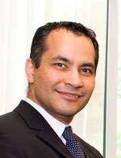 | Keynote Presentation Nano- and Microfabricated Hydrogels for Regenerative Engineering
Ali Khademhosseini, Professor, Department of Medicine, Brigham and Women’s Hospital; Wyss Institute for Biologically Inspired Engineering, Harvard University, United States of America
Engineered materials that integrate advances in polymer chemistry, nanotechnology, and biological sciences have the potential to create powerful medical therapies. Our group aims to engineer tissue regenerative therapies using water-containing polymer networks, called hydrogels, that can regulate cell behavior. Specifically, we have developed photocrosslinkable hybrid hydrogels that combine natural biomolecules with nanoparticles to regulate the chemical, biological, mechanical and electrical properties of gels. These functional scaffolds induce the differentiation of stem cells to desired cell types and direct the formation of vascularized heart or bone tissues. Since tissue function is highly dependent on architecture, we have also used microfabrication methods, such as microfluidics, photolithography, bioprinting, and molding, to regulate the architecture of these materials. We have employed these strategies to generate miniaturized tissues. To create tissue complexity, we have also developed directed assembly techniques to compile small tissue modules into larger constructs. It is anticipated that such approaches will lead to the development of next-generation regenerative therapeutics and biomedical devices. |
|
13:00 | Networking Lunch in the Exhibit Hall: Visit Exhibitors and Engage with Your Fellow Delegates |
13:30 | Luncheon Presentation: 3D Tissue Printing for Clinical Cardiovascular Applications
Jonathan Butcher, Associate Professor, Biomedical Engineering, Cornell University, United States of America
|
|
Session Title: The Convergence of 3D-Bioprinting and Tissue Engineering Fields and Synthetic Biology Applications Development |
| |
14:00 | 3D Bioprinting of GelMA Scaffolds Triggers Mineral Deposition by Primary Human Osteoblasts
Christine McBeth, Senior Research Scientist, Fraunhofer Center for Manufacturing Innovation, United States of America
We discuss our bioprinter that can generate osseoinductive structures that are directly grafted to implants destined for orthopedic applications. We further describe our novel hybrid bioinks that are specifically designed to meet both the demands of bioprinting and downstream applications. |
14:30 | Toward Building Complex Biomaterial Matrix Microenvironments by Low-Voltage Continuous Electrospinning Patterning
Yan Yan Shery Huang, Professor of BioEngineering, University of Cambridge, United Kingdom
The creation of more complex tissue models in vitro points to the need for controlled assembly of artificial and biological material architectures in two dimensional (2D) and three dimensional (3D) space. Existing biomaterial fabrication techniques (e.g., 3D printing, electrospinning, and templating) fall short in building the complex combinations of chemical and structural elements, with limited feature resolution. Our recent development in low-voltage electrospinning patterning (LEP), and its combination with additive manufacturing, opens up new avenues in the creation of geometrically defined biomaterial matrices. This presentation will demonstrate LEP’s applications in fabricating bio-scaffolds, single-fibres with multi-scale morphologies, and the printing of microelectrodes onto flexible substrates. |
15:00 | Perfusion Directed 3D-Bone Mineralization
Pranav Soman, Professor, Biomedical and Chemical Engineering; IPA Program Director, Advanced Manufacturing (AM), National Science Foundation (NSF), Syracuse University, United States of America
An inherent challenge in conventional tissue engineering strategies is the ability to efficiently deliver nutrients throughout the thickness of a complex, physiologically relevant biomimetic construct. In lieu of adequate interstitial perfusion, cellular viability and physiological function is compromised. In this work, we will present the creation of structurally supported, perfusable hydrogels capable of growing bone in user defined directions. Briefly, bone-like human osteosarcoma cells were encapsulated inside UV cross-linkable gelatin methacrylate (GelMA) hydrogels, and this cell-hydrogel mixture was casted onto a 3D printed poly(vinyl alcohol) (PVA) structure. PVA serves as a sacrificial material and was dissolved away to obtain hollow channels to facilitate the perfusion of media using a custom-made acrylonitrile butadiene styrene (ABS) bioreactor. Osteogenic media was perfused through the channels, and the radial zones of bone mineralization surrounding the channels were quantified. This study demonstrates that user-defined 3D printed channels can be used to spatially control bone mineralization. |
15:30 | Modification of Bacterial Nano-cellulose with IKVAV Peptide for Tissue Engineering Applications
Luismar Porto, Associate Professor, Federal University of Santa Catarina (UFSC) Brazil , Brazil
The fabrication of suitable porous polymer scaffolds which mimic natural tissues is still one of the main challenges of tissue engineering. Bacterial nanocellulose (BNC) hydrogels possesses a nanofiber network that resembles the native extracellular matrix. The immobilization of bioactive molecules into its microstructure may add functionality and improves support for adhesion and proliferation of human cells. The main goal of this work was the functionalization of BNC hydrogel membrane with IKVAV peptide (BHMI) to enhance the biomaterial. BNC membranes were produced by Gluconacetobacter hansenii. BHMIs were prepared by oxidation reaction of BNC followed by chemical derivatization with EDC (1-ethyl-3-(3-dimethylaminopropyl) carbodiimide) and functionalization with IKVAV. Scanning electron microscopy imaging of BNC before oxidation showed a clear 3D nanofiber network structure; after IKVAV immobilization, granules of the peptide were formed on the nanofiber surface. Functionalization was confirmed by Fourier Transformed Infrared Spectroscopy. Human umbilical endothelial cells (HUVECs) were aseptically seeded on BHMI and, after 48 hours, the functionalized scaffold was able to promote HUVEC adhesion and proliferation. Cell alignment, spreading and endothelial tube organization was also observed. Our results showed that BNC-IKVAV hydrogels supports tubulogenesis in vitro showing that they are a very versatile biomaterial. |
16:00 | Harnessing the Inflammatory Response for Tissue Regeneration
Kara Spiller, Assistant Professor, Drexel University, United States of America
The inflammatory response plays a major role in the body’s response to injury, disease, or implantation of a biomaterial. When the inflammatory response functions normally, it can be a powerful force that promotes tissue repair and regeneration, but when it goes awry, disease takes hold and healing is impaired. The goal of the Biomaterials and Regenerative Medicine Laboratory at Drexel University is to understand the mechanisms by which the inflammatory response orchestrates successful tissue regeneration and to develop novel biomaterial strategies that apply these principles to situations in which tissue regeneration is impaired. In particular, we focus on the behavior of the macrophage, which can rapidly change behavior in response to environmental stimuli to promote inflammation (M1), tissue deposition (M2a), or remodeling (M2c). Through their dynamic phenotypic changes, macrophages function as major regulators of healing. Current emphasis is on tracking macrophage phenotype changes in the healing (or lack thereof) of human chronic diabetic foot ulcers, which holds potential to allow a personalized medicine approach to wound care. We will also discuss novel drug delivery strategies that harness macrophage behavior to promote tissue regeneration and healing in a diverse array of tissues. |
16:30 | 3D Printing of Patient-Specific Brain Cancer Model
Hee-Gyeong Yi, Researcher, Pohang University of Science and Technology, Korea, Korea South
Demand for a patient-specific cancer model is critical to predicting the valuable clinical benefit of new drug combinations for personalized medicine. Since there are large numbers of drug candidates, the test solely relying on animals requires the prohibitively expensive and time-consuming procedures. Although the ex vivo cancer models are favorable to conduct high-throughput screening, their translational applications have been hindered by the inability to reflect the tumor micro-environment including three-dimensional (3D) extracellular matrix (ECM) and the cancer-associated cells. A challenge is to simultaneously mimic the dimensionality, complexity, and heterogeneity of the original environment. Here, we describe 3D printing technology for generating a highly biomimetic platform that replicates the heterogeneous tumor micro-environment and thereby can be a patient-specific model of human brain cancer. Our approach is demonstrated to have a promise to replicate the patient's brain cancer. We expect that our approach is probably applicable to other cancers.
|
17:00 | Engineering Lungs for Transplantation: Harnessing the Regenerative Potential of Human Airway Stem Cells
Sarah Gilpin, Massachusetts General Hospital (MGH), Harvard Medical School, Instructor, Department of Surgery, United States of America
This presentation will discuss our progress toward bioengineering transplantable human lungs, and describe the use of tissue-derived airway stem cells for ex vivo lung regeneration. |
17:30 | Origami-Enabled Tissue Engineering
Carol Livermore, Associate Professor, Department of Mechanical and Industrial Engineering, Northeastern University, United States of America
Replicating liver structure in engineered tissue is challenging because of liver’s dependence on the effective diffusion of nutrients and metabolic byproducts and because of the massive parallelism of its fine structure. Origami-based microfluidics offers a new paradigm for addressing these challenges. Folding offers a low-cost, rapid means of creating larger scale fluidic structure to mimic vasculature; co-folding of porous and nonporous materials permits diffusion between hepatocytes and “engineered sinusoids”; and directed cell assembly offers control of cell organization at smaller length scales for the creation of hierarchical architectures. This talk will present the enabling tools of origami tissue engineering and their demonstration through the design, fabrication, and biological characterization of origami liver tissue units. |
18:00 | Close of Day 2 of the Conference. |
