08:00 | Conference Registration, Materials Pick-Up, Morning Coffee and Tea |
|
Session: Conference Opening Session and Cell Therapy Update |
| |
08:30 | 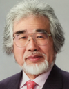 | Conference Chair Conference Chairman Welcome and Opening Remarks and Introduction to the Cell Therapy Space, circa 2018
Norio Nakatsuji, Chief Advisor, Stem Cell & Device Laboratory, Inc. (SCAD); Professor Emeritus, Kyoto University, Japan
|
|
09:00 | 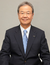 | Conference Chair Conference Chairman Welcome and Opening Remarks and State of Regenerative Medicine Industry
Yuzo Toda, President, FIRM (Forum for Innovative Regenerative Medicine, Japan), Japan
|
|
09:30 | 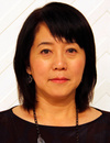 | Keynote Presentation Retinal Cell Therapy Using iPS Cells
Masayo Takahashi, Professor, RIKEN Center for Biosystems Dynamics Research, Japan
The first in man application of iPS-derived cells started in September 2014, targeted age-related macular degeneration (AMD). AMD is caused by the senescence of retinal pigment epithelium (RPE), so that we aimed to replace damaged RPE with normal, young RPE made from iPS cells. We judged the outcome 1 year after the surgery. Primary endpoint was the safety, mainly the tumor formation and immune rejection. The grafted RPE cell sheet was not rejected nor made tumor after two years. The patient’s visual acuity stabilized after the surgery whereas it deteriorated before surgery in spite of 13 times injection of anti-VEGF in the eye. Since autologous transplantation is time consuming and expensive, it is necessary to prepare allogeneic transplantation to establish a standard treatment. RPE cells are suitable for allogeneic transplantation because they suppress the activation of the T-cell. From in vitro and in vivo study, it is possible that the rejection is considerably suppressed by using the iPS cell with matched HLA. Our new protocol has accepted by ministry in Feb 2017. We are planning transplantation using allogeneic iPS-RPE cell suspension & sheet, and also autologous iPS-RPE. For the cell suspension transplantation we will not combine CNV removal and apply to milder cases than sheet transplantation. In Japan, pharmaceutical law has been changed and a new chapter for regenerative medicine was created for clinical trial. Also the separate law for safety of regenerative medicine for clinical research (study) was enforced in 2015. These laws made the suitable condition for the brand new field of regenerative medicine. We are making regenerative medicine in co-operation with ministry & academia. |
|
10:00 |  | Keynote Presentation Regenerative Medicine in Astellas
Yoshitsugu Shitaka, President, Astellas Institute for Regenerative Medicine, Japan
Astellas Institute for Regenerative Medicine (AIRM) was established in May 2016 following Astellas’ acquisition of Ocata Therapeutics. AIRM is an indirect, wholly owned subsidiary of Astellas and serves as Astellas’ global hub for RM and cell therapy in ophthalmology and other therapeutic areas that have few or no available treatment options. The outline of Astellas RM will be reviewed in this presentation. |
|
10:30 | Morning Coffee and Tea Break and Networking |
11:00 | Industrialization of Regenerative Medicine - Standards are the Key
Tatsuo Heki, Senior Expert, Regenerative Medicine Division, Fujifilm Corporation, Japan
Industrialization of a particular field means making its product/service available anytime, anywhere and to anybody. Written standards in the field of regenerative medicine will form a common international “language” for use by the stakeholders including manufacturers, suppliers and regulators to support effective delivery of cell-based medicines, and is the centerpiece of industrialization. ISO/TC 276 (International Organization for Standardization Technical Committee 276) Biotechnology. is developing standards in the areas of terms and conditions, biobanks and bioresources, analytical methods, bioprocessing and data processing in the field of biotechnology processes. ISO/TC 276 will contribute to regenerative medicine by establishing a common language for analytical methods and bioprocessing so that there is a more harmonized understanding by the stakeholders in the efforts from translation of research to transportation of cells for therapeutic use.
|
11:30 | Derivation of Clinical-Grade hESC in Japan
Hirofumi Suemori, Associate Professor, Laboratory of Embryonic Stem Cell Research, Institute for Frontier Life and Medical Sciences, Kyoto University, Japan
After obtaining governmental permission to derive clinical-grade hESC lines in 2017, we have been trying to produce hESC in compliant with relevant regulations and guidelines. Progress of our project will be presented.
|
12:00 | Networking Lunch |
|
Session Title: Cellular Classes for Cell Therapy and Current Research |
| |
13:00 |  | Keynote Presentation Translating Stem Cell Research: Challenges At Front Line
Hiromitsu Nakauchi, Prof of Genetics, Institute of Stem Cell Biology and Regenerative Medicine, Stanford University School of Medicine Project Professor, The Institute of Medical Science, Division of Stem Cell Therapy, Distinguished Professor Unit, The University of Tokyo, United States of America
The recent development of induced pluripotent stem cell (iPSC) technology opened the way to regenerative medicine using a patient’s own PSC-derived cells. In this presentation, I describe some of our iPSC technology-based projects aiming at clinical use. |
|
13:30 | Early Onset Preeclampsia (EOPE) in a Model For Early Stage Human Placental Trophoblast
Toshihiko Ezashi, Research Associate Professor, The University of Missouri, United States of America
We have established a model for early onset preeclampsia (EOPE) that uses induced pluripotent stem cells (iPSC) generated from umbilical cords of infants born to mothers who experienced EOPE. These iPSC lines were converted to placental trophoblast (TB), thus recapitulating early stage aspects of the pregnancies they represented. Eight control (CTL) iPSC-TB and 14 EOPE iPSC-TB were tested to assess their abilities to invade through Matrigel. Under 5 % O2, CTL-TB and EOPE-TB lines did not differ, but, under the more stressful 20 % O2 conditions, invasiveness was lower in EOPE-TB than in CTL-TB (P = 0.024). Although invasiveness of CTL-TB was not influenced by 20 % O2, that of EOPE-TB was markedly reduced (P = 0.008) compared to 5% O2. Placental growth factor (PGF) production also decreased (P < 0.05) in EOPE cultures under 20 % O2. RNAseq analysis revealed only two differentially expressed genes (RPS17, FDR = 0.0005; MTRNR2L2, FDR = 0.005) significantly down-regulated in EOPE-TB under 20 % O2. A weighted correlation network analysis (WGCNA), revealed ten gene modules in CTL lines, three of which were correlated with TB invasion. Of these three, two were also linked to O2 responsiveness and were enriched in ontology terms related to cell migration and angiogenesis. Of the four gene modules identified in EOPE-TB, none were correlated with invasion, and two were correlated with O2 responsiveness. These results indicate that, under high O2 and possibly other stressful conditions, invasion by EOPE-TB becomes dysregulated, which helps to reveal the underlying pathology of shallow invasion of TB in the EOPE placenta. The results also indicate the value of the iPSC approach to studying EOPE. |
14:00 | Afternoon Coffee and Tea Break and Networking |
|
Session Title: Multidisciplinary Research Update |
| |
15:00 |  | Keynote Presentation Cell Fiber Technology For 3D Tissue-on-a-Chip Application
Shoji Takeuchi, Professor, Center For International Research on Integrative Biomedical Systems (CIBiS), Institute of Industrial Science, The University of Tokyo, Japan
The talk describes a 3D cell culture method using core-shell hydrogel microfibers (cell fibers). The core is filled with cells and ECM proteins, and the shell is composed of calcium alginate. Since the core diameter is about 100 microns, oxygen and nutrients can be diffused into the central area of the 3D tissue; therefore this culture system allows us to culture the tissue for a long period without central necrosis. Using this culture system, fiber-based tissues such as blood vessels, nerves, and muscles can be formed with in the core. Here, I will discuss the application of the cell fiber technology for various tissue-on-a-chip studies including cardiac tissue, neuro-muscular junctions, and vascularized skin etc. |
|
15:30 |  | Keynote Presentation Intelligent Image-Activated Cell Sorting
Keisuke Goda, Professor of Chemistry, University of Tokyo and Adjunct Professor, Wuhan University, Japan
A fundamental challenge of biology is to understand the vast heterogeneity of cells, particularly how cellular composition, structure, and morphology are linked to cellular physiology. Unfortunately, conventional technologies such as fluorescence-activated cell sorting are limited in uncovering these relations. In this talk, I introduce our machine intelligence technology known as “Intelligent Image-Activated Cell Sorting” [Cell 175, 1 (2018)] that builds on a radically different architecture that realizes real-time image-based intelligent cell sorting at an unprecedented rate. This technology integrates high-throughput cell microscopy, focusing, and sorting on a hybrid software-hardware data-management infrastructure, enabling real-time automated operation for data acquisition, data processing, decision-making, and actuation. I show the application of the technology to real-time image-activated sorting of microalgal and blood cells based on intracellular protein localization and cell-cell interaction from large heterogeneous populations for studying photosynthesis and atherothrombosis, respectively. The technology is highly versatile and expected to enable machine-based scientific discovery in biological, pharmaceutical, and medical sciences including cell therapy.
|
|
16:00 |  Bioprinting of 3D Tissue Constructs for Cell Therapy Applications Bioprinting of 3D Tissue Constructs for Cell Therapy Applications
Matthew Mail, Scientific Application Specialist, CELLINK
Cell behavior and orientation in vitro depends on the microenvironment of the cells. CELLINK’s cutting edge 3D Bioprinting technology can be used to make 3D tissue constructs that encapsulate cells, allowing them to act as they would in vivo. One such application is for diabetes modelling, using beta cells.
|
16:30 | Creation of Functional Tissue with a Novel 3D Printing Technology
Shizuka Akieda, Chief Executive Officer, Cyfuse Biomedical K.K., Japan
Cyfuse is a regenerative medicine start-up focused on creation of functional 3D tissues and organs. The proprietary “Kenzan method” skewers multiple cellular aggregates with fine needles until cells fuse entirely in a few days. Our Bio 3D Printer, “Regenova” automated this skewering process and is designed to help researchers try various cell populations and culture conditions to discover a protocol of manufacturing functional organs. The system was launched commercially in Japan and US. The examples of cellular products include Blood Vessels, Peripheral Nerve Regeneration, and functional liver for drug discovery. Further applications with neural cells and cardiomyocytes are currently being explored by Japanese and US academia. |
17:00 | Close of Day 1 of the Conference. |


