08:00 | Conference Registration and Materials Pick-Up + Coffee in the Exhibit Hall |
09:00 |  | Conference Chair Welcome and Introduction by Conference Chairperson -- Scope of the Conference and Topics Covered + Modular Design Workflows for 3D Printed Microfluidics
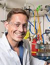
Noah Malmstadt, Professor, Mork Family Dept. of Chemical Engineering & Materials Science, University of Southern California, United States of America
3D printing brings with it a plethora of advantages for microfluidic applications. Principle among these are rapid prototyping, iterative design, and the ability to avoid the cost and overhead of cleanrooms. However, there is also an inherent advantage in being able to design and build devices in a truly three-dimensional, rather than layer-by-layer, geometry. One simple domain in which the advantages of true 3D routing are clear is in mixing. Control over a 3D geometry allows for multiple complex mixing configurations--herringbones, relamination mixers, chaotic advection--to be trivially constructed and recombined.
We have used these principles of 3D design to construct devices and systems for bioanalytical assays, for manufacturing biomaterials, and for industrial-scale manufacturing of novel materials. This talk will examine all of these applications and the manner in which 3d-centric microfluidic design can enable them. |
|
09:30 |  | Keynote Presentation Exploring the Potential of Microfluidics for Lab Automation
Burcu Gumuscu, Assistant Professor Biosensors and Devices Lab, Eindhoven University of Technology, Netherlands
Digital microfluidics (DMF) chips have garnered increasing attention over the past decade thanks to their ability to address individual droplets. These chips consist of an array of mm-sized electrodes to manipulate liquid-based, individually addressable droplets through applied voltages. Programmed sub-microliter scale droplets performing basic pipetting operations paved the way for the automation of laborious assays. Automated biological assays are an exciting application of DMF, including DNA-based analysis, electroanalysis, and short-term cell culture experiments. However, there are still limitations to be overcome. DMF chips are not yet able to excel in (1) multiplexed operations due to the 2D planar fabrication of operational units, limiting the number of operation units per chip, (2) long-term cell studies due to the typical mismatch of electronics devices and high humidity conditions in typical cell-culture incubators. In this talk, I will discuss these challenges and propose potential solutions to enhance the capabilities of DMF chips for automated biological assays. |
|
10:00 | Computer Simulations of Microfluidics – What Can We Learn, and How Can They Help Us?
Timm Krüger, Professor of Fluid and Suspension Dynamics, The University of Edinburgh, United Kingdom
Microfluidics has become ubiquitous in the past decades. Exploiting various physical and chemical mechanisms, microfluidics offers fast and affordable solutions to pressing problems in industry and healthcare. However, due to the complexity and interplay of the involved mechanisms and geometries, it is notoriously difficult to reliably predict outcomes and design microfluidic devices. Computer simulations can help shed light on the underlying mechanisms and facilitate the design process. I will highlight the opportunities for experimental/numerical synergies to advance microfluidics further. |
10:30 | Mid-Morning Coffee Break and Networking in the Exhibit Hall |
11:00 | Ultrasound – A New Tool for Biofabrication
Kai Melde, Group Leader, Heidelberg University, Germany
Biofabrication includes methods that print bioinks or directly assemble biological components (e. g. cells, microgels or spheroids) with the goal of creating functional tissues. Most of these methods require direct mechanical access (e. g. extrusion printing or aspiration-based pick & place) and work serially (point-by-point or layer-by-layer), which scales poorly to volumetric shapes and applies undue stress on biological cells. Over the last decade, ultrasound emerged as a tool for contactless manipulation of matter, including microparticles and biological cells. Its ease-of-use and favorable operating parameters led to a wide adoption especially in the microfluidics community (e. g. for cell sorting). However, the effects of ultrasound can act over much larger length scales, provided that there is sufficient control over the sound field. In this talk, I will present how ultrasound combined with holographic beamforming enables us to create very complex sound fields, which can direct the parallel assembly of matter to arbitrary shapes in 3D. Our technique is compatible with standard labware (such as sample tubes, culture inserts or cuvettes) and shown to work with solid microparticles, hydrogel beads and mammalian cells. As such, ultrasound promises to become a new tool for biofabrication, enabling the formation of cell aggregates without contact. |
11:30 |  | Keynote Presentation Prototyping of Microphysiologic Cell Culture Systems That Mimic Key Aspects of the Human Body
Mandy Esch, Project Leader, National Institute of Standards and Technology (NIST), United States of America
Single and multi-organ microphysiologic systems (MPS) can be used to detect secondary drug toxicities stemming from drug metabolites. Here we describe how to design and prototype such systems to replicate key aspects of the human body that influence the concentration of drug metabolites within the system. Using 3D printing we have prototyped and tested several microfluidic MPS that can recirculate near-physiological amounts of cell culture medium. For example, the body cube represents a part of the human body that is scaled down by a factor of 73000. It can recirculate 80 µL of medium (the equivalent of 5L to 6L of blood scaled down by a factor of 73000). We have also developed several devices that recirculate small amounts of cell culture medium in a way that makes it feasible to culture mechanosensitive cells such as HUVEC or GI tract epithelial cells within the system. The talk given here is a summary of our efforts in this area. |
|
12:00 |  | Keynote Presentation 3D Printing – Implementation and Integration of 3D Printing in the Development of Microreactors and their Applications
Stephen Hilton, Associate Professor, University College London School of Pharmacy, United Kingdom
3D printing is a key enabling technology that allows scientists to enhance the research that they do at low cost. In this talk, we will describe our journey into the 3D printing of microreactors and the development of key printing techniques to enable this along with novel materials that are chemically resistant. The talk will focus on the enabling potential of 3D printing and highlight key digital developments by the group to enable other researchers to easily implement this key technology in their laboratories. |
|
12:30 | Networking Buffet Lunch in the Exhibit Hall -- Networking with Colleagues, Engage with Exhibitors and View Posters |
14:00 | SOIL-ON-A-CHIP: Deciphering the Secret Life of Soil Microbes Using Novel Microfluidic Platforms
Claire Stanley, Senior Lecturer in Bioengineering, Imperial College London, United Kingdom
Soil is one of the most complex systems on Earth, governed by numerous physical, geochemical and biological processes, and provides the ecosystem services vital for all forms of terrestrial life. This ‘material’ supports a myriad of plants, microorganisms and microfauna and hosts a complex array of interactions taking place between these living elements at the cellular scale. Microbes play a crucial role in the ecosystem services provided by soils to humans and provide several important ecosystem functions that include nutrient cycling, the biocontrol of pathogens and regulation of greenhouse gas emissions. However, despite the importance of microbes in soil functioning, there exists a major knowledge gap concerning the function and dynamics of the soil microbiome and influence of the physio-chemical environment upon microbial interaction and communication at the cellular level. The ability to untangle microbial interaction and communication networks in soil is central to gaining an enhanced understanding of soil microbiome and ecosystem function. In recent years, it has been demonstrated that microfluidic technology offers new opportunities to study whole living organisms and their interactions at cellular level, affording precise environmental control, high-resolution imaging and the simulation of environmental complexity. Several microfluidic systems have been developed to probe interactions between fungi, bacteria and nematodes, as well as the interaction of plant roots with their environment. My lab is now developing new microfluidic tools to investigate the cell biology and physiology of microbial spore germination and arbuscular mycorrhizal fungi hyphal growth dynamics. |
14:30 | Engineering Synthetic Cells from the Bottom-Up
Tom Robinson, Lecturer in Chemical Engineering, University of Edinburgh, United Kingdom
One of the aims of synthetic biology is the bottom-up construction of synthetic cells from non-living components. Building biomimetic cells and controlling each aspect of their design not only provides the opportunity to understand real cells and their origins, but also offers alternative routes to novel biotechnologies. Giant unilamellar vesicles (GUVs) are commonly used as scaffolds to construct synthetic cells owing to their compatibility with existing biological components, but traditional methods to form them are limited. Microfluidic-based approaches for GUV production show great potential for encapsulating large biomolecules required for mimicking life-like functions (Yandrapalli et al. Micromachines, 11, 285, 2020; Love et al. Angew Chemie, 59, 5950–5957, 2020). First, I will present a microfluidic platform that is able to produce surfactant-free pure lipid GUVs in a high-throughput manner (Yandrapalli et al. Commun Chem, 4, 100, 2021). The major advancement is that the lipid membranes are produced in the absence of block co-polymers or surfactants that can affect their biocompatibility - which is commonly overlooked. The design can produce homogenously sized GUVs with tuneable diameters from 10 to 130 µm. Encapsulation is uniform and we show that the membranes are oil-free by measuring the diffusion of lipids via FRAP measurements. Next, I will present how we modified this device to encapsulate two sub-populations of nano-sized vesicles for the purpose of establishing enzymatic cascade reactions across membrane-bound compartments, therefore mimicking eukaryotic cells (Shetty et al. ACS Nano, 15, 15656, 2021). The final synthetic cell comprises three coupled enzymatic reactions, which propagate across three separate compartments in a specific direction due to size-selective membranes pores. Not only does microfluidics provide a high degree of control over the intra-vesicular conditions such as enzyme concentrations, buffers, and the number of inner compartments, but the monodispersity of our synthetic cells allows us to directly compare the effects that compartmentalization has on the biochemical reaction rates and product yields. This work demonstrates the effectiveness of microfluidics for the bottom-up assembly of synthetic cells, and paves the way for novel biotechnologies in areas such as compound production, sensing, and drug delivery. |
15:00 | A Microfluidics-based Approach to Tackle Bone Tumors: Towards New Materials for Bone Metastases
Gabriela Graziani, Assistant Professor, Politecnico di Milano, Italy
Tumor relapse after surgical excision of bone metastases poses a key unmet challenge in orthopedic oncology due to its high incidence rate and potentially fatal outcome. Even in advanced cancer state, patients undergo treatment with bone substitutes/implants, to fill gaps resulting from excision surgery. In this groundbreaking study, we propose novel antitumor metal-based coatings to functionalize implanted devices and prevent tumor relapses. For their design and validation, we use a microfluidic-based approach, where chips are designed to select the optimal concentration of metal. To achieve this, we designed and realized a gradient generator microfluidic device, to be used for both tumor (breast cancer cells that metastasize in bone, MDA-MB-231) and healthy cells (mesenchymal stem cells, MSCs). This enables the detection of the optimal concentration for antitumor efficacy while avoiding cytotoxicity. In the chip, cell chambers are designed for cell seeding in a medium (2D configuration) and in a biomimetic gel (3D configuration) for pre-screening and validation, respectively. Coatings, manufactured by Ionized Jet Deposition, are dissolved in a simulated medium to obtain the solution to inject in the device. Cell viability, proliferation and migration are measured by calcein staining and fluorescence microscopy analyses in 6 parallel columns of chambers, each receiving a different dilution of the active compound. Three chambers per column allow for obtaining replicates simultaneously. Results demonstrate that the gradient generator microfluidic can detect the effect of different concentrations of the eluates on cells viability, migration and proliferation, with the latter parameters significantly more affected by metals. Hence, by merging results on MSCs and MDA, the device enables a fine selection of the metal concentration to be used. Cellularised biomimetic gels (collagen and Matrigel based) can be injected into the chambers without creating defects or hampering cell visualization. This new approach appears promising for the design of coatings, and can be easily transferred to validate different biomaterials. |
15:30 | Mid-Afternoon Coffee Break and Networking in the Exhibit Hall |
16:00 | Discussion with Professor Malmstadt on Emerging Themes and Topics in Microfluidics |
18:00 | Networking Reception with Dutch Beer - Network and Engage with Colleagues in a Social Setting |
19:00 | Close of Day 1 of the Conference |


