Alexander Schreiner,
Team Leader Biological Applications,
PerkinElmer
As Team Leader of the Biological Applications Team at PerkinElmer in Hamburg Germany, Dr. Alexander Schreiner is responsible for developing applications and providing application related support for PerkinElmer’s high content screening systems. He holds a doctorate in Biology (molecular cell biology) from the Goethe University in Frankfurt. During his PhD he worked on the identification and analysis of protein-protein interactions. Following a postdoc period in the lab of Prof. Starzinski-Powitz in Frankfurt where he worked on the analysis of unusual long signal peptides he moved to the UK to work as a postdoc at the Cancer Research UK Cambridge Research Institute in the group of Prof. Fiona Watt, investigating the proliferative effect of aberrantly expressed integrins in the interfollicular epidermis. After this he worked in in the Light Microscopy Facility of the same Institute where he provided support and training for the different microscope systems within the facility.
More Content from High-Content Screening: Analyzing Specimen in 3D
Friday, 25 May 2018 at 09:00
Add to Calendar ▼2018-05-25 09:00:002018-05-25 10:00:00Europe/LondonMore Content from High-Content Screening: Analyzing Specimen in 3DHigh-Content and Phenotypic Screening Europe 2018 in Cambridge, UKCambridge, UKSELECTBIOenquiries@selectbiosciences.com
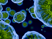
When developing a screening assay one of the most critical aspects is the choice of cellular model including the decision to choose between 2D or 3D approaches. Multicellular “oids” (tumoroids, spheroids, organoids) bear the potential to better predict drug candidate effects during preclinical screening. However, 3D models require more effort and compared to 2D cultures, 3-dimensional assays tend to be more complex across all steps of the workflow. Reliably generating sufficiently large numbers of uniform spheroids for screening and imaging of 3D volumes at high quality are among the challenges. Using 3D spheroids and cysts as examples, we will show experimental workflows, explaining how to effectively grow these models using ULA coated U-bottom plates or low concentration gels. The careful selection of dyes and clearing strategies can improve the image quality while targeted imaging of spheroids helps to significantly shorten imaging times and to minimize the data volume. Nevertheless, being able to effectively analyze spheroids is still one of the main bottlenecks. Dedicated high-content software tools can help users to, speed up 3D image acquisition by targeted imaging of objects of interest, better understand the spatial context of your 3D cell models through different visualization methods, measure volume and morphology changes in 3D to characterize your specimen in detail and export 3D movies to share and publish your results.
Add to Calendar ▼2018-05-24 00:00:002018-05-25 00:00:00Europe/LondonHigh-Content and Phenotypic Screening Europe 2018High-Content and Phenotypic Screening Europe 2018 in Cambridge, UKCambridge, UKSELECTBIOenquiries@selectbiosciences.com


 Add to Calendar ▼2018-05-25 09:00:002018-05-25 10:00:00Europe/LondonMore Content from High-Content Screening: Analyzing Specimen in 3DHigh-Content and Phenotypic Screening Europe 2018 in Cambridge, UKCambridge, UKSELECTBIOenquiries@selectbiosciences.com