Pinar Zorlutuna,
Sheehan Family Collegiate Professor of Engineering,
University of Notre Dame
Pinar Zorlutuna is the Sheehan Family Collegiate Professor of Engineering in Aerospace and Mechanical Engineering Department, Chemical and Biomolecular Engineering Department, and in Bioengineering Graduate Program at the University of Notre Dame. Her research explores designing biomimetic environments for understanding and controlling cell behavior, and cell-cell and cell-environment interactions using tissue engineering, genetic engineering and micro- and nanofabrication approaches. Dr. Zorlutuna received her PhD in Biotechnology Program from a joint project between Middle East Technical University (Ankara, Turkey) and Interdisciplinary Research Center in Biomedical Materials, Queen Mary University of London (London, UK). Her PhD work focused on biomimetic tissue engineering towards fabricating a functional blood vessel tissue through 3D tubular co-culture of vascular cell types using nanopatterned scaffolds. In her first postdoctoral fellowship with Rashid Bashir at UIUC, she worked on utilization of stereolithography for engineering microfabricated 3D neuro-muscular tissue as a first step towards engineering cell-based soft robots or “Bio-bots”. After that, she led Khademhosseini Lab’s Tissue Engineering Subgroup at the joint Harvard-MIT Division of Health Sciences and Technology and Center for Biomedical Engineering at Harvard Medical School, working on various projects and supervising a group of about ten researchers of different educational levels and backgrounds. Her research has been published in high impact journals such as Advanced Science, Advanced Materials, ACS Nano, Science Advances and Circulation Research. She received various awards including NSF CAREER Award and PECASE.
3D Bioprinting Physiologically Relevant Human Infarct Models and Cardiac Patches
Wednesday, 6 October 2021 at 11:00
Add to Calendar ▼2021-10-06 11:00:002021-10-06 12:00:00Europe/London3D Bioprinting Physiologically Relevant Human Infarct Models and Cardiac PatchesBioprinting and Bioink Innovations for 3D-Tissues in Virtual Event - Eastern Daylight Time (EDT) ZoneVirtual Event - Eastern Daylight Time (EDT) ZoneSELECTBIOenquiries@selectbiosciences.com
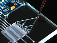
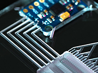
In the modern world, myocardial infarction (MI) is one of the most common cardiovascular diseases (CVDs) which are responsible for nearly 32% of all deaths causing almost 18 million people to die every year. Even though MI remains one of the leading CVDs, limited progress has been achieved with human MI models and therapeutic treatment options. 3D bioprinting has become one of the most powerful tools used to fabricate mimetic cardiac tissues, capturing the complexity of the native cellular composition and matrix structure. However, the generation of 3D tissue constructs with multiple cell types, matching mechanical properties, the ordered structure of the native extracellular matrix and the electroconductivity of the human heart remains a challenge in cardiac tissue engineering. To address the first two challenges, we developed novel bioinks combining gelatin methacryloyl (GelMA) or GelMA-methacrylated hyaluronic acid (MeHA) hydrogels with decellularized human cardiac extracellular matrix (dhECM), and characterized them in terms of mechanical, rheological, swelling, printability, and biocompatibility properties. Composite GelMA–MeHA–dhECM (GME) hydrogels demonstrated improved mechanical properties by an order of magnitude compared to the GelMA–dhECM (GE) hydrogels, which corresponds to the difference between the stiffness of healthy cardiac tissue (8–12 kPa) and scar tissue (>150 kPa) formed after myocardial infarction (MI). Knowing that, we printed an infarct region model using a dual printhead by mixing iCMs with GE to model the healthy tissue and hCFs with GME to represent the scar tissue. To address the latter challenges, we developed a new composite construct that can provide both conductive and topographical cues for iCMs by 3D printing conductive titanium carbide (Ti3C2Tx) MXene in pre-designed patterns on polyethylene glycol (PEG) hydrogels, using aerosol jet printing, at a cell-level resolution and then seeded with iCMs and cultured for one week with no signs of cytotoxicity.
Add to Calendar ▼2021-10-06 00:00:002021-10-06 00:00:00Europe/LondonBioprinting and Bioink Innovations for 3D-TissuesBioprinting and Bioink Innovations for 3D-Tissues in Virtual Event - Eastern Daylight Time (EDT) ZoneVirtual Event - Eastern Daylight Time (EDT) ZoneSELECTBIOenquiries@selectbiosciences.com


 Add to Calendar ▼2021-10-06 11:00:002021-10-06 12:00:00Europe/London3D Bioprinting Physiologically Relevant Human Infarct Models and Cardiac PatchesBioprinting and Bioink Innovations for 3D-Tissues in Virtual Event - Eastern Daylight Time (EDT) ZoneVirtual Event - Eastern Daylight Time (EDT) ZoneSELECTBIOenquiries@selectbiosciences.com