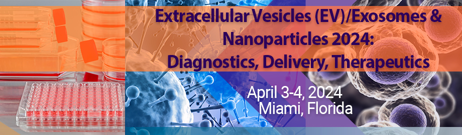
Co-Located Conference AgendasExtracellular Vesicles (EVs) & Nanoparticles 2024: Diagnostics, Delivery, Therapeutics | The Space Summit 2024 | 

Wednesday, 3 April 202407:30 | Conference Registration and Materials Pick-Up | 08:15 |  | Conference Chair Welcome and Introduction to the Conference by Conference Senior Co-Chairperson
Michael Graner, Professor, Dept of Neurosurgery, University of Colorado Anschutz School of Medicine, United States of America
|
| |
Session Title: Conference Opening Session - Extracellular Vesicles 2024 |
| | 08:30 |  | Conference Chair Extracellular Vesicle-Based Gene Therapy
Mei He, Associate Professor, University of Florida, United States of America
Genome editing is an emerging and powerful therapeutic tool for treating diverse diseases. However, clinical translation has been challenging, due to tremendous limitations in current delivery vehicles such as traditional viral vectors for carrying CRISPR Cas9 systems. Alternatively, extracellular vesicles (EV) based gene delivery is emerging as a safe and highly biocompatible approach for addressing current challenges in gene therapy. We developed a novel Microfluidic Droplet-based EV Electroporation System (µDES), which can handle various cargos loaded into EVs in large throughput and high efficiency. We achieved 10-fold enhancement of loading efficiency and more than 1000-fold increase in processing throughput on loading CRISPR RNP complexes into EVs compared with conventional bulk electroporation. In the Shaker-1 mouse model of dominant progressive hearing loss, we demonstrated the effective delivery of RNP-EVs into inner ear hair cells, with a clear reduction of Myo7ash1 mRNA expression compared to RNP-loaded lipid-like nanoparticles (RNP-LNPs), leading to significant hearing recovery for future clincial translation. |
| 09:00 |  | Keynote Presentation Integrating Microphysiological Systems and Extracellular Vesicle-Based Technologies to Advance Regenerative Medicine
Danilo Tagle, Director, Office of Special Initiatives, National Center for Advancing Translational Sciences at the NIH (NCATS), United States of America
Microphysiological systems are microfluidic cell culture chips capable of recapitulating key functional aspects of physiological human tissue and organ response. MPS have many contexts of use including evaluation of toxicity/safety, and efficacy of promising therapeutic compounds, disease modeling of both rare and common diseases, as well as within the regenerative and precision medicine space. Extracellular vesicles (EVs) are nano-sized, membrane-enclosed carriers of bioactive lipids, protein, and nucleic acids that are used for intracellular communication. Extracellular vesicles (EVs), membrane-bound particles containing a variety of RNA types, DNA, proteins and other macromolecules, are now appreciated as an important means of communication between cells and tissues, both in normal cellular physiology and as a potential indicator of cellular stress and other environmental exposures and early disease pathogenesis. EVs have pleiotropic actions in physiological and pathological conditions. EVs are commonly heterogeneous in size, ranging from 20 to 1,000 nm in diameter depending on their origin and mechanism of release, direct shedding or budding from the plasma membrane. Exosomes are vesicles with a diameter of 20–100 nm formed by the inward budding of endosomal membranes to form large multivesicular bodies (MVBs) and released extracellularly when MVBs fuse with the plasma membrane. Exosomes have recently been studied for their potential use in therapy as a 1) targeted and non-immunogenic delivery system for drugs or biological molecules, and 2) in the maintenance of tissue homeostasis and their contribution to tissue repair and regeneration. For the past few years, MPS and EV-based technologies have been combined within the regenerative medicine space to find safer, more efficacious patient therapies, as well as to probe for non-invasive diagnostic biomarkers. Combination of these technologies could potentially help address a key drug development challenge, i.e., on-target delivery without off-tissue toxicity by delivering therapeutics (small molecules, macromolecules, nucleic acids, etc.) via EVs that only act at the diseased tissue, regardless of whether a target is expressed elsewhere. This presentation will summarize NIH-funded activities in exploring the therapeutic applications of exosomes along with application of new experimental models, including organ-on-chip (OOC) systems and in vitro approaches to extend findings. |
| 09:30 |  | Keynote Presentation Plasma Extracellular Vesicles for Cardiovascular Disease
Dominique PV de Kleijn, Professor Experimental Vascular Surgery, Professor Netherlands Heart Institute, University Medical Center Utrecht, The Netherlands, Netherlands
Cardiovascular Disease (CVD) is with the cardiovascular events of Ischemic Heart Disease and Stroke, the number 1 and 2 cause of death in the world and expect to increase especially in Asia. Ischemic heart disease (IHD) comprises 2 entities: Chronic Coronary Syndrome (CCS) and Acute Coronary Syndrome (ACS). Because CCS is associated with 6-8 times increased risk of adverse cardiovascular events like myocardial infarction and death, early recognition of CCS. Blood markers for CCS do not exist, resulting in that 80-90% of chest pain patients undergoing costly imaging do not have CCS. We use plasma extracellular vesicle protein content of vesicles from plasma subfractions as an accurate source for early diagnosis of CCS better than a clinical risk model. Using automated plasma EV fraction analysis and CD9 as an internal marker, we hope with the plasma EV test to reduce the numbers of patients without CCS that are referred for costly imaging. |
| 10:00 |  | Keynote Presentation Moving Neural Stem Cell Derived Exosome into Clinical Trials: Manufacturing and Mechanistic Considerations
Steven Stice, Co-Founder and Chief Scientific Officer, Aruna Bio; DW Brook Distinguished Professor and Director of the Regenerative Bioscience Center, Georgia Research Alliance Eminent Scholar, University of Georgia, United States of America
Neural stem cell EVs (NSC EVs) derived in bioreactors have therapeutic potential for treating neurological disease and acute ischemic stroke (AIS). New FDA Investigational New Drug (IND) applications are being filed and specifically, we have an open IND for AIS. As the field grows, new INDs will be filed for various other therapeutic indications. In order for EV therapeutics to move efficiently through the regulatory process to approval, there is a need for more emphasis on and development of analytical assays directly related to complex, and likely multimodal, mechanisms of action. Research focused on this area will lead to new disease-specific potency assays and identification of critical quality attributes. Beyond AIS, one of the most promising applications of NSC EVs is in the treatment of amyotrophic lateral sclerosis (ALS). ALS is a neurodegenerative disease that affects the motor neurons in the brain and spinal cord. In a preliminary study, we have shown that NSC EVs significantly preserved motor function, decreased serum neurofilament light chain, and prolonged survival in ALS mice. NSC EVs also reduced inflammatory mediators TNFa, IL-1ß, IL-6, RIPK1, and NLRP3 in the lumbar spinal. These results suggest that NSC EVs have the potential to be developed as a therapeutic for ALS. The complex pathogenesis in the central nervous system during ALS suggests the need to develop drugs with multimodal therapeutic action and will likely require the development of multiple potency assays relevant to the active agents in and on the surface of the NSC EVs. |
| 10:30 | Mid-Morning Coffee Break and Networking in the Exhibit Hall | 11:00 | EVs in Treatment of Lung Diseases and Other Targets
Marcin Jurga, Chief Scientific Officer, EXO Biologics SA, Belgium
| 11:30 |  | Keynote Presentation High-Resolution Analysis of Single Extracellular Vesicles and Particles with Digital Flow Cytometry and Super-Resolution Imaging
Daniel Chiu, A. Bruce Montgomery Professor of Chemistry, University of Washington, United States of America
We have developed a multi-parametric high-throughput flow-based method for the analysis of individual extracelluar vesicles and particles (EVPs), and a super-resolution method for sizing individual EVPs in a high-throughput fashion. EVPs are highly heterogeneous and comprise a diverse set of surface protein markers as well as intra-vesicular cargoes. Yet, current approaches to the study of EVPs lack the necessary sensitivity and precision to fully characterize and understand the make-up and the distribution of various EV subpopulations that may be present. Digital flow cytometry (dFC) provides single-fluorophore sensitivity and enables multiparameter characterization of EVPs, including single-EVP phenotyping, the absolute quantitation of EVP concentrations, and biomarker copy numbers. dFC has a broad range of applications, from analysis of single EVPs such as exosomes or RNA-binding proteins to characterization of therapeutic lipid nanoparticles, viruses, and proteins. dFC also provides absolute quantitation of non-EVP samples such as dyes, beads, and Ab-dye conjugates. |
| 12:00 |  Characterization of Extracellular Vesicles and Other Biological Nanoparticles Using Nanoparticle Tracking Analysis (NTA) Characterization of Extracellular Vesicles and Other Biological Nanoparticles Using Nanoparticle Tracking Analysis (NTA)
Sven Rudolf Kreutel, Chief Executive Officer, Particle Metrix GmbH and CEO, Particle Metrix Inc., USA
Nanoparticle Tracking Analysis (NTA) has emerged as a fast and vital characterization technology for Extracellular Vesicles (EVs), Exosomes and other biological material in the size range from 30 nm to 1 µm. While classic NTA scatter operation feeds back the size and total particle concentration, the user typically cannot discriminate whether the particle is a vesicle, protein aggregate, cellular trash or an inorganic precipitate. The fluorescence detection capabilities of f-NTA however enables the user to gain specific biochemical information for phenotyping of all kinds of vesicles and viruses. Alignment-free switching between excitation wavelengths and measurement modes (scatter and fluorescence) allow quantification of biomarker ratios such as the tetraspanins (CD63, CD81 and CD9) within minutes. Furthermore, specific colocalization studies using c-NTA gives a deeper understanding of the composition of biomarker on single particle.
| 12:30 | Networking Lunch Break in the Atrium -- Network with Colleagues, Visit Exhibitors and View Posters | |
Session Title: Extracellular Vesicles for Drug Delivery and Therapeutics Applications |
| | 13:30 | Large Scale Manufacturing, Cargo Profiling, and Functional Effects of hTERT MSC EVs
Heather Branscome, Senior Scientist, ATCC and Research Assistant, George Mason University, United States of America
Ocular diseases are a major cause of visual impairment and morbidity. Furthermore, exposure to ionizing radiation (IR) can cause direct damage to the eye. Therefore, there is an urgent need to explore novel ocular therapeutics. Extracellular Vesicles (EVs) from Mesenchymal Stem Cells (MSCs) have demonstrated widespread regenerative properties across multiple pathologies. However, the reparative effects of MSC EVs against ocular damage remains relatively unexplored. Here, we report a large-scale platform for manufacturing of EVs from hTERT-immortalized MSCs and evaluate their reparative properties on retinal cells before and after exposure to IR. Additionally, we evaluate the efficacy of a novel EV lyophilization buffer for improved EV storage, transport, and stability. Physical and biochemical properties of EVs were assayed using various techniques including NTA, western blot, mass spectrometry, RNAseq, and multiplex immunoassays. EV functionality was evaluated in vitro using a combination of assays to assess cell viability, cell migration, cell cycle, and apoptosis before and after exposure to IR. Collectively, our data suggests that hTERT MSC EVs are enriched with unique cargos and that these EVs exert reparative properties on retinal cells in vitro against irradiation-induced damage. Importantly, lyophilization of EVs further extended their shelf life without impacting their function. | 14:00 |  | Keynote Presentation Therapeutic Development and Use of Exosomes in Neurological Disease and Injury
Damien Pearse, Professor, Department of Neurological Surgery; The John M. and Jocelyn H.K. Watkins Distinguished Chair in Cell Therapies, University of Miami Miller School of Medicine, United States of America
Exosomal vesicles (EVs) derived from neural and non-neural sources have shown potential in limiting damage to the CNS as well as promoting neurorepair. Our laboratory is interested in the development and utility of glial cell-derived exosomes as therapeutic agents in models of spinal cord injury, multiple sclerosis, and amyotrophic lateral sclerosis, among other neurological conditions. Herein I will discuss our advances in characterizing EVs from different cell sources, examining their ability to alter cellular responses in vitro in neural cell assays as well as investigating their capacity to alter pathological processes in neurological injury and disease paradigms in rodents with the goal of moving this approach towards clinical evaluation in humans. |
| 14:30 |  Step up your EV Characterization with Leprechaun Step up your EV Characterization with Leprechaun
Alex Shephard, Market Manager, Unchained Labs
Accurately characterizing extracellular vesicles (EVs) can be challenging, even in highly purified cell culture samples. Interference from lipoproteins, cell debris and protein aggregates make it hard to be confident that you're counting the right stuff. Add in complex biofluids, limited sample volumes, and rare EV subpopulations and the task gets even tougher. Leprechaun lightens the load by isolating EVs on its Luni consumable, before measuring particle size, concentration, and analyzing EV phenotype for up to 4 surface or cargo markers simultaneously, from <25 uL of sample. Leprechaun jives with a range of materials, from crude cell culture media to murine cerebral spinal fluid, without the need for sample purification. With sensitivity down to 5x10^5 particles/mL, single particle analysis and the ability to size EVs as small as 35 nm, Leprechaun is ready to help you step up your EV characterization no matter how rare or small.
| 15:00 | Engineering EVs for Cardiovascular Diseases
Eun Ji Chung, Dr. Karl Jacob Jr. and Karl Jacob III Early Career Chair, Associate Professor of Biomedical Engineering, Chemical Engineering and Materials Science, Surgery, and Medicine, University of Southern California, United States of America
Extracellular vesicles derived from healthy sources contain natural homing properties and therapeutic cargo and can be leveraged as biomimetic carriers for targeted delivery. Additionally, extracellular vesicles represent an endogenous source of nanoparticles and can offer enhanced safety as a nanoparticle platform technology. In this presentation, EVs derived from healthy cells of the vasculature and their ability to be engineered as agents that inhibit vascular calcification and inflammation in atherosclerosis will be included. Strategies to enable EV surface modification and enhance cargo loading will be presented, and the potential of engineered EVs towards clinical applications will be discussed. | 15:30 | Mid-Afternoon Coffee Break and Networking in the Exhibit Hall | 16:00 |  Single vs Multi-Parameter EV Isolation Methods Single vs Multi-Parameter EV Isolation Methods
Amber Murray, Senior VP, Application, Exokeryx
The success of extracellular vesicles (EVs) in diagnostics and therapeutics depends on scalable isolation methods that produce high recovery of highly pure EVs. Current methods for EV isolation exploit one physical property at a time—density for ultracentrifugation, size for size exclusion chromatography, presence of a given surface marker for immunoprecipitation, etc. As such, isolated EVs exhibit high recovery at the expense of purity for the cruder techniques or high purity (and bias) at the expense of recovery for the more tailored techniques. In contrast, we introduce a new EV isolation technique called dielectrophoresis that exploits two physical properties at once—particle diameter and particle composition (dipole moment) in the presence of a radio frequency electric field.
| 16:30 | Delivery of Mitochondria-Containing Extracellular Vesicles to the BBB
Devika Manickam, Associate Professor, Duquesne University, United States of America
Extracellular vesicles (EVs) are natural, cell-secreted nanoparticles that have known roles in intercellular communication. Our work has demonstrated that the innate mitochondrial cargo in EVs can be transferred to recipient cells and tissues resulting in increased mitochondrial function. My talk will describe how delivery of innate mitochondrial cargo can be exploited for BBB protection in ischemic stroke. | 17:00 |  | Keynote Presentation Determining Critical Quality Attributes (CQAs) of Adeno-Associated Virus Gene Therapies using Resistive Pulse Sensing
Steve Soper, Foundation Distinguished Professor, Director, Center of BioModular Multi-Scale System for Precision Medicine, The University of Kansas, United States of America
Adeno-associated virus (AAV) vectors have been used to successfully introduce therapeutic gene fragments (i.e., gene therapy) into host cells and thus offer a significant tool for combating diseases that are unaffected by conventional drug therapy. This has led to a significant number of new clinical trials involving AAVs. However, broad application of AAV gene therapy across potential disease targets is hampered by a lack in definition of critical quality attributes (CQAs), analytics to measure CQAs, development of universal customizable vector cassettes, and affordable manufacturing methods. Presently, one of the most significant production and quality control issues facing AAV manufacturing is the presence of non-transducing viral particles (including empty particles) in the final vector preparation. Not only does this introduce errors and inconsistencies in the identification of actual titer delivered to the patient, but it also results in decreased infectivity of the dose due to increased host immune response to the defective virions. The issue of empty capsids is considered one of the top five major concerns in the production of AAVs today. The establishment of quantifiable traits at various points in the production process along with suitable analytical techniques are needed. Techniques to determine the full-to-empty capsid ratio include transmission electron microscopy (TEM) and analytical ultracentrifugation. Unfortunately, these techniques are fraught with challenges. There have also been a number of different chromatographic techniques to determine the full-to-empty ratio, but are challenged by inter-laboratory variability. All of the aforementioned techniques are batch-type processes and thus, cannot do real-time reporting to optimize the manufacturing process in-line. In this presentation, we will discuss the use of synthetic nanopore-based sensors capable of detecting and characterizing AAVs. Specifically, we will discuss the use of nanopore technology to detect capsids, characterize capsids as either full or empty, and to analyze capsids to determine if they contain full-length or foreshortened gene fragments. The sensors consist of dual in-plane nanopores that flank either end of a nano-column from which one can deduce the electrophoretic mobility of the target nanoparticle. We will show that empty and full capsids possess different mobilities and with the use of resistive pulse sensing (RPS) and machine learning, we can classify the particles as either full or empty quantitatively in near real-time. |
| 17:30 |  | Keynote Presentation Nucleic Acid Delivery with Biological and Synthetic Lipid Nanoparticles
Raymond Schiffelers, Professor of Nanomedicine, University Medical Center Utrecht, Netherlands
Nucleic acid nanomedicines gain momentum. Starting with Onpattro, delivering siRNA to the liver and followed by the local injection of COVID mRNA vaccines, we currently witness an avalanche of new therapeutic applications. Following the same lipid nanoparticle recipe as Onpattro and the vaccines, enzyme replacement therapy for rare metabolic disorders in the liver and CRISPR mRNA/sgRNA combinations for hepatic gene editing are being explored clinically, amongst many others. For other applications in new tissues, this recipe needs to be tweaked. In this lecture, applications to spleen targeting, heart delivery and immune cell targeting will be discussed, and biological and synthetic systems are compared. |
| 18:00 | Networking Prosecco, Beer and Wine Reception - Meet Exhibitors, View Posters, Network with Colleagues | 19:30 | Close of Day 1 of the Conference |
Thursday, 4 April 202408:00 | Morning Coffee in the Exhibit Hall | 08:30 | Discovering the Secrets of Extracellular Vesicles for Diagnostics and Therapeutics
Fei Liu, Faculty of Medicine, Brigham and Women's Hospital, United States of America
Extracellular vesicles (EVs), particularly exosomes, play a crucial role in cell-to-cell communication by facilitating the exchange of biological information and materials. EVs are abundantly found in various clinical samples, including blood, urine, saliva, tears, and cerebrospinal fluid. Due to their cargo of diverse biomolecules such as proteins, peptides, lipids, and nucleic acids derived from parent cells, EVs hold great potential as valuable biomarkers for clinical diagnosis and prognosis. In this presentation, I will discuss our research findings on EV isolation techniques, biomarker discovery strategies, detection methods, and translational applications. Firstly, I will introduce the Exosome Total Isolation Chip (EXOTIC) device developed for isolating and identifying EVs from lung cancer patients. This platform enables modular separation and analysis of EV subtypes secreted by different cells along with their respective size distribution. Additionally, I will present the Exosome Detection via the Ultrafast-isolation System (EXODUS) for various applications including characterizing EVs in different biological samples and tracing tissue and cell functions based on urine samples. Next, I plan to introduce iTEARS, which is used to discover the secrets of tear EVs as biomarkers for detecting ocular disorders and systemic diseases. Furthermore, iNEBULA will be discussed for investigating the biological profiles of tear EV subsets with different sizes from healthy individuals and exploring the origins of EV proteins. Finally, I will present the gold nano-dual probe technique (nPES) for quantitative detection of individual plasma exosomes from patients with pancreatic cancer. Simultaneously, a robust acute pancreatitis identification and diagnosis (RAPIDx) method will be demonstrated through proteomic fingerprinting analysis of intact nanoscale EVs from clinical samples. The goal of our work on EVs is to support fundamental research, promote clinical diagnosis, and facilitate the translation of therapeutics. | 09:00 |  Accurate Extracellular Vesicle (EV) Size, Concentration, and Payload with Spectradyne’s ARC Particle Analyzer Accurate Extracellular Vesicle (EV) Size, Concentration, and Payload with Spectradyne’s ARC Particle Analyzer
Lew Brown, Business Development, Spectradyne
Spectradyne’s ARC particle analyzer uses a unique combination of electrical and optical measurement techniques to accurately measure the size, concentration, and internal and external payload of nanoparticles as small as 50 nm in diameter. Learn how scientists are using the ARC to quantify single-particle encapsulation efficiency for LNPs and characterize subpopulations of extracellular vesicles based on surface marker expression profiles.
| 09:30 |  High-Efficiency Isolation of Exosomes and Its Application in Diagnosis and Treatment High-Efficiency Isolation of Exosomes and Its Application in Diagnosis and Treatment
Johnny Zhuang, Product Application Scientist, EXODUS BIO
Introducing EXODUS, our innovative automatic exosome isolation system, which employs advanced techniques such as periodic negative pressure oscillation and double-coupled harmonic oscillation. This unique integration ensures the high yield and purity of label-free exosomes. Widely utilized in research for disease biomarker discovery, EXODUS also offers a large-scale model, the EXODUS-T, specifically designed to cater to industrial production demands.
| 10:00 |  Flexible Solutions for Extracellular Vesicle Analysis Flexible Solutions for Extracellular Vesicle Analysis
Shawn Sternisha, Product Manager, Beckman Coulter Life Sciences
During this presentation, we will discuss some of the current challenges
with Flow Cytometry for Extracellular Vesicle (EV) analysis and what it
would take to make a purpose-built analyzer. With over 88 years of
experience, Beckman Coulter Life Sciences is dedicated to driving
innovation in research through improvements in EV analysis. We offer a
number of products and solutions, including the CytoFLEX Flow Cytometer
and the CytoFLEX SRT benchtop cell sorter, providing the sensitivity and
performance you need in an easy-to-use system, for your EV research
solutions.
| 10:30 | Mid-Morning Coffee Break and Networking in the Exhibit Hall | 11:00 |  Multicolor, High-Resolution Exosome Analysis with the Delaware Flow NanoCytometer Multicolor, High-Resolution Exosome Analysis with the Delaware Flow NanoCytometer
Giacomo Vacca, President & CEO, Kinetic River Corp
The Delaware Flow NanoCytometer® is a particle analyzer that can measure from EVs to cells. It has a resolution of 6 nm, it can detect 28-nm gold, 60-nm polystyrene, and 68-nm liposomes. We have characterized HansaBioMed FLuoEVs (~90 nm) by scattering, membrane stain, expressed EGFP, and PE-conjugated tetraspanins (CD9, CD63, and CD81). We have benchmarked the Delaware against Particle Metrix’s ZetaView NTA, with close agreement on fluorescent fractions. The Delaware is designed for multiparameter analysis, with up to 6 channels of fluorescence and three scattering channels.
| 11:30 |  | Keynote Presentation Acoustofluidics: Merging Acoustics and Fluid Mechanics for Biomedical Applications
Tony Jun Huang, William Bevan Distinguished Professor of Mechanical Engineering and Materials Science, Duke University, United States of America
The use of sound has a long history in medicine. Dating back to 350 BC, the ancient Greek physician Hippocrates, regarded as “the father of medicine”, devised a diagnostic method for detecting fluid in the lungs by shaking patients by their shoulders and listening to the resulting sounds emanating from their chest. As acoustic technology has advanced, so too has our ability to “listen” to the body and better understand underlying pathologies. The 18th century invention of the stethoscope allowed doctors to gauge the health of the heart; the 20th century invention of ultrasound imaging revolutionized the field of biomedical imaging and enabled doctors to diagnose a range of conditions in the fields of obstetrics, emergency medicine, cardiology, and pulmonology. In the last decade, a new frontier in biomedical acoustic technologies has emerged, termed acoustofluidics, which joins cutting-edge innovations in acoustics with micro- and nano- scale fluid mechanics. Advances in acoustofluidics have enabled unprecedented abilities in the early detection of cancer, the non-invasive monitoring of prenatal health, the diagnoses of traumatic brain injury and neurodegenerative diseases, and have also been applied to develop improved therapeutic approaches for transfusions and immunotherapies. In this talk, I summarize our lab’s recent progress in this exciting field and highlight the versatility of acoustofluidic tools for biomedical applications through many unique examples, ranging from the development of high-purity, high-yield methods for the separation of circulating biomarkers such as exosomes and circulating tumor cells, to highly precise, biocompatible platforms for manipulating cells and studying cell-cell communication, to strategies for high-resolution 3D bioprinting. These acoustofluidic devices can precisely manipulate objects across 7 orders of magnitude (from a few nanometers to a few centimeters). Thanks to these favorable attributes (e.g., versatility, precision, and biocompatibility), acoustofluidic devices harbor enormous potential in becoming a leading technology for a broad range of applications, playing a critical role for translating innovations in technology into advances in biology and medicine. |
| 12:00 |  Comprehensive Single EV Characterization with NanoFCM Comprehensive Single EV Characterization with NanoFCM
Clayton Deighan, North American Sales and Applications Manager, NanoFCM
In this presentation we will review the principles of operation and common protocols for analysis of extracellular vesicles on NanoFCM’s Flow NanoAnalyzer. Example data of scatter based triggering and particle counting, fluorescent detection of surface proteins and nucleic acid cargoes on single vesicles and how this data can inform your research questions will be discussed. Please join us to learn more about nano flow cytometry for extracellular vesicles.
| 12:30 | Filamin-A and a4ß1 Integrin are CD47-dependent Cargo Proteins in Extracellular Vesicles
Sukhbir Kaur, Staff Scientist, National Cancer Institute, National Institutes of Health, United States of America
CD47 is a ubiquitously expressed membrane protein that functions as a receptor for thrombospondin-1 and the counter receptor for signal regulatory protein alpha in phagocytes. CD47 is expressed on a subset of extracellular vesicles (EVs) that contain a distinct population of RNAs {Kaur, 2018 #1}. CD47 colocalizes predominantly with CD81 and a4ß1 integrin on Jurkat T cell-derived EVs but not with classical EVs bearing CD63 or CD9 {Kaur, 2022 #2}. CD47 and its cytoplasmic adapter ubiquilin-1 regulate which RNAs are packaged into T cell EVs via physical interactions with components of the exportin-1/Ran nuclear export complex and its known cargos {Kaur, 2022 #3}. Here, we report that disruption of CD47 in Jurkat T lymphoblast and PC3 prostate carcinoma cells impairs the sorting of filamin A and a4ß1 integrin into EVs. Targeted mass spectrometry and coimmunoprecipitation analyses indicate that CD47 indirectly interacts with filamin A either via ubiquilin-1/Exportin-1 or ß1 Integrin. Filamin A may thereby play an important role in CD47-dependent sorting of protein and RNA cargoes into specific subsets of EVs.
Conclusions: CD47 and ubiquilin-1 interact with filamin A, which is known to interact with the cytoplasmic domain of ß1 integrins to regulate integrin function. Less filamin A and a4ß1 integrin sort into EV in the absence of CD47, suggesting that CD47 promotes filamin A and integrin sorting into EV, mediated through ubiquilin1 and/or exportin-1. | 13:00 | Networking Buffet Lunch in the Atrium -- Network with Colleagues, Engage with Exhibitors and View Posters | 14:00 | Round-Table Open Discussion: How does a Scientist Send Their Experiment into Space On-Board International Space Station ? -- Q&A with Mike Roberts and Kristin Kopperud, ISS-National Laboratory
| 15:00 | Mid-Afternoon Coffee Break and Networking in the Exhibit Hall | |
Session Title: The Convergence of Virus Research and EV Research |
| | |
Session Chairperson: Professor Michael Graner |
| | 15:30 |  | Keynote Presentation Development of Circulating Extracellular Vesicles as Theranostic of HIV Neurodisease Progression
Andrea Raymond, Associate Professor, Herbert Wertheim College of Medicine, Florida International University, United States of America
Exosomal extracellular vesicles(xEVs) released by cells are detected in bodily fluids(blood, generally used for intercellular communication. However, xEVs also deliver cellular proteins, modify gene expression, and modulate immune responses in recipient cells. HIV-infected cells release xEVs containing the HIV Negative factor (Nef). The role of these xEVs and Nef+ exosomal EVs(Nef-xEVs) in HIV neuropathogenesis is unknown. Despite successful anti-retroviral therapy(ART), some aviremic people with HIV (PWH) develop HIV-associated neurocognitive disorders(HAND) that make it challenging to think, perform basic tasks, or work. Here, we show that changes in serum-derived xEVs cargo correlated with fluctuations in neurocognitive status over time in PWHs on ART. PWHs that maintain the same neurocognitive status from weeks 16, 48, and 96 have lowered CD8 T-cell, reduced xEV quantify, and decreased Nef-xEVs. In contrast, PWHs with fluctuating neurocognitive impairment(NCI) ranging from no NCI, NCI-moderate, to NCI-high exhibited elevated CD8+ T-cells counts and elevated serum-derived xEV Nef. Proteomic analysis revealed that specific proteins such as nebulin and neurexin-2 were up-regulated only in PWHs with fluctuating NCI, suggesting a potential role of these proteins in NCI. Results from this identify xEVs/Nef-xEVs cargo as potential biomarkers of NCI status in aviremic PWHs on ART. |
| 16:00 | Exploiting an Antiviral Response to Improve Drug Delivery
Tom Anchordoquy, Professor, University of Colorado Skaggs School of Pharmacy and Pharmaceutical Sciences, United States of America
Early studies in gene delivery observed a period of time (days to weeks) after an initial injection when a repeat injection of lipoplexes had a minimal effect on expression. This “refractory period” was originally attributed to viral promoters and CpG sequence that elicit the production of inflammatory cytokines which silence expression. However, subsequent studies with plasmids lacking these components still observed a refractory period. Ultimately, this effect is responsible for the inability to use repeat administration to achieve progressively greater levels of gene expression and obtain therapeutic efficacy. While relatively few studies have characterized delivery after repeat injection, our recent experiments have shown that the refractory response involves the inhibition of particle uptake in addition to the silencing of gene expression. Although this results in reduced delivery to normal tissues that respond to cytokines, the immunosuppressed state of established tumors allows unimpeded delivery upon subsequent injections. Our data suggest that this effect can be exploited to reduce off-target accumulation and improve delivery to the tumor during repeat injection. | 16:30 | Targeting Viral Genomic RNA by the Delivery of Extracellular Vesicles-Mediated CRISPR Machinery
Houjian Cai, Associate Professor, University of Georgia, United States of America
Extracellular vesicles (EVs) have recently been co-opted as vehicles for the delivery of therapeutics, including CRISPR-Cas9 (Cas9), and are now being modified for higher gene editing efficiency. N-myristoylation is known to translocate Src kinase to the membrane. We reasoned that fusion of the N-terminal of Src to Cas9 may increase localization with the membrane, and subsequently increase EV-loading and gene editing efficacy in EV-treated recipient cells. Our study demonstrate that fusion of the octapeptide to Cas9 induced N-myristoylation and encapsulation of the mCas9/sgRNA complex into EVs. We provide proof of concept for N-myristoylation as a method to increase EV-mediated delivery of therapeutics. The technology can be applied for targeting oncogenic genes in prostate cancer cells. | 17:00 | Extracellular Vesicle Isolation Methods Identify Distinct HIV-1 Particles Released from Chronically Infected T-cells
Fatah Kashanchi, Professor and Director of Research, Lab of Molecular Virology, George Mason University, United States of America
In 2022, 1.5 million people acquired Human Immunodeficiency Virus (HIV-1), and an estimated 37.7 million individuals lived with HIV-1 (PLWH) worldwide. While combination antiretroviral therapy suppresses viral replication, it does not silence viral transcription. We have identified presence of HIV-1 products, including non-coding viral RNA and proteins, within extracellular vesicles (EVs). These EVs are not infectious and can be isolated from cell culture supernatants of HIV-1 chronically infected cell lines and biofluids. Here we expanded upon a sequential differential ultracentrifugation (DUC) method by employing higher g-force with longer spin times to recover smaller EVPs (<100 nm) and have found presence of virus in both large and very small EVPs. Furthermore, we modified a virus recovery assay which indicates that these EVPs were infectious, including the novel EVPs under 100 nm. Standard assays for EV characterizations were used for validation. Data was further validated using filtrations and other methods of EV purification. Viral and EV markers were used to quantify each prep. Collectively, we identified unique, infectious particles smaller than the currently accepted size for HIV-1. This methodology may be employed for other viruses or infectious agents where EVPs may impact disease progression by transmitting highly replicating virulent nucleic acids. | 17:30 | Round-Table Discussion on Viruses and EVs Moderated by Professor Michael Graner | 18:00 | Close of Conference |
|


 Add to Calendar ▼2024-04-03 00:00:002024-04-04 00:00:00Europe/LondonExtracellular Vesicles (EVs) and Nanoparticles 2024: Diagnostics, Delivery, TherapeuticsExtracellular Vesicles (EVs) and Nanoparticles 2024: Diagnostics, Delivery, Therapeutics in Miami, FloridaMiami, FloridaSELECTBIOenquiries@selectbiosciences.com