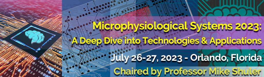
Co-Located Conference AgendasExtracellular Vesicles 2023: Drug Delivery, Biologics & Therapeutics | Extracellular Vesicles 2023: Technologies, Biomarker Cargo & Diagnostics | Microphysiological Systems 2023: A Deep Dive into Technologies & Applications | Space Summit 2023: Chips in Space | 

Wednesday, 26 July 202308:00 | Conference Registration and Materials Pick-Up + Coffee | |
Session Title: Emerging Trends and Themes in the Microphysiological Systems (MPS) Space |
| | |
Session Chairperson: Professor Mike Shuler, Cornell University and Hesperos |
| | 09:00 |  | Conference Chair Making Microphysiological Systems (MPS) Useful
Michael Shuler, Samuel B. Eckert Professor of Engineering, Cornell University, President Hesperos, Inc., United States of America
We will discuss briefly some of the origins and history of multiorgan MPS development as well as current applications of the technology. The differences between multiorgan (e.g., "body-on-a chip") and single organ models will be discussed. Examples of current uses will be described including the use of MPS data to gain FDA approval for clinical trials. Current "Body-on-a-Chip" systems combine organized tissue/organ mimics with the techniques of microfabrication based on PBPK (Physiologically Based?Pharmacokinetic) models. These systems are "self-contained" and do not require external pumps, leading to "pumpless systems" which reduce system cost and improve?operational reliability. While the fluid (or blood?surrogate) in these systems can be sampled directly to allow measurement of?concentrations of drug,?metabolites or biomarkers, they can also be interrogated in situ to determine functional responses such as electrical response, force generation, or barrier?integrity . The blood?surrogate can be made to be as a serum-free, chemically defined medium facilitating interpretations of responses that are more mechanistic than with serum?containing media. We have constructed models of barrier tissues and have integrated them into a multiorgan system. A key advantage of this "Body-on-a-Chip" approach is that we can predict both human efficacy and toxicity of a drug or drug combination in preclinical trails, A Hesperos system has been used as the sole source of efficacy data for an IND application by Sanofi for a drug currently in a Phase 2 clinical?trial?demonstrating that these systems are beginning to have direct commercial impact as well as helping companies evaluate drug leads. |
| 09:30 |  | Keynote Presentation An Outlook Towards Adoption of MPS in Drug Development
Jason Ekert, Head US Discovery Translational Technology, UCB Pharma, United States of America
- Outline where there are opportunities/challenges to implement MPS into the drug discovery pipeline
- Report on the use and uptake of MPS across the pharma industry plus where there is an opportunity when applicable to use animal MPS models.
- Provide examples of where MPS have been utilized in disease modeling and pharmacology to progress molecules in early drug discovery.
- Conclude on where there remains gaps and a future outlook for MPS in drug discovery.
|
| 10:00 |  | Keynote Presentation The Impact of Integrated Multi-Organ-Chip Systems in Substance Testing: Progress and Future Outlook
Reyk Horland, CEO, TissUse GmbH, Germany
Microphysiological systems (MPS) are complex cell culture models, which mimic an organ, or defined parts of an organ, in aspects of structure and function. MPS typically consist of several cell types, which are spatially and functionally organized similar to their organization in a normal organ. Integrated microfluidic MPS in particular have proven to be a powerful tool for recreating human tissue- and organ-like functions at research level, providing the basis for the establishment of qualified preclinical assays with improved predictive power. However, industrial adoption of microphysiological systems and respective assays is progressing slowly due to their complexity. In the first part of the presentation examples of established single-organ chip, two-organ and four-organ chip solutions are highlighted. The underlying universal microfluidic Multi-Organ-Chip (MOC) platform of a size of a microscopic slide integrating an on-chip micro-pump and capable to interconnect different organ equivalents will be presented. Issues to ensure long-term performance and industrial acceptance of MPS, such as design criteria, tissue supply and on chip tissue homeostasis will be discussed. The second part of the presentation focusses on the establishment of automated MOC-based assays as a robust tool for safety and efficacy testing of drug candidates. These automated assays will allow for increased throughput and higher inter-laboratory reproducibility thus eventually enabling broad industrial implementation. Finally, an outlook will be given on how these automated systems can be used for on-chip clinical trials as well as personalized medicine testing. |
| 10:30 | Mid-Morning Coffee Break and Networking in the Exhibit Hall | 11:15 |  | Keynote Presentation Human-on-a-Chip Systems as Pre-Clinical Models for Neurological Diseases and Disorders
James Hickman, Professor, Nanoscience Technology, Chemistry, Biomolecular Science and Electrical Engineering, University of Central Florida; Chief Scientist, Hesperos, United States of America
One of the primary limitations in drug discovery and toxicology research is the lack of good model systems between the single cell level and animal or human systems. This is especially true for neurodegenerative diseases such as ALS and Alzheimer’s as well as spinal cord injury. In addition, with the banning of animals for toxicology testing in many industries body-on-a-chip systems to replace animals with human mimics is essential for product development and safety testing. Our research focus is on the establishment of functional in vitro systems to address this deficit where we seek to create organs and subsystems to model motor control, muscle function, myelination and cognitive function, as well as cardiac and liver subsystems. The idea is to integrate microsystems fabrication technology and surface modifications with protein and cellular components, for initiating and maintaining self-assembly and growth into biologically, mechanically and electronically interactive functional multi-component systems. Specific embodiments of this technology are the creation of a functional human NMJ system to understand ALS and a long-term potentiation (LTP) for AD utilizing clinically relevant functional readouts. We have investigated four mutations found in ALS patients; SOD1, FUS, TDP43 and C9ORF72. The models have demonstrated variations of the disease phenotype compared to WT for NMJ stability and functional dynamics. We have also demonstrated the LTP model can reproduce ABeta pathology and tauopathy in human cortical neurons and demonstrated how AD therapeutics can recover these effects Examples will be given of some of the other human-on-a-chip systems being developed for CNS and PNS disease applications as well as the results Sanofi has used as efficacy data from one of our models to file the first IND only from MPS data that has enabled a clinical trial (#NCT04658472). |
| 11:45 |  | Keynote Presentation Design and Operation of Pumpless Multi-Organ Microphysiological Devices
Mandy Esch, Project Leader, National Institute of Standards and Technology (NIST), United States of America
We have developed pumpless multi-organ cell culture systems that can be used to co-culture interconnected human tissues to probe inter-organ interactions and secondary drug toxicities. Short connections among organ-chambers and pumpless operation using gravity-driven flow enable us to reduce the amount of liquid typically needed to operate the microfluidic cell culture systems. The devices can be operated with near-physiological amounts of recirculating blood surrogate (cell culture medium). We demonstrate the systems with heart and liver tissues scaled at 1/75,000. Our approach allows us to reduce the dilution of recirculating drug metabolites that cause acute toxicity and long-term toxicity. Better predictions of drug toxicity and efficacy with in vitro systems are needed, since clinical trials with humans often do not reproduce the results seen with animal models. |
| 12:15 | Networking Lunch - Network with Colleagues and Meet Exhibitors | 14:00 |  | Keynote Presentation Calcific Aortic Valve Disease on a Chip
Gretchen Mahler, Professor of Biomedical Engineering, Binghamton University, United States of America
The development of calcific aortic valve disease (CAVD) is an active process and ranges from mild valve thickening (aortic sclerosis) to severe leaflet calcification (aortic stenosis). Early CAVD is characterized by the infiltration of immune cells into the extracellular matrix (ECM), reorganization of the ECM, and the deposition of oxidized low-density lipoproteins (oxLDLs), while late CAVD includes severe calcification, collagen disorganization, and the deposition of glycosaminoglycans (GAGs) in the fibrosa layer. In the United States, the prevalence of moderate to severe calcific aortic stenosis in patients =75 years old is 2.8% and is projected to more than double by 2050. Current treatments for CAVD include surgical valve replacement and/or minimal drug interventions tailored to other cardiovascular diseases. Advances in valve disease research have relied on animals and static cell cultures, but engineered, dynamic models may help to further the understanding of CAVD onset and progression. We have created a microfluidic model of the aortic valve fibrosa that includes a dynamic, 3D culture environment, physiologically relevant extracellular matrix, and aortic valve and immune cells that remain viable for up to 21 days. Our previous work demonstrated that the addition of GAGs, particularly chondroitin sulfate, to 3D collagen I hydrogels promoted endothelial to mesenchymal transformation and calcification in a static cell culture. Our results show that after 14 days in dynamic culture both low (1 dyne/cm2) and high (20 dyne/cm2) shear stress induce significant calcifications in the microfluidic model, that the addition of GAGs to the 3D matrix only slightly advances pathogenesis, and that the presence of endothelial cells results in more significant calcification. SEM/EDX was used to analyze chemical composition, composition distribution, and composition concentration of the static or dynamic cell culture samples that formed calcified nodules. Our microfluidic device-derived calcifications are primarily composed of octacalcium phosphates (Ca/P = 1.33), while human valve calcifications are primarily composed of hydroxyapatite. These results were generated without osteogenic medium. The addition of 50 µg/mL oxLdLs to the collagen I ECM in the microfluidic device resulted in hydroxyapatite nodules forming within two days at high (20 dyne/cm2) shear rates, and differentiation of monocytes toward M1 macrophages. No other dynamic in vitro models have reported the natural development of hydroxyapatites. Given that CAVD has no targeted medical therapies, the creation of a physiologically relevant model of the aortic valve fibrosa may lead to contributions in preclinical studies for new therapeutic interventions. |
| 14:30 | Multiorgan Microphysiological Systems as Tools to Study Complex Diseases
Martin Trapecar, Assistant Professor, Johns Hopkins University, United States of America
Human multiorgan microphysiological systems (MOMPS) are engineered environments whose purpose is to mimic native cellular surroundings. This is accomplished with the use of tissue-specific biomaterials, machined or printed 3D architecture and perfusion. Controlled interaction of individual human tissues and the scalability of biological complexity in MOMPS, supported by advances in systems biology, might hold the key to identify novel relationships between interorgan crosstalk, metabolism, and immunity. The Trapecar lab is integrating donor-matched tissues into MOMPS to investigate how i) interorgan communication directs complex tissue development and organ-level renewal and how ii) a disruption thereof leads to emergence of immunometabolic pathologies. We show that tissue-level interaction between and within the three main germ layers ectoderm (neurons), mesoderm (lymphoid) and endoderm (gut and liver) leads to increased tissue maturation and increased in vivo-like functionality. In our approach we reconstruct donor-matched hepatic, gut-mucosal and neuronal tissue under fluidic communication and presence of the donors circulating immune cells. We further use the established system to derive how a metabolic disruption in immune-tissue signaling contributes to overlapping inflammatory disorders of the gut-liver-brain axis such as inflammatory bowel disease and neurodegeneration. Paired with multiomic analysis and resolution into molecular underpinnings of cellular and tissue homeostasis, MOMPS represent a unique opportunity to systematically dissect how interactions at a lower order inform new behavior at the macroscale within and between organ systems. Such scalable complexity might yield new insight into fundamental emergence of disease and tissue regeneration. | 15:00 | Intelligent Droplet Screen for High-Throughput Single Cell Level Tumor Profiling on Extracellular Matrix
Chia-Hung Chen, Associate Professor, City University of Hong Kong, Hong Kong
Precision cancer treatment refers to providing a personalized therapeutic strategy in time to maximize treatment efficiency and minimize the side effect of drugs. Individual cancer cells interact with the extracellular matrix (ECM), showing distinct endogenous drug resistance and therapeutic responses. The responses of cancer cells to mechanical cues from the ECM especially influence cancer progression and metastasis. It is therefore becoming clear that comprehensive approaches are needed to address single-cell functional profiling of patient tumor samples. The overall goals of this study are as follows: 1. develop an intelligent high-throughput single-cell screening platform integrated with smart hydrogel technology that allows the isolation of treatment-resistant cancer cells on ECM in patient tumors with an ultrahigh throughput of ~100 cells/second; and 2. identify and validate candidate therapeutic targets and biomarkers for rare critical cells representing drug treatment-resistant and/or metastatic subclones for therapeutic stratification. The hypothesis-driven therapeutics could be tested to provide a rationale for the inclusion of novel therapeutic targets and repositioning of existing drugs in cancer treatment regimens.
| 15:30 | Mid-Afternoon Coffee Break and Networking | 16:00 |  | Keynote Presentation Title to be Confirmed.
Matthias von Herrath, Vice President and Senior Medical Officer, Novo Nordisk, Professor, La Jolla Institute, United States of America
|
| 16:30 | Tissue Chips in Space: Modeling Human Disease States in Microgravity
Dmitriy Krepkiy, Program Officer, National Center for Advancing Translational Sciences (NCATS), United States of America
Several biological systems weaken or deteriorate in microgravity and these dysfunctions closely mirror some age-related disease states. These microgravity-induced phenomena provide the opportunity to model these diseases in space and gain insights into the mechanisms controlling age-related dysfunction. Through partnerships between NCATS, NASA and the Center for Advancement of Science in Space, the Tissue Chips in Space program was created (https://ncats.nih.gov/tissuechip/projects/space) to study the effects of microgravity environment on the human body at the International Space Station National Laboratory. The Tissue Chips in Space program supports development of tissue chip and organ-on-a-chip platforms that model physiological changes associated with aging and related diseases. This program enabled advances in the study of microgravity-associated age-related conditions mimicking accelerated aging pathophysiology in a relatively shorter period of time than it would take to undertake the same studies on Earth. We have learned how microgravity exerts a unique range of stresses and pathophysiological perturbations on the human body resulting in dramatic increase in oxidative stress and inflammation, muscle wasting, immune senescence, cardiovascular deconditioning and cardiomyopathy, alteration of gene expression and DNA damage. These findings have expanded our understanding of age-related conditions and will contribute to drug development that can slow the process of aging and lead to new interventions to improve human health. This program has also made key technological improvements in the tissue chips instrumentation systems towards automation and miniaturization required for space flight. These technological advances will be used for future engineering of tissue chip platforms for ease of use and broader accessibility of tissue chips in drug development and biomedical research here on Earth. | 17:00 | Close of Conference Programming + Networking Reception with Beer and Wine | 18:30 | Close of Conference Day |
Thursday, 27 July 2023 |
Please View Details of Programming under the Space Summit Website |
| |
|


 Add to Calendar ▼2023-07-26 00:00:002023-07-27 00:00:00Europe/LondonMicrophysiological Systems 2023: A Deep Dive into Technologies and ApplicationsMicrophysiological Systems 2023: A Deep Dive into Technologies and Applications in Orlando, FloridaOrlando, FloridaSELECTBIOenquiries@selectbiosciences.com