08:00 | Conference Registration, Materials Pick-Up, Morning Coffee and Breakfast Pastries |
|
Session Title: Emerging Approaches and Applications in the Microfluidics and Lab-on-a-Chip World |
| |
09:30 |  | Keynote Presentation Microfluidic Technologies for Liquid Biopsy & Precision Medicine
Chwee Teck Lim, NUS Society Chair Professor, Department of Biomedical Engineering, Institute for Health Innovation & Technology (iHealthtech), Mechanobiology Institute, National University of Singapore, Singapore
Capturing of circulating Tumor Cells (CTCs) from the peripheral blood of patients (a.k.a. liquid biopsy) can potentially lead to applications in cancer diagnosis and personalized medicine. Using a label-free approach via our developed microfluidic biochips, we not only show successful detection and retrieval of CTCs, but also how selective capturing of each single CTCs can lead to downstream single cell analysis and enable personalized treatment through the detection of specific actionable or druggable mutation. |
|
10:00 |  | Keynote Presentation Instrumented Tissue-in-a-Chip: A Bridge from In Vitro to In Vivo
Charles Henry, Professor and Chair, Colorado State University, United States of America
Microfluidic methods provide a promising path to mimicking human organ function with applications ranging from fundamental biology to drug metabolism and toxicity. The vast majority of these systems use dissociated, immortalized, or stem cells to create two and three-dimensional models in vitro. While these systems can provide valuable information, they are fundamentally incapable of recreating the three-dimensional complexity of real tissue. As a result, an important gap exists between in vitro models and in vivo systems. To address this gap, we have created instrumented Tissue-In-a-Chip (iTIC) system that combines microfluidic devices with ex vivo tissue slices or explants to recreate model systems that capture the cellular diversity of real tissue and bridge the gap between in vitro models and in vivo systems. In this presentation two systems will be discussed. The first uses a high-density electrode array equipped with microfluidic flow to image chemical release profiles from living adrenal slices. The second uses a 3D printed microfluidic device with removable inserts to hold and perfuse fluids over intestinal tissue, enabling generation of differential chemical conditions on either side of this important barrier tissue. |
|
10:30 | Coffee Break and Networking in the Exhibit Hall |
11:15 |  Microfluidic Mixing: Generic Solutions to a Ubiquitous Problem Microfluidic Mixing: Generic Solutions to a Ubiquitous Problem
Richard Chasen Spero, CEO, Redbud Labs
Some microfluidic challenges are common across applications. Perhaps none is more pervasive than microfluidic mixing, which is a necessary but confounding problem in many devices. Done effectively, microfluidic can dramatically accelerate molecular assays, improve analytic precision, and boost sensitivity. Here we survey applications where mixing is essential to assay success, including 10x faster microarray assays, 200x faster reagent dispersal, and 5x reduction in hybridization time. We will discuss the prognosis for achieving a generic solution for microfluidic mixing, with a focus on methods that can be combined with practical cartridge manufacturing and assembly methods.
|
11:45 | Constructing Microfluidic Analytical Systems from 3D-Printed Blocks with Integrated Functional Components
Noah Malmstadt, Professor, Mork Family Dept. of Chemical Engineering & Materials Science, University of Southern California, United States of America
The integration of functional components into microfluidic systems is necessary for building compact analytical devices. Optical detection and measurement, electromechanical fluid switching, active mixing, and magneto- and electrophoretic separations all require the integration of components on top of the underlying fluidic architecture. There has been significant work towards integrating such functional components by including them directly in the microfabrication process. This approach, however, leads to expensive fabrication workflows that are often limited to a narrow set of applications. To overcome these limits, we are designing a microfluidic architecture that is easily adaptable to a wide variety of applications. This architecture is based on 3D-printed elements that can be assembled into analytical devices. Functional components are integrated into these elements; the structures are printed to directly accommodate off-the-shelf components including photodiodes, heaters, sensors, and fiber optic fittings. We have demonstrated the utility of this approach by assembling a system capable of performing automated ELISA assays from 3D-printed blocks with integrated functional components. |
12:15 | Networking Lunch in the Exhibit Hall -- Meet the Exhibitors and View Posters |
|
Session Title: The Convergence of Microfluidics, 3D-Printing and Tissues-on-Chips |
| |
13:30 | Hydrogel Microarrays and Microfluidics – A Different Spin to Traditional SLA- and Extrusion-based 3D Printing Methods
Luiz Bertassoni, Associate Professor, Biomaterials and Biomechanics, School of Dentistry, Cancer Early Detection Advanced Research, Knight Cancer Institute, Oregon Health & Sciences University, United States of America
Fabrication of three-dimensional tissues with controlled microarchitectures has been shown to enhance tissue functionality. 3D printing can be used to precisely position cells and cell-laden materials to generate controlled tissue architectures. Therefore, it represents an exciting alternative for organ fabrication. Our group has been interested in developing innovative printing-based technologies to improve our ability to regenerate tissues with improved function, as well as to engineer hydrogel based microfluidic devices. In this seminar, we will present SLA/DLP-based 3D printing methods to fabricate high-throughput screening platforms to probe mechanotransduction and geometry-controlled stem cell differentiation. Further, we will discuss recent methods our lab has developed to engineer vascularized tissue constructs, and 3D printed magnetically-gated hydrogel microfluidic chips. The use of these technologies in various regenerative applications will be discussed. |
14:00 |  Latest Advancements in Microfluidic Flow Control and Microfabrication Technologies for Diagnostic, Organ-on-Chip and MEMS Applications Latest Advancements in Microfluidic Flow Control and Microfabrication Technologies for Diagnostic, Organ-on-Chip and MEMS Applications
Marko Blom, Chief Technical Officer, Micronit Microtechnologies
We will present the latest advances in flow control, together with commercially and scientifically relevant examples. Flow control will be discussed in a broad sense showing membrane-based valves and capillary flow control. We will show integrated membrane valve examples realized via adhesive-free techniques, combining a range of validated thermoplastic and elastomeric materials, with actuation via pneumatic but also via other means. Furthermore, we will present integration of the electrostatic triggering mechanism of a capillary burst valve in a low-cost, thermoplastic chip, enabling sequential capillary flow in fully autonomous Point-of-Care devices. On the chip design and fabrication side we will show workflow automation for the chip and process design process, partially enabled by recently initiated ISO standardization efforts. Combined with this we will present standardized top-down and edge-connect micro-to-micro and micro-to-macro fluidic and electrical interfacing. Finally, we will show new fabrication technologies using laser-based processing, next to integration techniques particularly relevant to the Organ-on-Chip field, integrating membranes and sensing elements, among others enabling TEER (transepithelial electrical resistance) and oxygen sensing.
|
14:30 |  | Keynote Presentation Digital Droplet Generation: From Digital PCR to Digital Pipetting
Tingrui Pan, Professor, Department of Biomedical Engineering, University of California-Davis, United States of America
Digital droplet generation enabled by microfluidic impact printing has been recently introduced, benefiting from the nature of simple device architecture, digital metering, low cost, non-contamination, scalable multiplexability and high throughput. In this talk, we will review this novel microfluidic-based droplet generation platform, utilizing modular microfluidic cartridges and expandable combinatorial printing capacity controlled by plug-and-play multiplexed actuators. Such a customizable microfluidic printing system allows for ultrafine control of the droplet volume from picoliters (~10pL) to nanoliters (~100nL), a 10,000 fold variation. The high flexibility of droplet manipulations can be simply achieved by controlling the magnitude of actuation (e.g., driving voltage) and the waveform shape of actuation pulses, in addition to nozzle size restrictions. Importantly, we will discuss the recent exciting developments of the microfluidic droplet generation into an array of emerging biomedical applications, including digital PCR and digital pipetting. |
|
15:00 | Coffee Break and Networking in the Exhibit Hall |
15:45 | Free-Surface Microfluidics and SERS for High Performance Sample Capture and Analysis
Carl Meinhart, Professor, University of California-Santa Barbara, United States of America
Nearly all microfluidic devices to date consist of some type of fully-enclosed microfluidic channel. The concept of ‘free-surface’ microfluidics has been pioneered at UCSB during the past several years, where at least one surface of the microchannel is exposed to the surrounding air. Surface tension is a dominating force at the micron scale, which can be used to control effectively fluid motion. There are a number of distinct advantages to the free surface microfluidic architecture. For example, the free surface provides a highly effective mechanism for capturing certain low-density vapor molecules. This mechanism is a key component (in combination with surface-enhanced Raman spectroscopy, i.e. SERS) of a novel explosives vapor detection platform, which is capable of sub part-per-billion sensitivity with high specificity. |
16:15 | 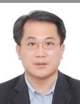 | Keynote Presentation Microfluidic Chip System Combined with Tetrahedron DNA Nanostructures For Circulating Tumor Cell Capture
Xianqiang Mi, Professor, Shanghai Advanced Research Institute Chinese Academy of Sciences, China
As the typical liquid biopsy technology for cancer prevention and control, circulating tumor cells (CTCs) analysis shows great prospects. However, the extremely low content of CTCs in blood brings great difficulty for its capture and detection. Microfluidic chip technology and DNA nano technology have made great progress in CTCs capture, but there are still some shortcomings, such as low capture efficiency, narrow spectrum for target and easily leading to cell inactivation. Here, based on the principle of microfluidic immuno-affinity, we propose a new CTCs capture method with the combination of microfluidic chip and DNA nanostructure technology. The performance of the developed microfluidic chip was verified by the tumor cell suspension and real blood samples. This new CTC capture method has the advantage of high capture efficiency, broad spectrum for target and good ability to keep cell viability. |
|
16:45 | Bioprinting Pancreas-on-a-Chip
Ibrahim Ozbolat, Hartz Family Associate Professor of Engineering Science and Mechanics, The Huck Institutes of the Life Sciences, Penn State University, United States of America
Despite the recent achievements in cell-based therapies for curing type-1 diabetes (T1D), vascularization of beta (ß)-cell clusters is still a major roadblock as it is essential for long-term viability and function of ß-cells in vivo. In this research, we report micro-vascularization within engineered pancreatic islets (EPIs) made of rodent cells. EPIs cultured in fibrin constructs maintained their viability and functionality over time while non-vascularized EPIs could not retain their viability nor functionality. We then bioprinted the EPIs along with bioprinted macro-vascular network in a pancreas-on-a-chip model. Here we demonstrate a proof-of-concept study for a vascularized pancreas-on-a-chip model for the first time, where patient specific stem cell-derived human beta cells can be vascularized in the near future for an effective treatment of T1D. |
17:15 |  | Keynote Presentation Bioprinting: The State of the Field
Gabor Forgacs, Professor, University of Missouri-Columbia; Scientific Founder, Organovo; CSO, Modern Meadow, United States of America
The modern era of bioprinting commenced in 2000 with the work of Thomas Boland and his re-engineered Hewlett Packard desktop inkjet printer. Since then a number of other technologies have been developed utilizing extrusion, acoustic waves, laser assisted delivery and lately “liquid bioprinting”. Meanwhile the field has also matured from its purely academic roots into successful commercial ventures. Overall bioprinting has seen spectacular progress in the past 17 years and a number of market analyses have predicted a bright future for the field. I will provide here a hype-free overview of the technology, where it stands today, what it has specifically accomplished and what can be expected in the years to come. |
|
17:45 | Networking Cocktail Reception with Beer, Wine and Appetizers in the Exhibit Hall. Engage with Colleagues and Visit the Exhibitors |
19:30 | Close of Day 2 of the Conference. |








.jpg)