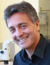
Albert Folch,
Professor of Bioengineering,
University of Washington
Albert Folch’s lab works at the interface between microfluidics and cancer. He received both his BSc (1989) and PhD (1994) in Physics from the University of Barcelona (UB), Spain, in 1989. During his Ph.D. he was a visiting scientist from 1990–91 at the Lawrence Berkeley Lab working on AFM/STM under Dr. Miquel Salmeron. From 1994–1996, he was a postdoc at MIT developing MEMS under Martin Schmidt (EECS) and Mark Wrighton (Chemistry). In 1997, he joined Mehmet Toner’s lab as a postdoc at Harvard-MGH to apply soft lithography to tissue engineering. He has been at Seattle’s UW BioE since June 2000, where he is now a full Professor, accumulating over 12,000 citations. In 22 years, he has supervised 19 postdocs (16% of whom have reached faculty rank), 36 graduate students (12 Ph.D. students, 25% of whom faculty rank, and 24 M.S. students), and ~43 undergraduates. In 2001 he received an NSF Career Award, and in 2014 he was elected to the AIMBE College of Fellows (Class of 2015). He served on the Advisory Board of Lab on a Chip 2010-2016 and serves on the Editorial Board of Micromachines since 2019. In 2022 he was elected a member of the Institute for Catalan Studies, one of the highest honors bestowed on Catalan scientists. He is the author of 5 books (sole author), including Introduction to BioMEMS (2012, Taylor&Francis), a textbook adopted by >103 departments in 18 countries, and Hidden in Plain Sight (MIT Press, 2022). Since 2007, the lab runs a celebrated outreach art program called BAIT (Bringing Art Into Technology), which has produced seven exhibits, a popular resource gallery of >2,000 free images related to microfluidics and microfabrication, and a YouTube channel that plays microfluidic videos with music which accumulate ~163,000 visits since 2009.
|

|

 Add to Calendar ▼2021-03-22 00:00:002021-03-22 01:00:00Europe/LondonTitle to be Confirmed.3D-Culture, Organoids and Organs-on-Chips 2021 in SELECTBIOenquiries@selectbiosciences.com
 Add to Calendar ▼2021-03-22 00:00:002021-03-23 00:00:00Europe/London3D-Culture, Organoids and Organs-on-Chips 20213D-Culture, Organoids and Organs-on-Chips 2021 in SELECTBIOenquiries@selectbiosciences.com