Co-Located Conference AgendasInnovations in Flow Cytometry 2023 | Innovations in Microfluidics 2023 | 

Thursday, 4 May 202308:00 | Conference Registration, Morning Coffee and Pastries in the Exhibit Hall | |
Session Title: Conference Opening Plenary Session |
| | |
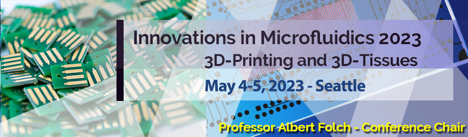
Conference Chairperson: Professor Albert Folch, Professor of Bioengineering, University of Washington |
| | 09:00 |  | Keynote Presentation New Flow-Cytometry Technologies: From Rare-Cell Isolation to the Analysis of Single Extracellular Vesicles and Particles
Daniel Chiu, A. Bruce Montgomery Professor of Chemistry, University of Washington, United States of America
This presentation will describe two flow-based technologies we developed
for the analysis of single rare cells and individual extracellular
vesicles and particles. The first flow platform is a rare-cell isolation
instrument, which is capable of operating in a rapid sequential sorting
mode for isolating single rare cells with exceptionally high
sensitivity and purity. The second flow instrument is a digital flow
cytometer, capable of high-sensitivity analysis of individual
extracellular vesicles and particles. I will outline the workings of
these two new flow cytometry platforms, describe their performance, and
discuss the clinical questions we are addressing with these
next-generation technologies. |
| 09:30 |  | Keynote Presentation Functional Flow Cytometry with Lab on a Particle Technologies
Dino Di Carlo, Armond and Elena Hairapetian Chair in Engineering and Medicine, Professor and Vice Chair of Bioengineering, University of California-Los Angeles, United States of America
The ultimate limits of measurement in biology are the “quantum” units that convey information, e.g. single nucleic acids, proteins, and cells. Microfluidics has emerged as a powerful tool to compartmentalize single cells and molecules into sub-nanoliter droplets as individual bioreactors to enable sensitive detection and analysis down to this quantum limit. However, the current systems for quantum assays have not been widely adopted, partly due to the requirement of specialized instruments and microfluidic chips to generate uniform droplets and perform adequate manipulations. I will discuss the platforms we are developing to perform quantum assays using standard flow cytometers and fluorescence activated cell sorters. “Lab on a particle” technology enables functional screening of T cell receptors based on the cytokine secretion of single T cells, the selection of colonies of yeast or bacteria based on growth and the production of high value bio-products, and the development of directed evolution workflows for protein biosensors. |
| 10:00 |  | Keynote Presentation Automated Flow Cytometric Analysis Compatible Microphysiological Systems
Shuichi Takayama, Professor, Georgia Research Alliance Eminent Scholar, and Price Gilbert, Jr. Chair in Regenerative Engineering and Medicine Wallace H. Coulter Department of Biomedical Engineering, Georgia Institute of Technology & Emory University School of Medicine, United States of America
Many organs-on-a-chip and microphysiological systems (MPS) are too small
to allow recovery of sufficient cells for convenient flow cytometric
analysis. This presentation will describe transitioning of microfluidic
organs-on-a-chip system to mesoscale systems compatible with automated
flow cytometry. The presentation will describe some of the underlying
engineering technologies along with accompanying biomedical applications
of the technologies. Additionally, this presentation will discuss some
of the standardization opportunities and challenges for these systems
with example applications to lung diseases. |
| 10:30 | Morning Coffee Break and Networking in the Exhibit Hall | 11:00 |  | Keynote Presentation A Miniaturized Intestine-on-Chip with Crypts, Microbes and Mucus
Nancy Allbritton, Frank and Julie Jungers Dean of the College of Engineering and Professor of Bioengineering, University of Washington in Seattle, United States of America
A 3D polarized epithelium using primary human gastrointestinal stem cells recapitulates gastrointestinal epithelial architecture and physiology. The intestine-on-chip supports chemical and oxygen gradients, a stem cell niche, differentiated cell zone, and mucus layer. An anaerobic luminal compartment permits co-culture of anaerobic intestinal bacteria. This in vitro human colon crypt array replicates the architecture, luminal accessibility, tissue polarity, cell migration, cell types and cellular responses of in vivo intestinal crypts. The microdevice possesses one hundred crypts providing exquisite control of the microenvironment for the investigation of organ-level physiology and disease. This bioanalytical platform is envisioned as a next-generation system for assay of microbiome-behavior, drug-delivery, and toxin-interactions with the intestinal epithelia. |
| 11:30 |  | Keynote Presentation Detection and Identification of Single Molecules using Nanoscale Electrophoresis and Resistive Pulse Sensing
Steve Soper, Foundation Distinguished Professor, Director, Center of BioModular Multi-Scale System for Precision Medicine, The University of Kansas, United States of America
We are generating a label-free single-molecule sensor that can detect and identify various molecules (small – ribonucleotides, deoxynucleotides, peptides – and large molecules – oligonucleotides, proteins) using a combination of nanoscale electrophoresis and resistive pulse sensing. The sensing technology employs a nanochannel to read the identity of individual molecules from their molecular-dependent electrophoretic mobility deduced from the travel of the molecule through a 2-dimensional (2D) nanochannel (~50 nm in width and depth; 5 – 10 µm in length) fabricated in a thermoplastic via nano-injection molding. The mold insert used for injection molding is made from a Si master that has undergone photolithography to build microstructures and focused ion beam milling to generate the nanostructures, which is used to produce resin stamps that serve as the mold insert. The 2D nanochannel is flanked on either end with an in-plane nanopore (effective diameter <10 nm) that can detect single molecules using resistive pulse sensing in a label free fashion. In this presentation, we will present our results using nanoscale electrophoresis to deduce the identity of deoxynucleotides, ribonucleotides, and peptides. The effect of material (type of plastic), scaling effects, and surface modifications of the 2D nanochannel on the performance of nanoscale electrophoresis will be discussed as well. We will also discuss the use of principle component analysis or machine learning to improve the identification accuracy of single molecules from not only their unique electrophoretic mobility, but also the molecular-dependent dwell time (current transient event width) and event amplitude generated from each in-plane pore. We will also discuss unique applications of this sensing platform including single-molecule DNA/RNA sequencing and protein fingerprinting using peptides produced from the solid-phase proteolytic digestion of single protein molecules. |
| 12:00 |  Fluorescence Microfluidic Resistive Pulse Sensing: A Rapidly Emerging Method for Flow Cytometry at the Nanoscale Fluorescence Microfluidic Resistive Pulse Sensing: A Rapidly Emerging Method for Flow Cytometry at the Nanoscale
Jean-Luc Fraikin, CEO, Spectradyne
Nanoparticle-based biotechnologies including virus-mediated cell and
gene therapies, lipid nanoparticles (LNPs), and extracellular vesicles
(EVs) hold enormous promise as vaccines and therapeutics. These
revolutionary technologies derive their potency from payload-carrying
particles as small as 20 nanometers in diameter. At this size scale,
basic physical properties of the particles directly dictate the efficacy
and safety of the overall product and are therefore critical parameters
that must be measured to effectively develop and produce products based
on these materials. The realization of the potential of these medicines
is limited, however, by a lack of practical analytical tools for
measuring three key particle properties that critically impact dose and
bioavailability: Particle size, concentration, and payload. Despite new
developments that stretch the limits of its sensitivity, optical flow
cytometry is fundamentally limited in its ability to accurately detect
and measure nanoscale particles in heterogeneous samples. Fluorescence
Microfluidic Resistive Pulse Sensing (F-MRPS) is rapidly emerging as a
powerful alternative to optical flow cytometry that accurately measures
nanoparticle concentration, size, and payload in complex samples. F-MRPS
combines two orthogonal analytical techniques into a single
measurement: A cartridge-based electrical method for counting and
sizing particles (MRPS), and simultaneous high sensitivity
single-particle fluorescence measurements for quantifying payload. In
contrast with purely optical flow cytometry methods, the detection limit
of F-MRPS is not limited by light scattering intensity, and
measurements of particle size are completely independent of the
particle’s optical properties. F-MRPS is therefore uniquely suited to
analyzing complex heterogeneous samples and is rapidly being adopted by
industrial and academic users for quantification of EVs, virus and LNPs.
A technical overview of the F-MRPS technology will be presented,
together with measurement examples from key impact areas including virus
and gene therapy, extracellular vesicles, lipid nanoparticles (LNPs),
and liposomes. The fluorescence sensitivity of F-MRPS is demonstrated
with measurements of synthetic particles cross-calibrated to
NIST-traceable fluorescence intensity standards.
| 12:30 |  Improvements in Fluorescence Nanoparticle Tracking Analysis Improvements in Fluorescence Nanoparticle Tracking Analysis
Sven Rudolf Kreutel, Chief Executive Officer, Particle Metrix GmbH and CEO, Particle Metrix Inc., USA
| 13:00 | Networking Luncheon in the Exhibit Hall -- Network with the Exhibitors and Engage with Colleagues | |
Session Title: 3D-Printing in Microfluidics |
| | 13:30 |  | Keynote Presentation High Resolution 3D Printing for Microfluidics
Gregory Nordin, Professor, Brigham Young University, United States of America
While there is great interest in 3D printing for microfluidic device fabrication, a main challenge has been to achieve feature sizes that are in the truly microfluidic regime (<100 µm). A key issue is that microfluidic devices are comprised primarily of negative space features, which therefore dominate 3D printing resolution requirements, as compared to positive space features that are typical for many other 3D printing applications. Consequently, we have developed our own stereolithographic 3D printers and materials that are specifically tailored to meet these needs. We have shown that flow channels as small as 18 µm x 20 µm can be reliably fabricated, as well as compact active elements such as valves and pumps. With these capabilities, we demonstrate highly integrated 3D printed microfluidic devices such as a 10-stage 2-fold serial dilutor that simultaneously creates a 3 order of magnitude range of concentrations, high density chip-to-chip interconnects (53 interconnects per square mm) that are directly 3D printed as part of a device chip, and droplet-on-demand structures to efficiently entrain individual bacteria in relatively few droplets despite initial large sample volume. These advances open the door to 3D printing as a replacement for expensive cleanroom fabrication processes, with the additional advantage of fast (~5-15 minute), parallel fabrication of many devices in a single print run due to their small size. |
| 14:00 |  3D Printing Microfluidic Devices 3D Printing Microfluidic Devices
Phillip Manchik, 3D Printing Specialist, Ergometa
In this presentation Phillip Manchik and Sergei Chapek will cover how the Asiga 3D Printing technology is unique which leads to why Asiga is the trusted hardware for fabrication of custom microfluidic devices.
| 14:30 |  | Keynote Presentation Modular Design of 3D Printed Microfluidics for Bioprocess Applications
Noah Malmstadt, Professor, Mork Family Dept. of Chemical Engineering & Materials Science, University of Southern California, United States of America
As 3D printing replaces traditional clean room manufacturing for microfluidic engineering applications, it’s becoming clear that this transition offers not only lower cost and faster design iterations, but also new opportunities for fluidic routing and control that are only possible due to the inherent three-dimensional nature of these systems. Over the past several years, we have developed design principles that take advantage of this three-dimensionality, as well as demonstrating several applications that benefit from this approach. At the core of these design principles is the modularity of microfluidic unit operations. In addition to designing prototypical unit operations such as mixers, splitters, flow focusers, droplet generators, thermal and optical sensors, and world-to-chip interfaces, we have developed systematic approaches to combining these modules into microfluidic circuits with predictable behaviors. This approach can be used to rapidly prototype complex microfludic operations by assembling physically distinct modules as well as to design monolithic microfluidic devices which can be printed in a single run. We have demonstrated the power of this approach by building several micro- and milifluidic systems for bio-analysis and bioprocess applications. These include systems for biomarker diagnostics, automated high-throughput affinity screening, and rapid manufacturing of vaccine lipid nanoparticles. We have also demonstrated how entire systems can be treated as modules, allowing for scaling of bioprocess production lines by massive parallelization. |
| 15:00 | 3D-Printed Autonomous Microfluidics
Ayokunle Olanrewaju, Assistant Professor, Bioengineering and Mechanical Engineering, University of Washington, United States of America
We envision a fundamental change in the design, prototyping, and deployment of point-of-care diagnostics to transform disease treatment and prevention. We design autonomous microfluidics, self-powered and self-regulated by capillary forces encoded in surface geometry and chemistry, to orchestrate instrument-free liquid handling. We rapidly and inexpensively prototype these devices using desktop 3D printing. These devices have the potential to enable real-time automation of complex biological processes directly at the point of need. | 15:30 |  Microfluidics and Additive Manufacturing: Dilase 3D, The (R)evolution Microfluidics and Additive Manufacturing: Dilase 3D, The (R)evolution
Nicolas Brillouet, CTO, Kloé
Over the last 15 years, Kloé company developed a complete range of equipment dedicated to UV lithography applications, in perfect agreement with the microfabrication requirements in Microfluidics. Thus, Kloé company did the bet, in the early 2000, that the development of researches and industry in Microfluidics would rapidly grow. So that, over the same time, Kloé company continuously followed and exchanged with the Microfluidics community to first well understand and then anticipate its needs in terms of microfabrication techniques and performance, in order to enable fabricating from simple to more demanding microfluidic chips like Lab on a Chip/Organ on a Chip. Among a very large range of 12 different machines, covering from soft lithography / masking systems to very high resolution direct laser writers particularly suitable for fast prototyping, high aspect ratio as well as thick layers laser processing, Kloé introduces one of its latest innovations that is Dilase3D : a 3D-Printer specifically developed to meet the expectations for 3D-printing in Microfluidics. Typically elaborated from the specifications of researches in Microfluidics and Medical Sciences, that were looking for one tool enabling to both fabricate large volume pieces, but still with very high-resolution patterning capabilities, this equipment also demonstrated more recently its capability to combine different materials for the fabrication of one piece/object, that multiplies its capabilities to fabricate very demanding and ever more complex microchips/microstructures. This way, we ensure our partners to benefit from the one of the most performing and cost effective 3D-printing solution in that domain, in agreement with their expected level of performance and their available budget.
| 16:00 |  Semi-Permeable Capsules Enable a Highly Versatile and Robust Analysis of Single-Cell Genomes and Transcriptomes Semi-Permeable Capsules Enable a Highly Versatile and Robust Analysis of Single-Cell Genomes and Transcriptomes
Vaidotas Kiseliovas, Director of R&D, Atrandi Biosciences
Over the last decade we witnessed an explosion in single-cell -omic technologies using sequencing as readout, and keep observing the advances they brought in fundamental and applied research. To satisfy the need to study thousands of individual cells per sample, well-, droplet-, and fixation-based approaches keep evolving in parallel to provide single-cell compartmentalization required during sequencing library preparation. However, these approaches suffer from a fundamental trade-off between throughput and versatility. Being individually addressable, microwells enable multi-step processing but are not scalable. Droplet- and fixation-based methods offer a throughput of up to a million cells per experiment but only allow a limited number of processing steps to be performed. Our semi-permeable capsule (SPC) technology combines the throughput of droplets with the versatility of wells by enabling a virtually unlimited number of processing steps on genetic material from millions of individual cells in parallel. We will share our results from applying SPCs to study genomes and transcriptomes of millions of individual cells, and demonstrate their suitability for multi-omic approaches.
| 16:30 |  | Keynote Presentation Custom-Built Artificial Cells and Tissues for Drug Discovery
Katherine Elvira, Associate Professor, Canada Research Chair, Michael Smith Health Research BC Scholar, University of Victoria, Canada
Cells are complex and it is not always possible to know what effect each component of the cell has on drug transport. The goal of my research is to use microfluidic technologies to build bespoke artificial cells and tissues from the bottom up, starting with the cell membrane and then adding other cellular components such as transporter proteins and the cell microenvironment. This allows us to quantify how each component of a cell affects the uptake of drugs. We use these artificial cells to mimic how an orally administered drug moves from the intestine into an intestinal cell, and then from the cell into the blood stream, to mimic how the cell membrane changes during cancer and to build artificial tissues such as the blood brain barrier on a chip. We want our new in vitro models to help us predict the in vivo drug behavior. |
| 17:00 |  A Single-Cell pDEP Capture Array for Nanoinjection Applications A Single-Cell pDEP Capture Array for Nanoinjection Applications
Amir Tahmasebipour, Senior Microfluidics Scientist, Mekonos Inc.
Mekonos is developing a nanoinjection platform using a scalable array of MEMS-driven nanoneedles and a microfluidic cell capture array separated by a porous membrane. This system can precisely deliver genetic material if capable of on-demand capture and release of cells.
| 17:30 |  Rapid Bacterial Count, Viability, and Gram Typing on Microcapillary Flow Cytometry Rapid Bacterial Count, Viability, and Gram Typing on Microcapillary Flow Cytometry
Stephanie Brunelle, Senior Product Manager, Cytek BioSciences
Evaluation of bacterial concentrations, viability, and Gram typing are frequently needed measurements in microbiological research. Characterization of bacteria has remained complex, cumbersome and labor-intensive with dependence on long culture times when using traditional colony counting plate-based methods and with limitations on daily throughput. Here, we present results from two quick and robust methods for bacterial analysis using combinations of microcapillary flow cytometry and dedicated kits based on pre-blended fluorescent dyes for count and viability determination and for Gram typing. The kits are mix-and-read, enabling easy assay preparation steps. Samples are analyzed on the easy to use Cytek® Guava® easyCyte™ Systems which provide absolute counts of population, percent viability and fluorescence measurements. Assays include high precision Guava Bacterial Count & Viability kits that provide concentration and viability information simultaneously, and easy to interpret Bacterial Gram Typing kits that provide information on gram type of bacteria. The data demonstrate the performance of these assays on a range of bacterial strains. For bacterial Gram typing, our assay was applied to 10 different strains including Gram-positive and Gram-negative strains and showed results corresponding well with crystal violet staining. Results could also provide information on whether individual or mixed strain types were present in the sample. The analysis can be performed in tubes or plates. Our approach in evaluating concentration and viability allowed for the determination of dead and viable bacteria in samples in mixed culture or monoculture. Approaches were applied to 10 different strains including both Gram-negative and Gram-positive strains. High precision was observed in both concentration and viability determination with average %CV of <12%. The combination of microcapillary cytometry and dedicated kits gives bacterial researchers new robust analytical tools for easy and rapid quantification and characterization of bacteria with reduced process times.
| 18:00 | Networking Reception with Beer and Wine in the Exhibit Hall | 19:00 | Close of Day 1 Conference Programming |
Friday, 5 May 202308:00 | Morning Coffee, Pastries and Networking in the Exhibit Hall | |
Session Title: Single Cells, 3D-Tissues -- Emerging Themes |
| | 09:00 |  | Conference Chair TME-Friendly Microfluidic Platforms for High-Fidelity Cancer Drug Testing
Albert Folch, Professor of Bioengineering, University of Washington, United States of America
There is a lack of confidence in present in vitro drug efficacy tests, as they do not properly recapitulate the dynamic physiology and pathophysiology of the human organism. This challenge is particularly acute in oncology: present tools to study drug responses fail to faithfully mimic the patient’s tumor microenvironment (TME) and thus have not kept up with drug testing needs. As a measure of this problem, on average less than 4% of oncology drugs in clinical trials end up being FDA-approved, a dismal approval rate that has dire social repercussions such as high cancer drug prices and difficult accessibility. We have developed a suite of microfluidic platforms that address this problem by multiplexing the delivery of drugs to intact-TME human biopsies, altogether bypassing animal testing. The first of these platforms (called OncoSlice) allows for delivering drugs to live tumor slices. We have demonstrated the use of Oncoslice for the delivery of small-molecule cancer drug panels to xenograft slices as well as to slices from patient solid tumors (GBM and colorectal liver metastasis). In addition, we have developed a precision slicing methodology that allows for producing large numbers of cuboidal micro-tissues (“cuboids”) from a single tumor biopsy. We have been able to trap cuboids in arrays of microfluidic traps in a 96-well platform and we are exploring robotics for very high-throughput automated placement of mouse cuboids in 384-well plates. We believe that with these approaches it will soon be possible to bypass animal testing and perform direct testing of drugs using only human tumors. Since these new-generation tests preserve the TME intact, we envision that they will minimize FDA failure rates and could contribute to alleviate the cost of cancer drugs. |
| 09:30 | High-Throughput Spheroid and Organoid Culture Standardization and Analysis
Shuichi Takayama, Professor, Georgia Research Alliance Eminent Scholar, and Price Gilbert, Jr. Chair in Regenerative Engineering and Medicine Wallace H. Coulter Department of Biomedical Engineering, Georgia Institute of Technology & Emory University School of Medicine, United States of America
This presentation will describe transitioning of microfluidic 3D cultures to high-throughput (96 and 384) droplet, microwell, and filter insert type 3D microscale models of the lung, kidney, and some cancers. The presentation will describe some of the underlying engineering technologies along with accompanying biomedical applications of the technologies. Additionally, this presentation will discuss some of the standardization challenges for these systems and some materials science and informatics solutions. | 10:00 | Sculpting Hydrogels and 3D Tissues with Open Microfluidics
Ashleigh Theberge, Associate Professor, University of Washington, United States of America
This talk will highlight advances in open microfluidics—fluid flow in spaces with one or more air-liquid interfaces—to pattern cells and extracellular matrix materials for regenerative medicine and cell signaling studies. Our methods are tunable and general, enabling 3D patterning of biomaterials that cannot be patterned using conventional 3D bioprinters. | 10:30 |  Ease of Developing 40+ Marker Flow Panels Ease of Developing 40+ Marker Flow Panels
Anita Kant, Senior Field Application Scientist, Standard BioTools, Inc.
CyTOF technology provides distinct advantages over other single-cell technologies for highly multiplexed analysis of cell phenotype and function. Mass cytometry uses metal conjugated antibodies for flexible and simple panel design and a streamlined workflow to optimize a 40+ marker panel and significantly reduce time from planning to results. In this master class, we will introduce the principles of panel design with our panel design tool and a universal ten-tube titration method to titrate and validate a 40+ marker mass cytometry panel.
| 11:00 |  | Keynote Presentation Node-Pore Sensing: A Label-Free Method to Immunophenotype and Mechanophenotype Single Cells
Lydia Sohn, Almy C. Maynard and Agnes Offield Maynard Chair in Mechanical Engineering, University of California-Berkeley, United States of America
We have developed an electronic method to screen cells for their phenotypic profile, which we call Node-Pore Sensing (NPS). NPS involves using a four-terminal measurement to measure the modulated current pulse caused by a cell transiting a microfluidic channel that has been segmented by a series of inserted nodes. Previously, we showed that when segments between the nodes are functionalized with different antibodies corresponding to distinct cell surface antigens, immunophenotyping can be achieved. In this talk, I will show how we have significantly advanced NPS by simply inserting between two nodes a straight “contraction” channel through which cells can squeeze “Mechano-NPS”, as we now call our method, can simultaneously measure a cell’s size, resistance to deformation, transverse deformation, and ability to recover from deformation. When the contraction channel is sinusoidal in shape, resulting in cells being periodically squeezed, mechano-NPS can also measure the viscoelastic properties of cells. I will describe how we have used mechano-NPS to distinguish chronological age groups and breast-cancer risk groups of primary human mammary epithelial cells and identify drug-resistant acute promyelocytic leukemia cells—all based on mechanical properties. I will also describe the development of the next-generation NPS platform which utilizes advanced signal processing algorithms—Barker and Gold codes—directly encoded in the NPS channels to thus achieve multiplexing. |
| 11:30 | Paper-Based Microfluidic Devices Using Commercially Available Printers
Andres Martinez, Professor, California Polytechnic State University, United States of America
Paper-based microfluidic devices, also known as microPADs, are an emerging platform for the development of low-cost point-of-care diagnostics. Like conventional microfluidic devices, microPADs can manipulate and analyze small volumes of fluids. Paper-based devices are also portable, inexpensive to fabricate, simple to operate, and can complete an assay without relying on electrical power or supporting equipment. This talk will describe our work developing techniques for fabricating paper-based microfluidic devices, and for harnessing evaporation-driven capillary flow in paper-based devices to enable more sophisticated, multi-step assays with minimal user input. | 12:00 |  | Keynote Presentation Real-Time Biosensors: Continuous Measurements of Biomolecules in Live Subjects
H Tom Soh, Professor, Stanford University, United States of America
A biosensor capable of continuously measuring specific molecules in vivo would provide a valuable window into patients’ health status and their response to therapeutics. Unfortunately, continuous, real-time molecular measurement is currently limited to a handful of analytes (i.e. glucose and oxygen) and these sensors cannot be generalized to measure other analytes. In this talk, we will present a biosensor technology that can be generalized to measure a wide range of biomolecules in living subjects. To achieve this, we develop synthetic antibodies (aptamers) that change its structure upon binding to its target analyte and produce an electrochemical current or emit light. Our real-time biosensor requires no exogenous reagents and can be readily reconfigured to measure different target analytes by exchanging the aptamer probes in a modular manner. Using our real-time biosensor, we demonstrate the first closed loop feedback control of drug concentration in live animals and discuss potential applications of this technology. Finally, we will discuss methods for generating the aptamer probes which are at the heart of this biosensor technology. |
| 12:30 |   | Keynote Presentation In Vitro Models for Drug Development in Diabetes
Johnna Wesley, Vice President, Global Drug Discovery for T1D & CKD, Novo Nordisk
Matthias von Herrath, Vice President and Senior Medical Officer, Novo Nordisk, Professor, La Jolla Institute, United States of America
The presentation will discuss improving disease model systems and using human data to guide the in vitro assay development to aid drug development. This includes a discussion of 3D islet-immune model and relevance to human Type 1 diabetes. |
| 13:00 | Networking Luncheon in the Exhibit Hall -- Network with the Exhibitors and Engage with Colleagues | 14:00 |  | Keynote Presentation Spatial Multi-Omics Sequencing via Microfluidic Deterministic Barcoding in Tissue
Rong Fan, Harold Hodgkinson Professor of Biomedical Engineering, Yale University, United States of America
Despite latest breakthroughs in single-cell sequencing that revealed cellular heterogeneity, differentiation, and interactions at an unprecedented level, the study of multicellular systems needs to be conducted in the native tissue context defined by spatially resolved molecular profiles to better understand the role of spatial heterogeneity in biological, physiological, and pathological processes. In this talk, I will begin with discussing the emergence of a whole new field – “spatial omics”, and then focus mainly on a new technology platform called microfluidic Deterministic Barcoding in Tissue (DBiT) for spatial omics sequencing developed in our laboratory over the past years. We conceived the concept of “spatial multi-omics” and demonstrated it for the first time by co-mapping whole transcriptome and proteome (~300 proteins) pixel-by-pixel directly on a fixed tissue slide in a way compatible with clinical tissue specimens including FFPE. It has been applied to the study of developing mouse brain, human brain, and human lymphoid tissues associated with normal physiology, disease, or aging. Recently, our research enabled another new field – “spatial epigenomics” – by developing multiple DBiT-based spatial sequencing technologies for mapping chromatin accessibility (spatial-ATAC-seq), histone modification (spatial-CUT&Tag), or further combined with transcriptome or proteins for spatial co-profiling. These new technologies allow us to visualize gene expression regulation mechanisms pixel by pixel directly in mammalian tissues with a near single cell resolution. The rise of NGS-based spatial omics is poised to fuel the next wave of biomedical research revolution. Emerging opportunities and future perspectives will be discussed with regard to clinical biomarker discovery and therapeutic development. |
| 14:30 |  | Keynote Presentation Leaf Vein-Inspired Microfluidic Platform to Study Cancer Metastasis
Xin Zhao, Associate Professor, Department of Biomedical Engineering, The Hong Kong Polytechnic University, Hong Kong
Vascular network is a central component of organ-on-a-chip system to build a 3D physiological microenvironment with controlled physical and biochemical variables. Inspired by ubiquitous biological systems such as leaf venation and circulatory systems, a fabrication strategy is devised to develop a biomimetic vascular system integrated with freely designed chambers, which function as niches for chamber-specific vascularized organs. As a proof of concept, human-on-leaf-chip system with biomimetic multiscale vasculature systems connecting the self-assembled 3D vasculatures in chambers is fabricated, mimicking the in vivo complex architectures of the human cardiovascular system connecting vascularized organs. Besides, two types of vascularized organs are built independently within the two halves of the system to verify its feasibility for conducting comparative experiments for organ-specific metastasis study in a single chip. Successful culturing of human hepatoma G2 cells (HepG2s) and mesenchymal stem cells (MSCs) with human umbilical vein endothelial cells (HUVECs) shows good vasculature formation, and organ-specific metastasis is simulated through perfusion of pancreatic cancer cells and shows distinct cancer encapsulation by MSCs, which is absent in HepG2s. Given good culture efficacy, study design flexibility, and ease of modification, these results show that the bioinspired human-on-leaf chip possesses great potential in comparative and metastasis studies while retaining organ-to-organ crosstalk. |
| 15:00 | Mid-Afternoon Coffee Break | 15:30 | Microfluidic Models for Studies of Single Cell and Tissue Mechanics in 3D
Mingming Wu, Professor, Biological and Environmental Engineering Department, Cornell University, United States of America
Microfluidics together with the advancement of the field of biomaterials present an unprecedented opportunity to recreate physiologically realistic 3D environment for mechanistic understanding of human cell and tissue mechanics. In this presentation, I will discuss about efforts from my lab in developing microfluidic models for recreating tumor microenvironment, with an emphasis on elucidating how physical forces regulate tumor cell/tissue mechanics and invasion in a 3D environment. Our work highlights enabling capabilities of microfluidic platform for providing well defined physical and chemical environment for cells, advancing frontiers of in vitro human tissue models. I will also briefly talk about our recent work on a nano-scribed 3D microswimmer, its ability to be steered remotely by ultrasound waves, and potential use in targeted drug delivery. | 16:00 | Integrating Flow Cytometry with Next Generation Sequencing to Find Determinants of Protein Secretion
Richard James, Principal Investigator and Associate Professor, Seattle Children’s Research Institute and University of Washington, United States of America
Protein secretion drives many functions in vivo; however, methods to
link secretions with surface markers and transcriptomes have been
lacking. By accumulating secretions close to secreting cells held within
cavity-containing hydrogel nanovials, we demonstrate workflows to
analyze the amount of IgG secreted from single human antibody-secreting
cells and link this information to surface marker expression and
transcriptional profiles from the same cells. Measurements using flow
cytometry and imaging flow cytometry corroborated an association between
levels of IgG secretion and CD138 expression. Using
oligonucleotide-labeled antibodies and droplet-based sequencing, we show
that pathways encoding protein localization to the endoplasmic
reticulum, NADH complex assembly, and mitochondrial respiration were
most associated with high IgG secretion. Altogether, this method links
secretion information to cell surface and single-cell sequencing
information (SEC-seq) and enables exploration of links between genome
and secretory function, laying the foundation for numerous discoveries
in immunology, stem cell biology, and beyond. | 16:30 | The OptiDrop Microfluidic Analyzer and Sorter: Multiplexed Fluorescence and Scatter Detection with Single Cell Resolution for Droplet Microfluidic Applications
Preksha Gupta, Senior Scientist, Centre for Cellular and Molecular Platforms, India
The OptiDrop platform is a novel optofluidic technology enabling multiparametric optical analysis and sorting of microfluidic droplets. Current droplet-based assays for multi-omic applications rely on expensive imaging-based methods or complicated flow cytometry based workflows for optical analysis of droplet contents. In a first, the OptiDrop platform combines the simplicity of on-chip fiber optics with the power of flow cytometry to provide multiplexed fluorescence and scatter signals from droplet contents with single cell resolution. | 17:00 | Detection of Cytochrome C from Micro-Dissected Tumors in Microfluidic Arrays using Aptamer-Based Electrochemical Sensors
Tran Ngoc Huyen Nguyen, Postdoctoral Scholar, University of Washington, United States of America
Functional assays on intact tissues can potentially complement and extend genomics-based approaches for precision oncology by capturing key determinants of therapeutic response, such as tissue architecture, tumor heterogeneity, and the tumor microenvironment (TME). The Folch lab has recently developed a platform for drug development and cancer precision therapy based on a multi-well microfluidic device that can selectively treat a large array of regularly sized, cuboidal-shaped microdissected tissues (referred to as “cuboids”). The tissues are never dissociated and retain much of the native TME. However, the present platform only provides fluorescent readouts, which is a semi-quantitative method best suited to be a single-time-point terminal assay or labor-intensive terminal immunostaining analysis. Thus, we seek to develop a novel platform based on aptamer-based electrochemical sensing, which enables the periodic monitoring of cytochrome c, a cell death indicator. Using the platform, we demonstrate the ability to measure cytochrome c from live cuboids cultured with different drug conditions on our platform. | 17:30 | DLP-based Stereolithography For Rapid-Prototyping of Microfluidic Actuators
Alireza Ahmadianyazdi, Postdoctoral Scholar, University of Washington, United States of America
The process of stereolithographic 3D-printing (SLA) can produce highly precise microfluidic and lab-on-a-chip devices, but the resulting plastics often lack the mechanical flexibility of elastomers like poly(dimethyl siloxane) (PDMS), which hinders the ability to create efficient microfluidic actuators. In this presentation, I introduce a new photopolymer resin, PEGDA-co-PEGMEMA, which is a blend of two monomers that can be tuned to adjust the elastic modulus of the printed plastic. This allows the plastic to reach the same level of flexibility as PDMS while retaining desirable properties of SLA-printed poly(ethylene glycol) diacrylate (PEGDA), such as low drug absorption and cytocompatibility. I will then demonstrate the potential of PEGDA-co-PEGMEMA for creating functional 3D-printed microfluidic devices by using it to create high-performance microvalves, micropumps, and microregulators with a custom multimaterial printing protocol with a desktop DLP-based SLA. | 18:00 | Close of Day 2 Conference Programming |
|


 Add to Calendar ▼2023-05-04 00:00:002023-05-05 00:00:00Europe/LondonInnovations in Microfluidics 2023Innovations in Microfluidics 2023 in SeattleSeattleSELECTBIOenquiries@selectbiosciences.com