Tuesday, 1 March 202208:00 | Conference Registration, Materials Pick-Up, Continental Breakfast and Networking | |
Session Title: Conference Opening Session -- Extracellular Vesicles 2022 |
| | 09:00 |  | Conference Chair Welcome and Introduction by Conference Chairperson and an Overview of the Field of Extracellular Vesicles, circa 2022
Dominique PV de Kleijn, Professor Experimental Vascular Surgery, Professor Netherlands Heart Institute, University Medical Center Utrecht, The Netherlands, Netherlands
|
| 09:30 |  | Keynote Presentation Affinity Selection of Extracellular Vesicles using Plastic-based Microfluidic Devices for the Management of Different Diseases
Steve Soper, Foundation Distinguished Professor, Director, Center of BioModular Multi-Scale System for Precision Medicine, The University of Kansas, United States of America
We have been developing tools for the diagnosis of a variety of diseases. The commonality in these tools is that they consist of microfluidic devices made from plastics via injection molding. Thus, our tools can be mass produced at low-cost to facilitate bench-to-bed side transition and point-of-care testing (PoCT). We have also been generating novel assays focused on using liquid biopsy samples that are enabled using microfluidics. In this presentation, I will talk about the evolution of our fabrication efforts of plastic-based microfluidic and nanofluidic devices as well their surface modification to make the devices biocompatible for in vitro diagnostics. One tool that we have generated is a plastic device (38 × 42 mm) that consists of 1.5M pillars, which are surface decorated with affinity agents targeting certain disease-associated extracellular vesicles (EVs). The affinity agents are covalently attached to the surface of the microfluidic device using a bifunctional linker, which consists of a coumarin moiety to allow for the photolytic release of the captured EVs using a blue-light LED to minimize photodamage to the EVs’ molecular cargo. We have also developed a high-throughput nano-Coulter counter (nCC) made from a plastic via injection molding for the counting of captured EVs from clinical samples to allow their enumeration. The nCC consists of multiple pores that are ~350 nm to allow for high throughput counting with exquisite LODs (500 EVs/mL). In this presentation, I will discuss the utility of these microfluidic and nanofluidic devices in several diseases, for example, using EVs as a source of mRNAs for molecular sub-typing of breast cancer patients. EVs were affinity selected from breast-cancer patients’ plasma by searching for both epithelial and mesenchymal expressing EVs to allow for highly efficient sub-typing using the PAM50 gene panel. In an addition, the microfluidic and nanofluidic devices were integrated into a single platform (modular-based system) for PoCT to screen for early stage ovarian cancer. Affinity probes were used to target EVs specifically generated from tumor cells that signal early-stage ovarian cancer disease with the nCC used for enumerating the number of EVs captured. Finally, the modular system was used for the detection of COVID-19 at the PoC by affinity selecting SARS-CoV-2 viral particles. The integrated system could process saliva samples to search for the viral particles and count them in <20 min. |
| 10:00 |  Utilize Smart Microfluidics to Support Liquid Biopsy Technologies in Detecting Rare Biomarkers for Neurodegeneration in Serum Utilize Smart Microfluidics to Support Liquid Biopsy Technologies in Detecting Rare Biomarkers for Neurodegeneration in Serum
Magdalena Schimke, Business Development and Sales, Single Cell Diagnostics, STRATEC Consumables GmbH
Neurodegeneration affects 37 million people annually. Until now, most biomarkers to detect the early onset or progresses of neurodegenerative diseases were retrieved by either imaging technologies or liquor analysis – both eventually very invasive procedures for patients. Smart consumables play a critical role in novel liquid biopsy technologies and market value as they support the finding of novel biomarkers and the possibility to find and characterize rare biomarkers in peripheral body fluids. Thereby, the burden for the patient can be reduced, monitoring tightened and potential treatments initiated earlier, personalized and hence more efficiently applied.
| 10:30 | Mid-Morning Coffee Break and Networking in the Exhibit Hall | 11:00 | Investigating Extracellular Vesicle-based Therapeutic Options Using Alternatives For Animal Models
Bas WM van Balkom, Assistant Professor, University Medical Center Utrecht, Netherlands
Mesenchymal stromal cell (MSC)-derived small EVs (sEVs) show therapeutic potential in multiple disease models, including kidney injury. Clinical translation of sEVs requires further preclinical and regulatory developments, like elucidation of the mode of action (MoA) and formulation of safety and release criteria. sEVs tend to accumulate in the liver and at sites of injury. Bio-distribution knowledge is crucial to assess MoA, efficacy and safety, and can be obtained using labelled sEVs in animal models, which come with ethical concerns, are time-consuming and expensive, and do not represent all human physiological processes equally well. Our models recapitulate the efficacy and bio-distribution of MSC-sEVs as observed in animal models and provide alternatives for animal experiments. Their systemic or human background allows for in-depth analysis of the MoA and identification of potential side effects and accelerated development of EV-based therapeutics. | 11:30 | Are Extracellular Vesicles In Liquid Biopsies the Source of My Signals?
Edwin van der Pol, Assistant Professor, Amsterdam University Medical Center, Netherlands
To discover new biomarkers in blood plasma, bulk assays are frequently applied after particle isolation. Particles of interest include extracellular vesicles (EVs), EV-associated miRNA, and circulating cell-free DNA. Blood plasma, however, is a complex body fluid that contains many other particles, such as lipoprotein particles, protein aggregates, and residual platelets, which may have similar physical properties. Therefore, isolation methods neither enrich nor recover 100% over the particles of interest. Moreover, signals of bulk assays do not necessarily originate from the particles of interest. By combining physical models with new isolation methods and a flow cytometer of which all aspects are calibrated, we gained new insights in the efficacy of methods to isolate and detect EVs and other particles in plasma. | 12:00 | Networking Lunch, Meet Exhibitors and Engage with Colleagues | 13:00 | | Keynote Presentation Plasma Extracellular Vesicles for Cardiovascular Disease
Dominique PV de Kleijn, Professor Experimental Vascular Surgery, Professor Netherlands Heart Institute, University Medical Center Utrecht, The Netherlands, Netherlands
Cardiovascular Disease (CVD) is with the cardiovascular events of Ischemic Heart Disease and Stroke, the number 1 and 2 cause of death in the world and expect to increase especially in Asia. In this presentation we use plasma extracellular vesicle (EV) protein content of vesicles from plasma subfractions on plasma of stroke and TIA patients after carotid atherectomy (CEA). AtheroExpress the large ongoing CEA biobank gives us the unique possibility to compare the different biomarker sources of EV, plasma and carotid plaque in association with onset symptoms and risk of a second Major Adverse Cardiovascular Event (MACE: myocardial infarction, stroke or cardiovascular death). We also evaluated if EV proteins could contribute to identification of patients at high risk for MACE or Major Adverse Limb Event (MALE: repeated surgery or limb amputation) after surgery in Peripheral Artery Disease (PAD) patients. Identification of such high risk patients is very important for possible (expensive) add-on pharmaceutical therapy or the decision to operate or not. |
| 13:30 |  Need For Workflow Solutions From Sample Collection to Liquid Biopsy Assay Need For Workflow Solutions From Sample Collection to Liquid Biopsy Assay
Andrea Huxhold, Senior Scientific Marketing Manager, QIAGEN/PreAnalytiX
From sample collection, through stabilization and storage, to nucleic acid isolation, there are critical parameters to consider to enable sensitive and reproducible liquid biopsy assay analysis. In this presentation, the new ISO Standards for molecular in vitro diagnostic examinations with liquid biopsies, Circulating cell free DNA whole blood collection, preservation and analyte purification and workflow performance evaluation according to new CE-IVD Regulation (IVDR 2017/746) will be addressed.
| |
Session Title: The Therapeutic Opportunities -- Extracellular Vesicles-based and microRNA, small RNA-based |
| | 14:00 | Delivery of a small RNA to Tumors: From Bench to Bedside
Roel Schaapveld, Chief Executive Officer, InteRNA Technologies BV, Netherlands
Preclinical and early clinical evaluation of an LNP-formulated, chemically-modified tumor suppressor microRNA mimic. | 14:30 | Clinical Potential of MSC-EVs and Translational Challenges
Bernd Giebel, Group Leader, Institute for Transfusion Medicine, University Hospital Duisburg-Essen, Germany
Small (exosome-sized) extracellular vesicles (sEVs) harvested from supernatants of human mesenchymal stromal cells (MSCs) exert therapeutic functions in various disease models. Furthermore, they have been successfully applied in a steroid-refractory Graft-versus-Host Disease patient and in a single centre, randomized, placebo-controlled phase II/III clinical pilot study on chronic kidney disease patients without revealing any site effects. Apparently one mode of action is their ability to modulate immune responses from the pro-inflammatory into the regulatory state. At the example of an ischemic stroke model, we demonstrate that systemically applied MSC-sEV preparations, considered as therapeutically active, attenuate stroke induced lymphopenia and neutrophil immigration into the lesion site and thus importantly contribute to the MSC-sEVs’ therapeutic effect. Notably, independent MSC-sEV preparations can differ in their immunomodulatory and therapeutic activity, with a proportion of them appearing therapeutically inactive. Thus, according to our understanding it is a challenging but essential task during the translation process into the clinics to develop appropriate potency assays allowing discrimination of therapeutically active and therapeutically inactive MSC-sEV preparations. | 15:00 | EV-microRNAs as Liquid Biopsy Tool for Therapy Response Prediction in Hematological Malignancies
D. Michiel Pegtel, Associate Professor, Amsterdam University Medical Center, Netherlands
In this lecture Dr Pegtel will talk about a novel small RNA sequencing method developed in his Lab called Isoseek. Isoseek accurately identifies the full composition of microRNAs in plasma EVs from cancer patients which may be leveraged for predictive non-invasive diagnostics using machine learning in patients with hematological malignancies including Multiple Myeloma ad diffuse large B cell Lymphoma. | 15:30 | Afternoon Coffee Break and Networking in the Exhibit Hall | 16:00 |  Trends in EV Characterization Technologies Trends in EV Characterization Technologies
Jean-Luc Fraikin, CEO, Spectradyne
Methods for characterizing EVs are evolving, and new technologies are being developed that deliver size, concentration, and fluorescent phenotyping, each to a varying degree of success. Learn about how some of these new and cutting-edge technologies work, their strengths and weaknesses, and where we at Spectradyne see the technology landscape moving next.
| 16:30 |  EV Analysis Using Imaging Flow Cytometry EV Analysis Using Imaging Flow Cytometry
Matthew Rodrigues, Instrument Field Application Scientist, Luminex Corporation
This presentation demonstrates the advantages to be had when using Flow Cytometry, in particular Imaging Flow, to detect small particles including Extracellular Vesicles.
| 17:00 |  Improvements in fluorescent Nanoparticle Tracking Analysis (f-NTA): Reliable Characterization of Extracellular Vesicles Improvements in fluorescent Nanoparticle Tracking Analysis (f-NTA): Reliable Characterization of Extracellular Vesicles
Sven Rudolf Kreutel, Chief Executive Officer, Particle Metrix GmbH and CEO, Particle Metrix Inc., USA
During the last decade, Nanoparticle Tracking Analysis (NTA) has emerged as a vital and fast characterization technology for Extracellular Vesicles, Exosomes and Microvesicles for concentration and size. While classic NTA scatter operation feeds back total particle concentration, the fluorescence detection capability enables the user to gain specific biochemical information for phenotyping of bio nanoparticles for estimation of sample purity. Here we report on latest improvements in size resolution and multi-channel fluorescent colocalization studies on standard material as well as selected biological vesicles.
| 17:30 |  Purification-Free Measurement of Exosomes and Viruses Down to 20nm Purification-Free Measurement of Exosomes and Viruses Down to 20nm
Jan Brants, Regional Sales Director - France Benelux, NanoView Biosciences
Current analytical tools have detection limitations when used for exosome characterization due to their small size. Specifically, exosomes have fewer surface epitopes available and low-signal relative to background levels, especially in complex samples. Current workflows often require multiple measurement technologies that result in the inability to link phenotypic and physical characterization on individual EVs across a biologically relevant size range and down to the smallest of sizes present within many samples. In addition, sample purification often results in loss of sample and technique dependent biases. NanoView Biosciences has developed an imaging technique that allows individual EVs and viruses as small as 20nm to be characterized direct from sample with no purification in most cases. Physical characteristics such as size and EV count can be presented alongside phenotypic information at the single EV level. Low volumes of sample can be probed and a panel of up to 10 surface markers can be interrogated in parallel, in a single measurement. Lastly, through the addition of fluorescent labeling, three additional markers can be probed and colocalized across the existing panel of 10 markers. These capabilities make the tool ideally for basic EV research, biomarker discovery and the development of EV based liquid biopsies and therapeutics.
| 18:00 |  | Keynote Presentation Engineered Synthetic Bacterial Vesicles as Cancer Immunotherapy and as Vaccines against Bacteria and SARS-CoV-2
Jan Lötvall, Professor, University of Gothenburg; Founding President of ISEV; Editor-in-Chief, Journal of Extracellular Vesicles, Sweden
We are developing a novel platform of “exosome-like” synthetic bacterial vesicles (SyBV) that can be utilized as cancer immunotherapy, and as preventive vaccines against viral or bacterial infection. Briefly, the SyBV are produced from isolated bacterial membranes, and exhibit many of the features of natural bacterial veicles (OMV), but have been detoxified efficiently by removal of multiple bacterial components that activate pattern recognition receptors (PRR), including Toll Like Receptors (TLR). This strongly reduces the dose-limiting side effects of the SyBVs, but retains their stimulatory effects on adaptive immunity. The SyBV efficiently induce specific immunity via T-cell and humoral responses in both cancer models, and against bacterial components. SyBV combined with tumor exosomes induce immune responses in models of malignant melanoma and colon cancer, and synergize with checkpoint inhibitors to reduce or stop tumor growth. Further, SyBV can function as adjuvant in virus vaccine candidates, including SARS-CoV-2. SyBV is a safe and highly efficient platform with potentially very wide clinical applications in both cancer immunotherapy, as well as preventive vaccines against both bacterial and viral infections. |
| 18:30 | Close of Conference Programming For Day 1 of the Conference |
Wednesday, 2 March 202208:00 | Morning Coffee and Pastries in the Exhibit Hall | |
Session Title: Technology Developments in the Circulating Biomarkers Space |
| | 09:00 |  | Keynote Presentation What Microfluidics Can Do For Liquid Biopsy: Enabling the Accessibility and Clinical Utility of Circulating Biomarkers
Lorena Diéguez, Leader of the Medical Devices Research Group, INL- International Iberian Nanotechnology Laboratory, Portugal
Early dissemination of cancer is difficult to detect by traditional imaging and pathological methods. While the presence of cancer material in body fluids is well known, current techniques for the isolation, analysis and characterization of these biomarkers are not efficient enough to be fully applied in clinical routine. Microfluidics presents numerous advantages for the handling of biological samples, as it provides careful control of fluids in the microscale. When it comes to biomarkers enrichment, microfluidics has demonstrated superior sensitivity and enhanced recovery compared to traditional methods. In this talk, we present our most recent work for integrated isolation and analysis of multiple circulating biomarkers and their validation in clinical settings. |
| 09:30 |  | Keynote Presentation Cell Free Circulating DNA For Cancer Patient Follow-up: From Technology Developments to Clinical Validations
Valérie Taly, CNRS Research Director, Professor and Group leader Translational Research and Microfluidics, Université Paris Cité, France
|
| 10:00 |  Moving Urine as a Liquid Biopsy into Clinical practice: Bridging the Gap Moving Urine as a Liquid Biopsy into Clinical practice: Bridging the Gap
Stephanie Jordaens, Biomedical Scientist, University of Antwerp and Novosanis
Urine is a promising alternative liquid biopsy, harboring valuable biomarkers, for detecting and monitoring treatment of urological and other cancer types. In terms of analysis, the isolation of various urinary analytes (such as DNA/RNA, cfDNA/cfRNA, EVs, proteins) shows great potential due to the ease of sampling and high acceptability compared to blood and tissue. Initial results of urinary biomarkers are presented for the use in multi-omic applications in prostate, breast and cervical cancer research. Here, we introduce Colli-Pee® containing UASTM preservative (RUO), as a self- sampling device for the standardization of urine collection, handling and storage to potentially bridge towards clinical applications.
| 10:30 | Mid-Morning Coffee Break and Networking in the Exhibit Hall | 11:00 | Multi-Cancer Early Detection Based on a Classifier for Extracellular Vesicle Biomarkers
Jean Lewis, Associate Director, Research, Biological Dynamics, United States of America
Simultaneous detection of multiple, early-stage cancers was tested using extracellular vesicle (EV) bound protein markers from plasma purified by an alternating current electrokinetic (ACE) microarray. In a case-control study, this method detected 95.7% of pancreatic, 75.0% of ovarian, and 43.8% of bladder cancers at 99.5% specificity, all at stage I. The initial results suggest that this liquid biopsy screen may have great promise for early stage, multi-cancer screening. | 11:30 |  NanoAnalyzer: Combining Flow Cytometry and Particle Analysis to Meet the Challenges of EV Analysis NanoAnalyzer: Combining Flow Cytometry and Particle Analysis to Meet the Challenges of EV Analysis
Dimitri Aubert, Managing Director, NanoFCM Co., Ltd
| 12:00 |  Accelerate Assay Development and Commercialization Using Reference Materials Accelerate Assay Development and Commercialization Using Reference Materials
Ronald Vos, Business Development Manager, SensID
Assay development in clinical genomics is a demanding process especially when complying correctly to all rules and regulations. When assays are becoming even more complex, like in liquid biopsy, where multiplexing becomes the standard and low to ultra low AF frequencies are demanded, the task gets near impossible. Add IVD or FDA demands on top of that and the complexity is that of a giant puzzle with many different aspects. Obtaining the correct and the right amount of clinical samples while facing all these demands is where you can expect friction. For these reasons, there is an acceptance for “contrived” materials but how does it all work. What are the official guidelines?
| 12:30 | Networking Lunch in the Exhibit Hall with Exhibitors and Conference Sponsors | |
Session Title: Harvesting Value from EVs: Diagnostics and Therapeutics Development |
| | 13:30 | A Development and Analysis Pipeline for microRNA Biomarker Signatures in Clinical and Molecular Diagnostics Based on Circulating EVs
Michael Pfaffl, Professor, Technical University of Munich, Germany
Extracellular vesicles (EVs) circulate in body liquids and are involved in the intercellular communication. They have significant regulative functions in almost any physiological or pathological process. Especially exosome-like small EVs have gained huge scientific and clinical interests because of their specific microRNA biomarker signature and hence future potential in molecular diagnostics. The past decade has brought about the development and commercialization of a multitude of extraction methods to isolate EVs, primarily from blood compartments. The EV purity and which EV subpopulations are captured strongly depend on the applied isolation method, which in turn determines how suitable resulting samples are for potential downstream applications and microRNA biomarker discovery. Hence, a development and analysis pipeline was developed, optimized and validated on various clinical cohorts. Applied isolation methods were benchmarked regarding their suitability for microRNA biomarker discovery as well as biological characteristics of captured vesicles, according to the latest MISEV 2018 guidelines. To deeply analyze the small-RNA deep sequencing results various self-established bioinformatical tools were used: microRNA analysis pipeline (based on R), analysis of microRNA isoforms (via isomiRROR), identification of stable microRNA references (via miREV). Differential expressed microRNAs candidates were identified by multivariate statistics (HCA, PCA, PLS-DA) to find reliable biomarkers. Final goal was the development of microRNA biomarker signatures for an early diagnosis and a valid classification of critical ill patients. Various independent patient cohorts were investigated: healthy volunteers, mild- or severe pneumonia, acute pulmonary failure (ARDS), septic shock, and recently Covid-19 patients. Distinct miRNA signatures could be identified, which are applicable to indicate disease progression from limited inflammation present in pneumonia, to severe inflammatory changes as seen in ARDS, septic shock or Covid-19. The study results indicate that EV microRNA biomarkers have high future potential for early diagnosis of pneumonia and to indicate disease progression towards severe inflammation events. Furthermore, the methodological findings provides guidance for navigating the multitude of EV isolation methods available, and helps researchers and clinicians in the field of molecular diagnostics to make the right choice about the optimal isolation strategy to get the most valid EV biomarker signature. | 14:00 | IVDR Applicable as of 26 May 2022 - What Changes?
Erik Vollebregt, Partner, Axon Lawyers, Netherlands
| 14:30 | Panel Discussion: Regulations in the EU Relating to Circulating Biomarkers, Diagnostics Tests | 15:00 | Function and Clinical Utility of Cancer-derived Extracellular Vesicles
Andreas Möller, Associate Professor, Group Leader and Head, Tumour Microenvironment Laboratory, QIMR Berghofer Medical Research Institute, Australia
Late diagnosis and lack of reliable prognostic outcome predictions are important unmet clinical needs. Reasons for our inability to generate reliable diagnostic, prognostic and/or predictive biomarkers are the incomplete understanding of cancer progression, especially metastatic spread. For a cancer to spread from the primary site to distant organs, a large number of essential steps have to be overcome. We, and others, showed that cancer-secreted factors, including extracellular vesicles (EVs), are mediators of creating immune-suppressed, permissive environments (pre-metastatic niches) at distant organs before the arrival of cancer cells. In this presentation, I will summarize the research approaches in the area of cancer-derived EVs undertaken by my group. I will discuss novel data on how cancer-derived EVs can specifically distribute to certain organs in the body, depending on the tumor microenvironment of the primary tumors. These modifications increase pre-metastatic niche formation and metastatic spread. Analyzing the content of cancer-derived EVs, I will additionally show intriguing clinical data on how the content of plasma EVs can be used to diagnose cancer in the first place and then to predict the outcome of a patient at baseline. | 15:30 | Afternoon Snack and Coffee Break | 16:00 |  Extracellular Vesicles: Paving the Way to Automation and Standardization in Precise Isolation and Single-particle Characterization Extracellular Vesicles: Paving the Way to Automation and Standardization in Precise Isolation and Single-particle Characterization
Stephane Mazlan, Regional Director (EMEA), Izon Science
The focus on Izon Science lies in the standardization of extracellular vesicle (EV) isolation and characterization. The addition of automation brings much needed efficiency and precision in this already complex field of research. Used in conjunction with the Automatic Fraction Collector (AFC), our qEV columns allow efficient EV isolation by not only preserving EV morphology but also providing higher EV purity. In terms of EV measurement, the level of detail and certainty that Tunable Resistive Pulse Sensing (TRPS) offers allow for high resolution especially so where size heterogeneity is an aspect. Capable of measuring EVs as small as 40 nm, our current TRPS instrument, The Exoid, provides not only the accuracy and resolution needed in EV measurements, but it also automizes them, contributing to efficiency in data collection. The EV research community is beginning to make advances in developing EVs as a new class of biological therapeutics. As the potential for EV therapeutics grows, so does the need for precise isolation and measurement of these complex nano biological particles – a challenge that Izon strives to achieve.
| 16:30 | BloodPAC: Establishing Standards and Aggregating Data to Accelerate Development, Approval and Accessibility of Liquid Biopsy Technology
Lauren Leiman, Executive Director, Blood Profiling Atlas in Cancer (BloodPAC), United States of America
The Blood Profiling Atlas in Cancer (BloodPAC) looks to improve outcomes for patients with cancer. To address this challenge, BloodPAC established a collaborative infrastructure to develop standards and best practices, organize and coordinate research studies through its members and operate the BloodPAC Data Commons (BPDC) to support the exchange of raw and processed data generated by the liquid biopsy research community. Data from retrospective basic, clinical and regulatory member studies, as well as projects BloodPAC has prospectively organized since its inception, is aggregated and contributed to the BPDC to establish an open, publicly accessible data commons for the global liquid biopsy community. Today, the BloodPAC is entirely member funded and member driven. In addition to developing standards and aggregating data, BloodPAC works collaboratively with all stakeholders in the field to broaden awareness and implementation of the suggested guidelines and establish a wider chain of feedback and discussion in the community. BloodPAC’s unique approach to collaboration in the field has led to the organization’s success and helps to guide our work into the future. | 17:00 |  | Keynote Presentation Developing Technologies for Single Vesicle Isolation and for Regenerative Medicine
Danilo Tagle, Director, Office of Special Initiatives, National Center for Advancing Translational Sciences at the NIH (NCATS), United States of America
Extracellular vesicles (EVs) are lipid membranous vesicles released from almost all cell types, and they provide a tremendous opportunity as sources of novel biomarkers from liquid biopsies, as well as agents for tissue repair and wound healing in regenerative medicine. EVs carry complex molecular cargoes, such as proteins, RNAs [e.g., mRNA and noncoding RNAs (microRNA, transfer RNA, circular RNA and long noncoding RNA)], and DNA fragments; these cargoes are delivered to recipient cells and serve as a cell-to-cell communication system. The molecular contents of EVs largely reflect the cell of origin and thus show cell-type specificity. Exosomes are endogenous nanoparticles that constitute a fraction of extracellular vesicles that are secreted by all cell types into the extracellular environment, and play an important role in intercellular signallng. Exosomes are being utilized in a variety of biomedical applications, including targeted drug delivery, gene therapy, diagnosis, and tissue regeneration. Despite significant efforts made in this relatively new field of research, progress has been held back by challenges such as inefficient separation methods, difficulties in characterization, and lack of specific biomarkers. This presentation will elaborate on the NIH support for exosome isolation, as well as its potential use in regenerative medicine. |
| 17:30 |  | Keynote Presentation Mechanism of Action of a MSC Exosome-mediated Therapeutic Activity: Inhibition of IL-17 in Psoriasis
Sai Kiang Lim, Research Director, Institute of Medical Biology, A*STAR, Singapore
Mesenchymal stem/stromal cell (MSC) exosomes are widely known for their potent immunomodulatory properties. Recently, we reported that when topically applied, MSC exosomes could alleviate IL-17, a key disease driver of psoriasis in the skin of a mouse model of imiquimod (IMQ)-induced psoriasis. As topically applied MSC exosomes are confined to the stratum corneum, the targets of MSC exosome immunomodulating activity must reside in the stratum corneum. The psoriatic stratum corneum is known to be rich in two immune components, activated complements and infiltrating neutrophils in Munro microabscesses. To investigate if topically applied MSC exosomes reduce IL-17 through a mechanism involving complements and neutrophils, we observed for the first time that IL-17 secretion in neutrophils could be induced by C5b9 the terminal complement complex and this induction is inhibited by MSC exosomes. A neutralizing antibody against CD 59 abolished the exosome-mediated inhibition of IL-17 secretion with a concomitant reduction in C5b9 concentration. Together these observations demonstrated that topically applied MSC exosomes reduce IL-17 secretion in psoriatic skin by inhibiting formation of activated complement complexes through exosomal CD59 and thereby reducing the induction of IL-17 secretion by neutrophils. |
| 18:00 | Close of Conference |
|


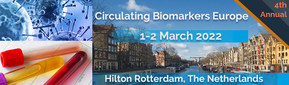
 Add to Calendar ▼2022-03-01 00:00:002022-03-02 00:00:00Europe/LondonCirculating Biomarkers, Exosomes and Liquid Biopsy Europe 2022Circulating Biomarkers, Exosomes and Liquid Biopsy Europe 2022 in Rotterdam, The NetherlandsRotterdam, The NetherlandsSELECTBIOenquiries@selectbiosciences.com