Other Track Agendas3D-Printing in Life Sciences Conference | Organ-on-a-Chip Conference |

Wednesday, 8 July 201508:00 | Conference Registration, Coffee, and Breakfast Pastries | |
Opening Plenary Session: Opportunities in 3D-Printing and Organ-on-a-Chip Technologies and Approaches |
| | |
Plenary Session Chairman: Jon Rowley, Ph.D., Founder and CEO, RoosterBio |
| | 09:00 |  | Keynote Presentation Bioprinting in Regenerative Medicine and Beyond
Gabor Forgacs, Professor, University of Missouri-Columbia; Scientific Founder, Organovo; CSO, Modern Meadow, United States of America
|
| 09:30 |  | Keynote Presentation Organs-on-Chips: In vitro Surrogates for Accurate Human Bioemulation
Geraldine A Hamilton, President/Chief Scientific Officer, Emulate Inc, United States of America
All too often conventional cell-culture methods and animal testing fail
to serve as true predictive models, unable to recapitulate human organ
function or physiology. These shortcomings compel us to pursue more
"humanized" alternatives to the traditional approaches, as we strive to
attain authentic human response. This discussion will explore the
bioemulation capabilities of our Organs-on-Chips technology and its
potential use within drug development and safety pharmacology.
Organs-on-Chips are in vitro surrogates of the human body that may
accelerate the identification of novel therapeutics, ensure safety and
efficacy, and reduce significant drug development costs. We review our
platform and its incorporation of tissue-tissue interfaces, biochemical
cues, mechanical forces and physiological perfusion in an organ-specific
context. Our experience from organs such as the lung, liver, intestine
and kidney consistently shows that these bioengineered microenvironments
lead human cells to assume in vivo function and effectively reproduce
organ-level physiology and disease responses. Based on experimental data
collected from this platform, it is evident that Organs-on-Chips stand
as a more predictive, human-relevant alternative to traditional drug
development methods. |
| 10:00 |  | Keynote Presentation Can Multi-Organ Chips Be Used for Drug Development Studies?
Michael Shuler, Samuel B. Eckert Professor of Engineering, Cornell University, President Hesperos, Inc., United States of America
Effective human surrogates, created from combining human tissue engineered constructs with microfabricated systems, could have a major impact on drug development, particularly in making better decisions on which drugs to take into human clinical trials. Early prediction of human response would be crucial to such decisions. Our designs are guided by physiologically based pharmacokinetic models. We will describe possible approaches to use “pumpless”, low cost platforms to build such human surrogates for evaluation of potential response to drugs. |
| 10:30 | Coffee Break and Networking | 11:00 | 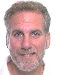 | Keynote Presentation Microengineered Cell and Tissue Systems for Drug Screening and Toxicology Applications: Evolution of In-Vitro Liver Technologies
Martin Yarmush, Founding Director of the Center for Engineering in Medicine, Massachusetts General Hospital and Harvard Medical School, United States of America
|
| 11:30 | The Patent Landscape of Organs-On-a-Chip
Robert Esmond, Director, Sterne, Kessler, Goldstein & Fox P.L.L.C, United States of America
Organs-on-a-chip have present-day real-world uses such as for the
preclinical testing of drugs. Thousands of patents have been filed on
various aspects of organs-on-a-chip, including their manufacture. A
company seeking to make, use or sell an organ-on-a-chip must consider
the patent landscape. This talk will focus on ways to protect
organs-on-a-chip innovation, patent filings directed to organs-on-a-chip
technology, certain exceptions to patent infringement, as well as the
possibility of future litigation. | 12:00 | Networking Lunch, Visit Exhibitors and View Posters | 13:30 |  Technology Spotlight: Technology Spotlight:
3D Bio-printer to Print Cornea and Meniscus
Caddie Wang, CEO, Qingdao Unique Product Develop Co., Ltd
The most difficulties to overcome in traditional preparation of artificial cornea are scaffold support strength and optical properties, as well as internal structure controllability and contour curvature of artificial cornea. Meanwhile, the orientation and arrangement of corneal cells on scaffolds are issues too. 3D printing technology offers the opportunity of molding complex organs based on its high precision and personalization. 3D printing based on different parameters in various ages, creates personalized mold and produces individualized simulated artificial cornea. As for the medical treatment of meniscus injury, the classic methods are repairing, resection or transplantation. However, 3D printing meniscal scaffolds, choosing biodegradable materials to meet biocompatibility and mechanical properties, can be well controlled the morphology, structure and porosity. Qingdao Unique Product Develop Co., Ltd developed this 3D Bio-printer which prints a variety of biomaterials and cells simultaneously. On the basis of different biomaterial and densities in human tissue structure, the build platform with four nozzles is designed to print multiple biomaterials in one object. Our 3D Bio-printer has the capacity of fabricating scaffolds using the widest range of materials, such as PCL (polycaprolactone), HA (hydroxyapatite), TCL (tricalcium phosphate), P(CL-LLC), collagen, fiber and chitosan etc.
| |
Session Title: Emerging Technologies and Trends in 3D-Printing |
| | 14:00 | 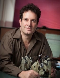 | Keynote Presentation The Next 25 Years of 3D Printing
Hod Lipson, Professor, Cornell University, United States of America
Additive manufacturing has evolved over the last three decades from limited and expensive prototyping equipment in the hands of few, to commodity production tools available to almost anyone. It’s been broadly recognized that this burgeoning industrial revolution will transform almost every industry, and every aspect of our lives. But where will this technology go next? This talk will describe the underlying disruptive future of 3D printing: From printing arbitrarily complex shapes to creating new kinds of materials, and ultimately, moving from fabricating passive parts to printing active, integrated systems, including electronics, actuators and sensors. Will we be able to print a robot that will walk out of the printer, batteries included? |
| 14:30 |  | Keynote Presentation Simple Approaches for Complex Structures: Biofabrication with Multi-Channel 3D Plotting
Michael Gelinsky, Professor and Head, Center for Translational Bone, Joint and Soft Tissue Research, Faculty of Medicine, Technische Universität Dresden, Germany
By means of 3D plotting scaffolds and cell-loaded constructs consisting of a variety of different biomaterials can be fabricated easily. With a novel biopolymer blend open porous, cell-loaded structures of clinically relevant size can be manufactured. Other developments describe the processing of hollow strands as artificial vascular structures and those consisting of two different biomaterials, organized in a core/shell fashion. |
| 15:00 | 3D Biofabrication of Complex Living Cardiovascular Tissues: Past, Present, Future
Jonathan Butcher, Associate Professor, Biomedical Engineering, Cornell University, United States of America
Cardiovascular disease remains a leading cause of death worldwide.
Despite their potential, achieving functional living tissue replacements
has been an elusive “Holy Grail” for the last two decades. Among the
challenges is that natural tissues are highly complex, hierarchical
structures that are difficult to replicate. Tissue biofabrication
technology, in particular 3D tissue printing, has made significant
strides over the past 5 years to close this gap. 3D tissue printing has
the capacity to prescribe both macro and microstructural environmental
cues that are essential for coordinating whole tissue function. We
discuss recent findings on two prominent applications: trileaflet heart
valves and vascularized tissue flaps. | 15:30 | Coffee Break and Networking | 16:00 | High-throughput Generation and Characterization of Cellular Aggregates for 3D Bioprinting
Jordan Miller, Assistant Professor of Engineering and Founder of the Advanced Manufacturing Research Program, Rice University, United States of America
| 16:30 | Print and Play Microfluidics
Albert Folch, Professor of Bioengineering, University of Washington, United States of America
Biologists and clinicians typically do not have access to microfluidic technology because they do not have the engineering expertise or equipment required to fabricate and/or operate microfluidic devices. Furthermore, the present commercialization path for microfluidic devices is usually restricted to high-volume applications in order to recover the large investment needed to develop the plastic molding processes. We are developing microfluidic devices through stereolithography, a form of 3D printing, in order to make microfluidic technology readily available via the web to biomedical scientists. The Folch Lab’s long-term mission is to make microfluidic devices as easy to use as smartphones. 3DSkema is a web marketplace founded by Albert Folch that will facilitate the exchange and evolution of digital designs for 3D-printing. | 17:00 | Engineering Tissue Microenvironments Informed by Development, Healing and Disease
Catherine Kuo, Assistant Professor, Tufts University, United States of America
Tendons are critical tissues of the musculoskeletal system that transfer forces from muscle to bone and stabilize joint structures. Despite surgical intervention, injured or diseased tendons are challenged by reparative wound healing responses that result in scar tissue and mechanical dysfunction. This significant problem has motivated efforts to enhance in vivo healing and also to generate tissue replacements. We are systematically characterizing structure-property relationships of these load-bearing tissues during development, healing, and disease progression in order to identify design parameters for engineered tissues. Specifically, we are designing scaffolds and mechanical loading bioreactors to present cells with healthy, injured, and diseased microenvironments. Our objectives are to engineer tissues as functional replacements and also as research platforms for the development of therapeutics to treat fibrosis and other aberrant musculoskeletal tissue conditions. | 17:30 |  | Keynote Presentation Microtissue Building Blocks for 3D Tissue Fabrication
Shoji Takeuchi, Professor, Center For International Research on Integrative Biomedical Systems (CIBiS), Institute of Industrial Science, The University of Tokyo, Japan
Large-scale 3D tissue architectures that mimic microscopic tissue structures in vivo are very important for not only in tissue engineering but also drug development. In this presentation, I will talk about a bottom-up tissue construction method using different types of cellular building blocks (eg. cell beads and cell fibers). For example, a cell-encapsulating core-shell hydrogel fiber was produced in a double coaxial laminar flow microfluidic device. When with myocytes, endothelial, and nerve cells, they showed the contractile motion of the myocyte cell fiber, the tube formation of the endothelial cell fibers and the synaptic connections of the nerve cell fiber, respectively. By reeling, weaving and folding the fibers using microfluidic handling, higher-order assembly of fiber-shaped 3D cellular constructs can be performed. Moreover, the fiber encapsulating beta-cells is used for the implantation of diabetic mice, and succeeded in normalizing the blood glucose level. |
| 18:00 | Universal Microfluidic Acoustic Printing (UMAP) for Life Science Research
Tingrui Pan, Professor, Department of Biomedical Engineering, University of California-Davis, United States of America
Microfluidic acoustic printing has been recently introduced, utilizing its nature of simple device architecture, low cost, non-contamination, scalable multiplexability and high throughput. In this talk, we will introduce this novel impact-based droplet printing platform utilizing a simple plug-and-play microfluidic cartridge driven by cantilever piezoelectric actuators. Such a customizable printing system allows for ultrafine control of the droplet volume from picoliters (~10nL) to nanoliters (~100pL), a 10,000 fold variation. The high flexibility of droplet manipulations can be simply achieved by controlling the magnitude of actuation (e.g., driving voltage) and the waveform shape of actuation pulses, in addition to nozzle size restrictions. Detailed printing characterizations on these parameters have been conducted consecutively. A multichannel impact printing system has been prototyped and demonstrated to provide the functions of single-droplet jetting and droplet multiplexing as well as concentration gradient generation. Moreover, several biological assays have been implemented and validated on this flexible printing platform. Therefore, the microfluidic acoustic printing system could be of potential value to establishing multiplexed micro droplet reactors for high-throughput life science applications. | 18:30 | Cocktail Reception: Premium Beers, California Wines, Appetizers and Networking on the 15th Floor of the Wyndham Boston Beacon Hill with a View of Boston and the Charles River Sponsored by MIMETAS | 20:00 | Close of Day 1 of the Conference |
Thursday, 9 July 201508:00 | Morning Coffee, Breakfast Pastries, and Networking | |
Session Title: Emerging Biofabrication Technologies and Applications |
| | 08:30 | Emulsion Templated Scaffolds with Tuneable Mechanical Properties for Bone-on-a -Chip Devices
Frederik Claeyssens, Senior Lecturer, Materials Science and Engineering, University of Sheffield, United Kingdom
An important self-assembly route for porous polymeric materials manufacture is emulsion templating via high internal phase emulsions (HIPEs) where typically a dispersed aqueous droplet phase is used with a curable pre-polymer continuous phase. During the curing process a foam is produced where the pores are interconnected (a polyHIPE). Here I will highlight our recent research efforts on using these polyHIPEs in combination with UV-based curing and stereolithography to make hierarchical structures where the internal porosity is governed by self-assembly and the macroscopic structure is constructed by additive manufacturing. These 3D structured scaffolds are produced with varying mechanical properties via changing the monomer mixing ratio to make the polyHIPE structures. These materials have an interconnected internal microporosity of ~10-50 µm, while the macroscale features are defined by the additive manufacturing process. Culturing of human embryonic stem cell-derived mesenchymal progenitors (hES-MPs) reveals that the scaffolds support osteogenic differentiation. Additionally, cells proliferate and penetrate into the micro- and macro-porous architecture. The presented hybrid manufacturing technique addresses the resolution trade-off between macro structuring with micro resolution, while maintaining user control over porosity throughout the process. This work illustrates the excellent potential of these hierarchical structured scaffolds for constructing bone-on-a-chip devices. | 09:00 |  | Keynote Presentation 3D Cell Printing for In Vitro Drug Model
Wei Sun, National “Thousand-Talent” Distinguished Professor, Tsinghua University; Albert Soffa Chair Professor, Drexel University, China
Applying 3D-Cell Printing technique to construct functional in vitro cell/tissue models for drug testing. Examples of printing HeLa cells for 3D cervical tumor model in vitro and printing of micro-organ devices will be presented. |
| 09:30 | 3D-Printing of Plant Cells in Gels
Paul Calvert, Professor,, New Mexico Institute of Mining and Technology , United States of America
A 3D printer has been used to make porous open gel “logpiles” with 200 micron bars containing embedded cells including yeast and algae. The activity of the cells has been studied as a function of gel composition. As expected, cells can metabolize and multiply within a 100 microns of the gel surface. Following the activity of a yeast membrane-bound invertase and comparing this with known diffusion coefficients allows us to quantitatively model metabolism. The gel formulations need to give good strength coupled with easy diffusion of nutrients and products will be discussed. Diffusion coefficients have also been measured for a number of macromolecules in different types of printed gels in order to determine the possibility of using encapsulated cells in 3D gel scaffolds as small bioreactors to produce proteins. | 10:00 | Bioprinting an In vitro System of the Microcirculation
Monica Moya, Research Engineer, Lawrence Livermore National Laboratory, United States of America
Essential to human organ function and survival is a microcirculation. An often oversimplified challenge in creating organ in vitro model systems for drug efficacy and toxicity studies is the development of a vascular supply. The ability to screen chemical toxins to determine the permeability across human capillaries or direct toxicity to the microcirculation is of enormous importance in understanding the integrated effect of an individual toxin. In this work, a direct ink write system is used to print an in vitro model system of human microvessels will be presented. | 10:30 | 3D Bioprinting of Human Cartilage and Skin with Novel Bioink
Paul Gatenholm, Professor, Director of 3D Bioprinting Center, Chalmers University of Technology, Sweden; CEO, CELLHEAL AS, Norway, Sweden
3D Bioprinting technology is expected to revolutionize the field of cell culturing, tissue engineering and regenerative medicine. High resolution 3D Bioprinters can reconstruct complex microarchitecture of living tissue and organs with a resolution of 10µm by dispensing a bioink and positioning several cell types and also provide the microenvironment for stem cell differentiation. We have developed a new hydrogel bioink, CELLINK, for printing living soft tissue like cartilage and skin, with cells. CELLINK is based on non-animal nanofibrillated biopolymers with unique shear thinning properties which provide high printing fidelity. The composition of CELLINK provides high cell viability and ideal conditions for cell proliferation, migration and production of extracellular matrix. CELLINK is easily crosslinked after printing to keep stable 3D structures with outstanding mechanical properties which are importance for use in bioreactors. The human chondrocytes have been successfully printed with CELLINK in complex 3D shape of human ear, meniscus and trachea. Long term study showed neocartilage formation in 3D Bioprinted constructs. | 11:00 | Coffee Break, View Posters, Visit Exhibitors and Networking | 11:30 | From Materials to Markets: Identifying Key Needs in Bioprinting
Anthony Schiavo, Research Associate, Lux Research Inc, United States of America
Bioprinting, the use of additive manufacturing to create structures from living or biological materials, is one segment of the increasingly hyped medical printing area. This presentation will take a step back from the hype to assess how bioprinting falls into the 3D printing market as a whole, and will identify key areas for innovation in materials, systems, and strategies needed to drive this technology from concept to market. | 12:00 | 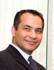 | Keynote Presentation Engineered Hydrogels for Regenerative Medicine Applications
Ali Khademhosseini, Professor, Department of Medicine, Brigham and Women’s Hospital; Wyss Institute for Biologically Inspired Engineering, Harvard University, United States of America
Engineered materials that integrate advances in polymer chemistry, nanotechnology, and biological sciences have the potential to create powerful medical therapies. Our group aims to engineer tissue regenerative therapies using water-containing polymer networks, called hydrogels that can regulate cell behavior. Specifically, we have developed photocrosslinkable hybrid hydrogels that combine natural biomolecules with nanoparticles to regulate the chemical, biological, mechanical and electrical properties of gels. These functional scaffolds induce the differentiation of stem cells to desired cell types and direct the formation of vascularized heart or bone tissues. Since tissue function is highly dependent on architecture, we have also used microfabrication methods, such as microfluidics, photolithography, bioprinting, and molding, to regulate the architecture of these materials. We have employed these strategies to generate miniaturized tissues. To create tissue complexity, we have also developed directed assembly techniques to compile small tissue modules into larger constructs. It is anticipated that such approaches will lead to the development of next-generation regenerative therapeutics and biomedical devices. |
| 12:30 | Networking Lunch, Visit Exhibitors and View Posters | 13:40 |  Technology Spotlight: Technology Spotlight:
The Latest Dispensing Technology for 3D-Bioprinting
Ryutaro Izumi, President, Izumi International Inc
Dispensing technology and automation has been available in the manufacturing industry for decades. This technology has been used for the high-tech electronics industry, automotive industry, and other various high volume industries for a long time, and therefore already been proven for speed and accuracy for these demanding applications. In this presentation, we will introduce our various dispensing technologies that can be used in bio-printing. One of these is non-contact air-jet dispensing, which is seeing a lot of interest in regenerative medicine and bioprinting.
| |
Session Title: Implementation of 3D-Printing in the Life Sciences using Various Bioinks |
| | 14:00 | 3D Bioprinting of Personalized Nasal Scaffolds
Jing Yang, Senior Research Fellow, University of Nottingham, United Kingdom
In the case of a particularly aggressive or mature nasal cancer, the surgical solution is to remove the whole nose from skin down to skeleton. A total nasal reconstruction is then undertaken if the defect site is confirmed to be cancer free. Current gold standard for reconstruction after rhinectomy or severe trauma includes transposition of autologous cartilage grafts in conjunction with autologous mucosal flaps and coverage using a skin flap. The difficulty of this approach is the limited amount of autologous cartilage and mucosa from donor sites to achieve a satisfactory aesthetic and functional reconstruction. Moreover, harvest of autologous cartilage requires a major additional procedure which creates donor site morbidity. We have developed a method for making scaffolds for nasal reconstruction using 3D printing. In particular, we have developed a printing method that allows us to control the shell porosity of the printed scaffolds. Cell penetration into the scaffolds has been found to be improved with higher shell porosity. The co-printing of a cell-laden hydrogel and a polymer into a scaffold has also been demonstrated. | 14:30 |  | Keynote Presentation 3D Bioprinting and the Cardiovascular System
Stuart Williams, Director, Bioficial Organs, University of Louisville, United States of America
|
| 15:00 | Why Manufacturing Matters: How Today’s Cell Therapy BioProcess Innovations are Laying the Foundation for a Sustainable Tissue Engineering Revolution
Jon Rowley, Chief Executive & Technology Officer, Rooster Bio Inc, United States of America
Technology is rapidly moving toward the integration of biologics into products like cell therapies, engineered tissues, bio-robotics, implantable devices, 3D printing, food, clothing, and even toys. This coming decade will see the incorporation of living cells into all these platforms and others not yet imagined. To expedite this biologics revolution, inventors, developers and suppliers will require a limitless, standardized, low-cost supply of living cells – and today’s cell therapy biomanufacturing innovations are laying the groundwork to make this a reality. | 15:30 | Coffee Break and Networking | 16:00 | From Bioprinting of Vascular Networks to Fabrication of Reconfigurable Hydrogel Microfluidics
Luiz Bertassoni, Assistant Professor, Oregon Health and Science University/The University of Sydney, Australia
Fabrication of three-dimensional (3D) organoids with controlled microarchitectures has been shown to enhance tissue functionality. Bioprinting can be used to precisely position cells and cell-laden materials to generate controlled tissue architecture. Therefore, it represents an exciting alternative for organ fabrication. Our group has been interested in developing innovative bioprinting-based microscale technologies, both to improve our understanding human tissues and to increase our ability to regenerate them with improved function. In this seminar, we will present novel bioprinting techniques to fabricate 3D cell-laden organoids. We will also demonstrate a new technique we developed to bioprint biomimetic microvascular networks embedded within macroscale cell-laden tissue constructs. These techniques have been used to engineer hepatocyte-laden hydrogels on-a-chip and to fabricate osteon-like structures mimicking vascularized bone. Additionally, we have adapted a bioprinting technique to fabricate hydrogel-based three-dimensional microchannel networks that can be dynamically reconfigured on demand for microfluidics applications. The use of these technologies in various regenerative applications will be presented. | 16:30 | Tissue Engineering, Bioprinting and The “Reconstructive Ladder”
Jason Spector, Professor of Plastic Surgery and Otolaryngology, Weill Cornell Medical College, United States of America
The ultimate purpose of nearly all tissue engineering and Bioprinting is the clinical translation thereof. However, the actual clinical needs and specific parameters required are not always clearly defined. As such, this talk provides an overview (from a surgeon’s perspective) of the clinical scenarios in which tissue engineering and bioprinting might have their greatest impact with a particular focus on the current clinical strategies utilized in reconstruction including the use of grafts (non-vascularized) and flaps (vascularized). The discussion will also include an overview of the shortcomings of currently available tissue engineered products as well as highlight strategies for achieving the “Holy Grail” of tissue engineering—constructs with their own inherent hierarchical vascularity. | 17:00 | Solving the Limitation for Functional Regeneration of Airway Epithelium by Development of Bioink derived from Mucosal Tissue and in vitro and in vivo Approaches
Ju Young Park, Graduate Student, Pohang University of Science and Technology, Korea South
We investigated a new bioink derived from mucosal tissue for solving the limitation of a functional regeneration of airway epithelium in vitro and in vivo. Using this bioink, the speed of mucociliary epithelial cells formation was accelerated with a significant increase of mucociliary functionality. We also applied the bioink for 3D printing of trachea and microchip fabrication, and their effects were verified in vitro and in vivo. | 17:30 | Decellularized Extracellular Matrix Biomaterials for Printing Engineered Tissues: Applications to Cardiac Repair
Jinah Jang, Associate Professor, Pohang University of Science And Technology (POSTECH), Korea South
Myocardial infarction (MI) is a major contributor to cardiovascular disease, and it is the leading cause of death in the world. Although much progress has been made in treating tissues via stem cell therapy, a major challenge now is regenerating the tissue architecture to integrate a microvascular circulation and afferent arterioles into such an engineered tissue. 3D cell printing technology is currently capable of directly embedding vasculature into the engineered tissue construct by depositing various cells with a flowable hydrogel called bioink. In this study, we used a bioink composed of heart decellularized extracellular matrix (hdECM) to enhance tissue functions and fabricate vascular network embedded 3D patch for grafting after myocardiac infarction. | 18:00 | Close of Day 2 of the Conference |
|

 Add to Calendar ▼2015-07-08 00:00:002015-07-09 00:00:00Europe/London3D-Printing in Life Sciences ConferenceSELECTBIOenquiries@selectbiosciences.com