Co-Located Conference Agendas3D-Culture & Organoids 2019 | 3D-Printing in the Life Sciences | Organ-on-a-Chip World Congress 2019 | Stem Cells in Drug Discovery Tox & Organoids 2019 | The Space Summit 2019 | 

Monday, 14 October 201907:30 | Conference Registration, Materials Pick-Up, Morning Coffee and Pastries in the Exhibit Hall | |
Welcome and Introduction to the Conference by Conference Chairpersons |
| | |
Plenary Session Venue: Coronado Ballrooms A & B |
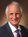
| | 08:15 |  | Conference Chair Conference Chairperson
Nancy Allbritton, Kenan Professor of Chemistry and Biomedical Engineering and Chair of the Joint Department of Biomedical Engineering, University of North Carolina and North Carolina State University, United States of America
|
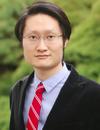
| 08:15 |  | Conference Chair Conference Chairperson
Y. Shrike Zhang, Assistant Professor of Medicine, Harvard Medical School, Associate Bioengineer, Division of Engineering in Medicine, Brigham and Women’s Hospital, United States of America
|
| 08:30 | Modeling Immune Mediated Beta Cell Destruction in Human Type 1 Diabetes with Organoids
Matthias von Herrath, Vice President and Senior Medical Officer, Novo Nordisk, Professor, La Jolla Institute, United States of America
In the past 15 years we have been studying the pathology of human type 1 diabetes with access to donor pancreata through the human pancreatic organ donor consortium (nPOD). These studies have led to several findings, for example that certain cytokines are generated by beta cells themselves, sometimes under stress, and also that there are probably key factors that render beta cells susceptible to immune attacks. Mechanistically, the importance and meaning of these observations needs to be addressed in a suitable and easily manipulable in vitro system consisting of human islets and immune cells. We have built such a system in collaboration with the company InSphero and will discuss emerging findings. | 09:00 |  | Keynote Presentation Pumps, Valves, and Fluidic Connectors for Automating and Integrating Microphysiological Systems
John Wikswo, Gordon A. Cain University Professor, A.B. Learned Professor of Living State Physics; Founding Director, Vanderbilt Institute for Integrative Biosystems, Vanderbilt University, United States of America
Much of the organ-on-chip and tissue-chip research to date has relied on
very simple perfusion schemes that include surface tension, hydrostatic
pressure, rocking plates, syringe pumps, pressurized reservoirs, and
on-chip pneumatic or electromagnetic peristaltic pumps. A number of
commercial microphysiological systems are moving towards high-throughput
well-plate formats. Each of these fluid handling technologies presents
different advantages in cost, convenience, and simplicity, and
disadvantages regarding reservoir filling, media change, recirculation
and volume minimization, organ-organ integration, bubble- and
microbe-free addition and removal of chips from multi-organ
microphysiological systems, temporal control of drug concentrations,
adjusting/balancing flow rates, and controlling sensors for long-term
use. While a common trend is towards high throughput and low cost, there
are critical applications where high content is more important than
high throughput, particularly those requiring multi-omic
characterization of physiological responses over time to drugs and
toxins for both single and multiple organs. We review how our computer
controlled, modular, rotary planar peristaltic micropumps and
twenty-five port rotary planar valves can be used to create
autocalibrating electrochemical MicroClinical Analyzers, multi-well
MicroFormulators that can create in vivo the pharmacokinetics observed
in animals and humans, and perfusion controllers for multiple
NeuroVascular Unit, Gut-in-a Puck, and Thick Tissue Mammary Bioreactors.
Pumps and valves are useful for compact perfusion controllers that
operate in microgravity environments. Miniature rotary valves will
enable bubble-free, sterile connection and disconnection of individual
organ chips to other organs and analyzers. Ongoing and future
applications include 100-port valves and systems for optimizing the
differentiation of induced pluripotent stem cells, a 10,000 channel
yeast MicroChemostat, and a MicroDialysis imager, all with direct
injection into a mass spectrometer. The cost and complexity of these
systems over present technologies is balanced by sophisticated computer
control and sensor integration, reduced technician effort, high content
results, and parallelization. |
| 09:30 |  | Keynote Presentation Intestinal Epithelium for Basic Physiology and Drug Assays
Nancy Allbritton, Kenan Professor of Chemistry and Biomedical Engineering and Chair of the Joint Department of Biomedical Engineering, University of North Carolina and North Carolina State University, United States of America
Organ-on-chips are miniaturized devices that arrange living cells to
simulate functional subunits of tissues and organs. These microdevices
provide exquisite control of tissue microenvironment for the
investigation of organ-level physiology and disease. Human
organ-on-chips are expected to transform biomedical research providing
platforms that accurately replicate human tissues, enable a better
understanding of human-to-human physiologic variations, and even permit
patient-specific organ mimics. These human organ facsimiles will
fundamentally alter drug discovery and development by providing human
constructs for screening assays, toxicity measurements and investigation
of molecular-level drug actions. Breakthroughs in stem-cell biology now
enable single stem cells or intestinal crypts isolated from primary
mouse or human intestine or differentiated from induced pluripotent stem
cells (iPSCs) to grow and persist indefinitely in defined 3D culture
conditions to form organotypic structures generically termed enteroids.
We have developed a long-lived, self-renewing monolayer culture format
from primary intestinal cells. A surface matrix and chemical factors
sustain the epithelial cell monolayers so that they possess all cell
repertoires found in vivo. This technical advance has made it possible
to create primary tissues on platforms that are compatible with
high-content screening strategies. For example, the screening of 77
dietary compounds revealed that these compounds altered proliferation to
increase stem cell numbers or increased cell differentiation with
formation of increased numbers of goblet cells or enterocytes.
Measurement of drug transport and metabolism has also been demonstrated
in these human intestinal monolayer systems as well as formation of
physiologic mucus layers many hundreds of microns thick. Culture of
these monolayers on a shaped scaffold under chemical gradients
replicates much of the cell compartmentalization and physiology observed
in vivo. This bioanalytical platform is envisioned as a next-generation
system for assay of microbiome-, drug- and toxin-interactions with the
intestinal epithelia. Finally, intestinal biopsy samples can be used to
populate these constructs with cells producing patient-specific tissues
for personalized medicine that can be applied to emerging areas of
disease modeling and microbiome studies. |
| 10:00 |  | Keynote Presentation Digital Manufacturing of Microfluidic Tissue Chips
Albert Folch, Professor of Bioengineering, University of Washington, United States of America
The microfluidics field is at a critical crossroads. The vast majority of microfluidic devices are presently manufactured using micromolding processes that work very well for a reduced set of biocompatible materials, but the time, cost, and design constraints of micromolding hinder the commercialization of many devices. PDMS, in particular, is extremely popular in academic labs, yet the fabrication procedures are based on cumbersome manual methods and the material itself strongly absorbs lipophilic drugs. As a result, the dissemination of many cell-based microfluidic chips – and their impact on society – is in jeopardy. Digital Manufacturing (DM) is a family of computer-centered processes that integrate digital 3D designs, automated (additive or subtractive) fabrication, and device testing in order to increase fabrication efficiency. Importantly, DM enables the inexpensive realization of 3D designs that are impossible or very difficult to mold. The adoption of DM by microfluidic engineers has been slow, likely due to concerns over the resolution of the printers and the biocompatibility of the resins. We have developed microfluidic devices by SL in PEG-DA-based resins with automation and biocompatibility ratings similar to those made with PDMS. I will also present our work on our microfluidics platform (digitally-manufactured in thermoplastics) for cancer diagnostics using live tumor biopsies and I will review the bright future ahead for the promising, fertile field of DM.
|
| 10:30 | Morning Coffee Break and Networking in the Exhibit Hall | 11:00 |  | Keynote Presentation Models of Neurological Disease
Roger Kamm, Cecil and Ida Green Distinguished Professor of Biological and Mechanical Engineering, Massachusetts Institute of Technology (MIT), United States of America
Many of the most debilitating and life-threatening diseases are
associated with the central nervous system. Some, such as
neurodegenerative diseases, predominately afflict our aging population.
Yet others such as brain cancers and ALS are prevalent among young and
old alike. In this presentation, models will be presented that attempt
to recapitulate certain aspects of the brain and these diseases, both to
probe the disease process, and as a platform to screen for new
therapeutics. Three examples will be presented. First, a model for the
healthy blood-brain barrier and neurovascular unit have been developed
in order to capture the essential aspects associated with these
diseases. Second, the blood-brain barrier model is used to study
metastasis of cancers to the brain, as well as primary glioblastoma. In
the third part of the talk, a model for the healthy and diseased
neuromuscular junction will be presented as a step toward developing
therapeutic strategies for treating ALS. |
| 11:30 |  | Keynote Presentation “Bone Marrow-on-a-Chip”: A Strategy to Replicate Hematopoiesis and Cancer Cell Dormancy
Steven C. George, Edward Teller Distinguished Professor and Chair, Department of Biomedical Engineering, University of California-Davis, United States of America
Tissue engineering holds enormous potential to not only replace or
restore function to a wide range of tissues, but also to capture and
control 3D physiology in vitro (e.g., microphysiological systems or
“organ-on-a-chip” technology). The latter has important applications in
the fields of drug development, toxicity screening, modeling tumor
metastasis, and repairing damaged cardiac (heart) muscle. In order to
replicate the complex 3D arrangement of cells and extracellular matrix
(ECM), new human microphysiological systems must be developed. The past
decade has brought tremendous advances in our understanding of stem
cell technology and microfabrication producing a rich environment to
create an array of “organ-on-a-chip” designs. Over the past six years
we have developed novel microfluidic-based systems of 3D human
microtissues (~ 1 mm3) that contain features such as perfused
human microvessels, primary human cancer, mouse tumor organoids, stem
cells, and spatiotemporal control of oxygen. The technologies are part
of two early start-up companies, Kino Biosciences and Immunovalent
Therapeutics, based in Irvine, CA and St. Louis, MO. This seminar will
describe our approach and early results on a strategy to create a 3D in
vitro model of human bone marrow with applications in hematopoiesis and
cancer cell dormancy. |
| 12:00 |  | Keynote Presentation Human Tissue Chips for Drug Development, Disease Modeling, and More…
Kevin Healy, Jan Fandrianto and Selfia Halim Distinguished Professorship in Engineering, University of California, Berkeley, United States of America
Our work has emphasized creating both healthy and diseased model organ systems, we call microphysiological systems or ‘tissue chips’, to address the costly and complex drug discovery process. The average time to develop and launch a new drug in the United States is 10-15 years, and costs ~ $2-5b. The poor efficiency and high failure rates are attributed to the heavy reliance on non-human animal models employed during safety and efficacy testing that poorly reflect human disease states. With the discovery of human induced pluripotent stem cells, we can now develop tissue chips to be used for high content drug screening, disease modelling, and numerous other applications. While tissue chips are poised to disrupt the drug development process and significantly reduce the cost of bringing a new drug candidate to market, tissue chip technology is much more robust and creates a whole new paradigm in how to conduct biological science, and advances medicine in revolutionary ways. Ultimately, the vision is to reduce or eliminate the use of animals in drug discovery, and conduct ‘clinical trials’ in patient-specific tissue chips that can accommodate variations in genetics, environment, and lifestyle. |
| 12:30 | Networking Lunch in the Exhibit Hall, Exhibits and Poster Viewing | 13:00 | Delegates Welcomed to Attend Afternoon Concurrent Sessions: The Space Summit (Ballroom C) or Organoids (Ballroom D) | 18:00 | Networking Reception with Beer, Wine and Dinner in the Exhibit Hall -- Sponsored by ATCC | 19:00 | Close of Day 1 of the Conference |
Tuesday, 15 October 201908:00 | Morning Coffee, Pastries and Networking in the Exhibit Hall | |
Session Title: Emerging Themes in 3D-Bioprinting, circa 2019 |
| | |
Venue: Coronado Ballroom B |
| | 08:30 | Integrating Multiple Biofabrication Technologies To Create Complex and Functional Musculoskeletal Tissue Grafts
Riccardo Levato, Associate Professor in Translational Bioengineering and Biomaterials, University Medical Center Utrecht, Netherlands
Major challenges in musculoskeletal tissue engineering revolve around recapitulating the architecture, and therefore the function, of native tissues. Present strategies to treat chondral and osteochondral defects, including tissue engineering and cell implantation, prevalently result in repair tissue with poor mechanical properties, which is prone to degeneration, and can only delay the insurgence of severe pathologies like osteoarthritis.
Biofabrication is opening new avenues for the restoration of impaired joints tissues. Multi-material bioprinting enables to fabricate composite structures combining cell-laden, soft hydrogels with mechanically strong polymers for structural support. By the accurate 3D patterning of stem and tissue-specific progenitor cells, salient features of the native zonal and depth dependent organization of articular cartilage can be replicated. Alongside hydrogel extrusion and bioprinting, different additive manufacturing technologies, such as melt electrowriting of polymeric microfibers, ceramic plotting and digital light processing lithographic printing of hydrogels, can be combined to create composite, cell-laden constructs that enable integration between engineered cartilage hydrogels and bone scaffolds. These bioartificial osteochondral grafts exhibit improved interfacial mechanical strength, favouring their integration in vivo.
Herein, the latest development in the field of bioink printing to create zonal-biomimetic cartilage constructs will be discussed, together with the integration of multiple (bio)printing strategies (i.e. co-fabrication of hydrogels, reinforcing polymers and bioceramics), and the impact of these technologies towards the generation of fully biofabricated, high-performance engineered osteochondral grafts, with potential application for regenerative medicine. Finally, technological advances and challenges towards the biofabrication of large, clinically-relevant multi-tissue constructs will be discussed. | 09:00 |  | Keynote Presentation Computed Axial Lithography For Volumetric Printing of Soft Structures
Hayden Taylor, Assistant Professor, Mechanical Engineering, University of California-Berkeley, United States of America
Volumetric additive manufacturing is defined as producing the entire volume of a component or structure simultaneously — rather than by layering — and has been envisioned as a possible way to increase the speed of additive manufacturing. Until recently, however, no practicable technique existed for creating arbitrary 3D geometries volumetrically. Recently, we have demonstrated Computed Axial Lithography (CAL) to meet this need. CAL essentially reverses the principles of computed tomography (widely used in imaging, but not previously in fabrication) to synthesize a three-dimensionally controlled illumination dose within a volume of photocurable resin. The photosensitive volume rotates steadily while a video projector illuminates the material from a direction perpendicular to the axis of rotation. The illumination pattern typically changes >1000 times per revolution, so that light from many different projection angles contributes to the cumulative dose. Where the dose exceeds a threshold, the resin solidifies and the part is formed. In this talk I will discuss the CAL process in the context of its possible bioprinting applications. The CAL printing technique has several potential advantages. Because there is no relative motion between the component being printed and the resin during printing, the printing speed is not limited by resin flow effects, as it can be in layer-by-layer photopolymerization-based printing. The absence of relative motion also allows highly viscous resins or soft, highly compliant materials such as hydrogels to be printed. Because layers are not used in CAL, the surfaces of printed components are very smooth (c. 1–4 µm roughness in preliminary experiments), which may open up new applications. Additionally, it is possible to print objects around pre-existing solid objects that could have been made using a different material or process. This ‘overprinting’ capability suggests applications in mass-customization for end users and also potentially in fabricating biological structures with highly heterogeneous mechanical properties. |
| 09:30 | Engineering Human Tissues Using 3D Bioprinting Technology
Jinah Jang, Associate Professor, Pohang University of Science And Technology (POSTECH), Korea South
Recent development in bioengineering enables us to create human tissues by integrating various native microenvironments, including tissue specific cells, biochemical and biophysical cues. A significant transition of 3D bioprinting technology into the biomedical field helps to improve the function of engineered tissues by recapitulating physiologically relevant geometry, complexity, and vascular network. Bioinks, used as printable biomaterials, facilitate dispensing of cells through a dispenser as well as supports cell viability and function by providing engineered extracellular matrix. Successful construction of functional human tissues requires accurate environments that are able to mimic biochemical and biophysical properties of target tissue. Formulation of printable materials with stem cells are critical process to guide cellular behavior; however, this is rarely considered in the context of bioprinting in which the tissue should be formed. This talk will cover my research interests in building 3D human tissues and organs to understand, diagnose and treat various intractable diseases, particularly for cardiovascular disease. A development of tissue-derived decellularized extracellular matrix bioink platform will be mainly discussed as a straightforward strategy to provide biological and biophysical phenomena into engineered tissues. I will also discuss about a development of 3D vascularized cardiac stem cell patch that is generated by integrating the concept of tissue engineering and the developed platform technologies. Combined with recent advances in human pluripotent stem cell technologies, printed human tissues could serve as an enabling platform for studying complex physiology in tissue and organ contexts of individuals. | 10:00 | Ultrafast 3D Printing of Large-scale Engineered Tissues
Ruogang Zhao, Associate Professor, SUNY Buffalo, United States of America
Large size engineered soft tissues hold great promise for tissue repair and organ transplantation, but their fabrication using 3D bioprinting is hindered by the cellular damage caused by the prolonged exposure to the unfavorable printing environment and the limited multiscale printing capability. Here we present a Fast hydrogeL prOjection stereolithogrAphy Technology (FLOAT) that can create a centimeter-sized, multiscale solid hydrogel tissue model within several minutes, thus significantly reducing environmental stress-induced cellular injuries. As a proof of principle, we rapidly print a centimeter-sized hydrogel liver model containing perfusable vascular channels. Media perfusion enabled by the channel network is shown to play a critical role in promoting cell survival and metabolic function in the deep core of the model over long term. We demonstrate the good compatibility of the FLOAT with multiple photocurable hydrogel materials and the strategies to optimize the printing material to improve tissue regeneration such as endothelialization of pre-fabricated vascular channels. Together, these studies show that FLOAT is a promising approach for the fabrication of vascularized engineered tissues of clinically-relevant size and potentially suitable for rapid injury repair and transplantation.
| 10:30 | Morning Coffee Break and Networking in the Exhibit Hall | 11:00 |  | Keynote Presentation Handheld Skin Printer: In-Situ Formation of Biomaterials and Skin Tissues
Axel Guenther, Professor and Co-Lead, Collaborative Centre for Research and Applications in Fluidic Technologies (CRAFT), University of Toronto, Canada
I discuss the in-situ delivery of biomaterials and cells for skin tissue repair. We developed a handheld skin bioprinter, specifically for intraoperative use. I will discuss unique features of the handheld bioprinter instrument, physical and in vitro characterization of in-situ deposited biomaterials and tissues, as well as in vivo results that were obtained in porcine wound in collaboration with Dr. Marc Jeschke’s team at Sunnybrook Health Sciences Center in Toronto. |
| 11:30 |  | Keynote Presentation Innovations in Bioprinting
Ali Khademhosseini, Professor, Department of Bioengineering, Department of Radiology, Department of Chemical and Biomolecular Engineering, University of California-Los Angeles, United States of America
|
| 12:00 | Networking Lunch in the Exhibit Hall and Poster Viewing | 13:00 |  3D Hydrogels and Bioinks for Realistic In-Vitro Modelling and Bioprinting 3D Hydrogels and Bioinks for Realistic In-Vitro Modelling and Bioprinting
Elia Lopez-Bernardo, Global Business Development Manager, Biogelx, Ltd.
Biogelx have developed innovative biomaterials offering artificial tissue environments for 3D cell culture and bioprinting applications. The technology platform is based on peptide hydrogels and bioinks which contain ECM-relevant ligands and are highly tunable. They are three-dimensional, 99% water and have the same nanoscale matrix structure as human tissue. Biogelx biomaterials are non-animal derived and their surface chemistry and mechanical properties can be tuned to meet the needs of any given cell or tissue type.
These novel hydrogels and bioinks form a nanofibrous network mimicking the extracellular matrix, which supports cell function, signaling, and proliferation. They have been developed and improved to ensure the rheological properties are optimal for 3D bioprinting. Additionally, they provide great 3D fidelity and do not require the use of support/sacrificial materials or curing agents. These are unique, versatile materials which offer important benefits for researchers by providing a base modular gel in which the stiffness and surface peptides can be adapted. They offer complete reproducibility, viscosity control, an easy crosslinking method and excellent printability within the same material.
These novel materials are creating new opportunities in the fields of cancer biology, stem cell research and tissue engineering by offering synthetic-yet-biologically-relevant alternatives to traditional, animal-derived matrices.
| 13:30 |  How 3D Bioprinting Begins To Take Shape In Industry How 3D Bioprinting Begins To Take Shape In Industry
Ricky Solorzano, CEO, Allevi
3D bioprinting has been a very promising technology to allow more and more research to be published and new discoveries made. However, how does bioprinting begin to penetrate the industrial sector? What specific areas should the field focus on? How can we measure impact?
| 14:00 |  Bioprinting of Human Soft Tissues Bioprinting of Human Soft Tissues
Nicole Diamantides, Bioprinting Field Application Scientist, CELLINK
Discussion of how bioprinting techniques can be utilized to enable the fabrication of human soft tissues.
| 14:30 |  | Keynote Presentation Putting 3D Bioprinting to the Use of Tissue Model Fabrication
Y. Shrike Zhang, Assistant Professor of Medicine, Harvard Medical School, Associate Bioengineer, Division of Engineering in Medicine, Brigham and Women’s Hospital, United States of America
The talk will discuss our recent efforts on developing a series of bioprinting strategies including sacrificial bioprinting, microfluidic bioprinting, and multi-material bioprinting, along with various cytocompatible bioink formulations, for the fabrication of biomimetic 3D tissue models. These platform technologies, when combined with microfluidic bioreactors and bioanalysis, will likely provide new opportunities in constructing functional microtissues with a potential of achieving precision therapy by overcoming certain limitations associated with conventional models based on planar cell cultures and animals. |
| 15:00 | Afternoon Coffee Break and Networking in the Exhibit Hall | 15:30 |  | Keynote Presentation Modulating Physical, Chemical, and Biological Properties of Tissue Constructs via Rapid 3D Bioprinting
Shaochen Chen, Professor and Chair, NanoEngineering Department, University of California-San Diego, United States of America
In this talk, I will present our laboratory’s recent research efforts in rapid 3D bioprinting to create 3D tissue constructs using a variety of biomaterials and cells. These 3D biomaterials are functionalized with precise control of micro-architecture, mechanical (e.g. stiffness), chemical, and biological properties. Such functional biomaterials allow us to investigate cell-microenvironment interactions at nano- and micro-scales in response to integrated physical and chemical stimuli. From these fundamental studies we have been creating both in vitro and in vivo precision tissues for tissue regeneration, disease modeling, and drug discovery. To vascularize these engineered tissues, we have also developed a pre-vascularization technique by using the rapid 3D bioprinting method. Multiple cell types mimicking the native vascular cell composition were encapsulated directly into hydrogels with precisely controlled distribution. |
| 16:00 | Nano to Microscale Engineering and Bioprinting of Vascularized and Innervated Bone-Like Microenvironments
Luiz Bertassoni, Associate Professor, Biomaterials and Biomechanics, School of Dentistry, Cancer Early Detection Advanced Research, Knight Cancer Institute, Oregon Health & Sciences University, United States of America
Bone tissue, by definition, is an organic-inorganic nanocomposite, where metabolically active cells are embedded three-dimensionally within a matrix material that is heavily calcified on the nanoscale. While the field of cell biology has evolved to culture cells three-dimensionally embedded in soft hydrogels, currently there are no strategies that replicate these definitive characteristics of bone tissue. Here, we describe a series of biomimetic approaches to mimic the 3D microenvironment of human bone. We discuss recently reported methods that we developed to encapsulate stem cells, vascular capillaries and nerve fibers within high density nanoscale mineralized materials that are inherently osteogenic. We also discuss digital light processing bioprinting strategies to mimic the structure, composition and function of the bone matrix. Ultimately, these approaches enable fabrication of bone-like tissue models with high levels of biomimicry with broad implications for disease modeling, drug discovery, and regenerative engineering. | 16:30 | The Bioprinter as a Platform for 3D Cell Biology Experimentation
Thomas Angelini, Professor, Department of Mechanical and Aerospace Engineering, University of Florida, United States of America
The remarkable differences between cells grown on plates and cells in vivo or in 3D culture are well-known. At the physical level, cell shape, structure, motion, and mechanical behavior in 3D are totally different from those in the dish and are far less explored. At the molecular level, cells grown in monolayers exhibit gene expression profiles that do not correlate or are anticorrelated with those of cells grown in 3D culture or xenograft animal models. However, our understanding of cell biology has been heavily shaped by the culture plate, whether viewed through the lens of gene expression profiles, signaling pathways, morphological characterization, or mechanical behaviors. Closing this major gap between 2D in vitro culture and in vivo biology requires a tunable and flexible method for creating 3D cell assemblies and performing experiments on cells in 3D environments. In this talk I will describe how we use a bioprinter in combination with a 3D culture medium made from jammed microgels to perform a wide range of 3D experiments. I will demonstrate this experimental platform’s ability to print structures made from multiple cell types or extracellular matrix with predictable feature sizes down to the scale of a few cell bodies. I will also present data from numerous types of experiments performed in 3D, designed to explore collective cell behavior and cell-cell interactions. For example, I recent results will be presented from a 3D immunotherapy model in which we investigate how immune-specific T cells attack 3D printed brain tumoroids. Our results demonstrate that, in parallel to pursuing the long-standing goals shared by those within the 3D bioprinting field, the current state of bioprinting technology can leveraged to facilitate the emergence and growth of new areas of scientific investigation. | 17:00 | Hybrid Laser Platform for Printing 3D Multi-Scale Multi-Material Hydrogel Structures
Pranav Soman, Professor, Biomedical and Chemical Engineering; IPA Program Director, Advanced Manufacturing (AM), National Science Foundation (NSF), Syracuse University, United States of America
Over the course of billions of years, nature has created and refined numerous elegant biosynthetic processes to make sophisticated functional structures. In contrast, current manufacturing techniques are still limited in their ability to fabricate 3D multiscale multi-material structures. Few research groups have utilized the ability of ultrafast lasers to shape hydrogel materials into complex 3D structures. However, current laser based methods are limited by scalability, types of materials, and incompatible laser and materials processing requirements, thereby preventing its widespread use in the field. In this work, we report the design and development of a Hybrid Laser Printing (HLP) technology, that combines the key advantages of additive stereolithography (quick on-demand continuous fabrication) and multiphoton polymerization/ablation processes (high-resolution and superior design flexibility). Using a series of proof-of-principle experiments, we show that HLP is capable of printing 3D multiscale multi-material structures using model biocompatible hydrogel materials that are highly difficult and/or extremely time consuming to fabricate using current technologies. | 17:30 | Decellularized Extracellular Matrix Bioinks and the External Stimuli to Enhance Cardiac Tissue Development in vitro
Sanskrita Das, Researcher, Pohang University of Science and Technology (POSTECH), Korea South
Heart-derived decellularized extracellular matrices, composed of an intricate network of multi-domain macromolecules, provide a comprehensive micro-environmental niche to recreate functional 3D cardiac tissue models. | 18:00 | Close of Conference |
|


 Add to Calendar ▼2019-10-14 00:00:002019-10-15 00:00:00Europe/London3D-Printing in the Life Sciences3D-Printing in the Life Sciences in Coronado Island, CaliforniaCoronado Island, CaliforniaSELECTBIOenquiries@selectbiosciences.com