Co-Located Conference AgendasLab-on-a-Chip and Microfluidics Europe 2018 | Organ-on-a-Chip, Tissue-on-a-Chip Europe 2018 | Point-of-Care Diagnostics & Biosensors Europe 2018 | 

Tuesday, 5 June 201807:30 | Conference Registration, Materials Pick-Up, Morning Coffee and Tea | |
Session Title: Conference Opening Plenary Session |
| | |
Plenary Session Chairman: Richard Spero, Ph.D., Co-Founder and CEO, Redbud Labs: Advanced Microfluidic Technologies |
| | 08:15 |  | Conference Chair Conference Chairman Welcome and Opening Remarks
Richard Chasen Spero, CEO, Redbud Labs, United States of America
|
| 08:30 | 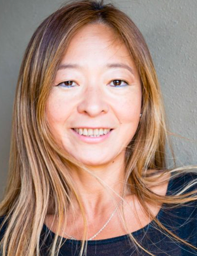 | Keynote Presentation Nanoplasmonic Biosensors: From Innovative Materials to Multimode Sensing with Integrated Microdevices
Amy Shen, Professor and Provost, Okinawa Institute of Science and Technology Graduate University, Japan
Gold nanostructures are a highly attractive class of materials with unique electrochemical and optical sensing properties. Recent developments have greatly improved the sensitivity of optical sensors based on metal nanostructured arrays. We introduce the localized surface plasmon resonance (LSPR) sensors and describe how its exquisite sensitivity to size, shape and environment can be harnessed to detect molecular binding events. We then describe recent progress in three areas representing the most significant challenges: integration of LSPR with complementary electrochemical techniques, long term live-cell biosensing and practical development of sensors and instrumentation for routine use and high-throughput detection. As an example we will demonstrate a novel refractive index and charge sensitive device integrated with nanoplasmonic islands to develop nano-metal-insulator-semiconductor (nMIS) junctions. The developed sensor facilitates simultaneous detection of charge and mass changes on the nanoislands due to biomolecule binding. A brief insight on microcontact printing to functionalize proteins on nanoplasmonic sensors will also be discussed. The developed nanosensors can readily be adopted for multiplexed and high throughput label-free immunoassay systems, further driving innovations in biomedical and healthcare research. |
| 09:00 |  | Keynote Presentation Towards Wearable Real-Time Clinical Monitoring Using Microfluidic Devices
Martyn Boutelle, Professor of Biomedical Sensors Engineering, Imperial College London, United Kingdom
Modern acute critical care medicine is increasingly seeking to protect vulnerable tissue from damage by monitoring the patterns of physical, electrical and chemical changes taking place in tissue – so called multimodal monitoring. Such patterns of molecular changes offer the exciting possibility of allowing clinicians to detect changes in patient condition and to guide therapy on an individualized basis in real time. Microfluidic lab-on-chip devices coupled to tissue sampling using microdialysis provide an important new way for measuring real-time chemical changes as the low volume flow rates of microdialysis probes are ideally matched to the length scales of microfluidic devices. In this presentation, I will describe the combination of miniature electrochemical sensors and biosensors with 3D printed microfluidic devices for transplant organ and patient monitoring. Concentrations of key biomarker molecules can then be determined continuously using either optically or electrochemically, using amperometric, potentiometic and array sensors. Wireless devices allow analysis to take place close to the patient. Droplet-based microfluidics, by digitizing the dialysis stream into discrete low volume samples, both minimizes dispersion allowing very rapid concentration changes to be measured, and allows rapid transport of samples between patient and analysis chip. This talk will overview successful design, optimization, automatic-calibration and use of both continuous flow and droplet-based microfluidic analysis systems for real-time clinical monitoring, using clinical examples from our recent work. |
| 09:30 | 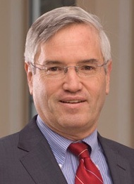 | Keynote Presentation Silicon Nanotechnology Meets Biology (Smaller and Wetter is Better)
Gregory Timp, Keough-Hesburgh Professor of Electrical Engineering & Systems Biology, The University of Notre Dame, United States of America
According to Moore’s law, the scaling of silicon integrated circuits is supposed to reach the 5 nm-node sometime after 2020, although the schedule is still problematic due to the astronomical cost and atomically precise line-rules. On the other hand, biology has been performing cost-effectively using proteins the size of 5 nm (and smaller) that fold with atomic precision for 4.28 billion years now—it is a robust and proven technology, albeit wet. In this talk, it is argued that there is still “plenty of room at the bottom” for improving performance if silicon nanotechnology is adapted to biology. With silicon nanotechnology it is now within our grasp to create an interface to biology on a nanometer-scale. Three examples of such interfaces are proffered. The first is a liquid flow cell that works like an envelope made from 30 nm-thick silicon nitride membranes, which can hold and sustain living cells in medium and yet fits inside a Scanning Transmission Electron Microscope (STEM). In a STEM, the liquid cell can be used to visualize and track live cell physiology like a phage infecting a bacterium with nucleic acids at 5 nm resolution. The second is a nanometer-diameter pore sputtered through a silicon nitride membrane 10-nm-thick that can be used to transfect cells precisely with nucleic acids to affect gene expression in them and, under different bias conditions, detect protein secretions from single cells with single molecule sensitivity. The secretions inform on the cell phenotype and offer a molecular diagnosis of disease. Finally, the third interface is a sub-nanometer-diameter pore, which is about the size of an amino acid residue, in either silicon dioxide or silicon nitride membranes ranging from 6 to 10 nm-thick. Sub-nanopores like this have been used to read the primary structure of a protein, i.e. the amino acid sequence, with low fidelity, but with single molecule sensitivity, vastly outstripping the sensitivity of conventional methods for sequencing such as mass spectrometry. Taken altogether, the prospects are dazzling for a new type of integrated circuit that incorporates biology with state-of-the-art silicon electronics. |
| 10:00 | Morning Coffee Break and Networking in the Exhibit Hall | 10:45 |  | Keynote Presentation Extracellular Vesicles (EVs) and Cell Free DNA (cfDNA) as Blood-based Biomarkers: Plastic-based Microfluidics for their Enrichment and Analysis
Steve Soper, Foundation Distinguished Professor, Director, Center of BioModular Multi-Scale System for Precision Medicine, The University of Kansas, United States of America
While there are a plethora of different blood-based markers, EVs are generating significant interests due to their relatively high abundance (~1013 particles per mL of blood) and the information they carry. EVs contain a diverse array of nucleic acids, such as mRNA, lncRNA, and miRNA that can be used for disease management. In addition to EVs, cfDNA also are biomarkers that can be used to help manage different disease states using the mutations they possess that can have high diagnostic value. In spite of the relatively high abundance of cfDNA in diseased patients (~160 ng/mL), the extraction and enrichment of cfDNA has been inefficient, even by commercial kits, due to the low abundance of the tumor bearing DNA fragments (<0.01%) and the short nature of these fragments, especially cancer-related cfDNA (as small as 50 bp). In this presentation, we will discuss the design, fabrication and analytical figures-of-merit of a microfluidic device that can serve the dual purpose for the affinity-based selection of EVs and the solid phase extraction of cfDNA directly from plasma using the same device. The microfluidic is made from a plastic that can be injection molded to produce high quality devices at low cost. For EVs, the device is made cyclic olefin copolymer (COC) is UV/O3 activated to allow for the efficient immobilization of affinity agents to the surface of the device. In the case of cfDNA, the device is made from COC as well, but is only UV/O3 activated (i.e., no affinity agents used). Information will be provided as to the ability to molecularly profile the cargo contained within the affinity-selected EVs, in particular mRNA expression profiling. We will also discuss the use of this microfluidic to isolate with high recovery cfDNA from plasma samples with size selection capabilities. The isolated cfDNA could be queried for mutations using an allele-specific ligation detection reaction at a mutant to wild-type ratio <0.1%. |
| 11:15 |  | Keynote Presentation Microphysiological Models Relying on Emergence of Multi-Cellular Engineered Living Systems
Roger Kamm, Cecil and Ida Green Distinguished Professor of Biological and Mechanical Engineering, Massachusetts Institute of Technology (MIT), United States of America
Recent work from many labs has demonstrated the unique capability of
cells placed in 3D culture to self-organize into functional units and
organ-like systems. In some cases, pluripotent cells can be induced to
differentiate down independent pathways, leading to an organoid. In
others, interacting units can be generated, often from iPS cells, and
‘engineered’ to interact in a way that recapitulates certain aspects of
in vivo function or disease. Such models have tremendous potential both
to gain new insight into disease processes and for moderate throughput
drug screening. In this talk I will describe several models developed
in our lab including a neuromuscular junction, blood-brain barrier, and
vascularized skeletal muscle, addressing some of the design principles
they have in common, the future potential, and barriers to progress. |
| 11:45 |  | Keynote Presentation Microscale Flow Patterning
Moran Bercovici, Associate Professor, Faculty of Mechanical Engineering; Head, Technion Microfluidic Technologies Laboratory, Technion, Israel Institute of Technology, Israel
The ability to manipulate fluids at the microscale is a key element of any lab-on-a-chip platform, enabling core functionalities such as liquid mixing, splitting and transport of molecules and particles. Lab-on-a-chip devices are commonly divided in two main families: continuous phase devices, and discrete phase (droplets) devices. While a large number of mechanisms are available for precise control of droplets on a large scale, microscale control of continuous phases remains a substantial challenge. In a traditional continuous-flow microfluidic device, fluids are pumped actively (e.g. by pressure gradients, electro-osmotic flow) or passively (e.g. capillary driven) through a fixed microfluidic network, making the device geometry and functionality intimately dependent on one another (e.g. DLD, inertial mixer, H-separator, etc.). The advent of on-chip microfluidic valves brought more flexibility in routing fluids through microfluidic networks, adding a dynamic dimension to the static geometrical network. However, the number of degrees of freedom of valve-based systems is restricted by their dependence on bulky pneumatic lines (regulators, pressure systems, controllers), which are difficult to scale down in size and cost. In this talk I will present our ongoing work leveraging non-uniform EOF and thermocapillary flows to control flow patterns in microfluidic chambers. By setting the spatial distribution of surface potential or a spatial temperature distribution, we demonstrate the ability to dictate desired flow patterns without the use of physical walls. We believe that such flow control concepts will help break the existing link between geometry and functionality, bringing new capabilities to on-chip analytical methods. |
| 12:15 | Networking Lunch in the Exhibit Hall -- Meet the Exhibitors and View Posters | |
Session Title: Emerging Themes in Microfluidics Technologies |
| | 13:30 |  | Keynote Presentation Acoustofluidics Enables Advanced Microfluidic Cell Handling and Enrichment of Extracellular Vesicles
Thomas Laurell, Professor, Lund University, Sweden
|
| 14:00 | Microchannel Mixing and Multifractal Dimensions
Miron Kaufman, Professor of Physics, Cleveland State University, United States of America
The geometry of mixing structures developed in several types of microchannels is quantified by using entropic and fractal measures. Insights into mixing mechanisms and on optimizing microchannels will be discussed. | 14:30 |  All in One – Advanced Technologies For Complex Low Cost Microfluidic Devices in Glass, Silicon and Quartz All in One – Advanced Technologies For Complex Low Cost Microfluidic Devices in Glass, Silicon and Quartz
Klaus Kadel, Business Development, Little Things Factory GmbH
The Little Things Factory covers the whole portfolio to set up new functionalities for microfluidic systems in glass and we describe recent innovations in this field. The talk will introduce new possibilities to combine microfluidics with hermetically sealed electrical connectors for various applications that needs electrodes in contact with fluids or electrodes isolated from fluids.
| 15:00 | Afternoon Coffee Break and Networking in the Exhibit Hall | 15:30 | Translating Customer Ideas from Prototyping to Mass Manufacturing
Iris Prinz, Head of Sales and Business Development, STRATEC Consumables GmbH, Austria
Maria Losurdo, Director of Research, CNR-NANOTEC, Italy
Since 2009 STRATEC Consumables (former Sony DADC BioSciences) started partnerships with Life Sciences and Diagnostics companies enabling the industrial manufacturing of smart polymer parts. Within the talk the successful transfer from prototypes made of PDMS, Glass or even Steel are shown into mass manufactured disposable polymer parts for different applications like digital PCR and MALDI. Also new innovations will be presented, e.g. the work done within the TWINFYSON EC Project as well as cooperation with Fianostics, realising plasmonic metal structures in order to raise the sensitivity of immonoassays. | 16:00 | Microbes in Microfluidics
Winnie Edith Svendsen, Professor, Technical University of Denmark, Denmark
In this talk I will address how we work with microorganism such as bacteria and yeast in microfluidic devices. I will address the challenges of handling bacteria in microfluidic systems and controlling yeast reproduction for aging studies. | 16:30 | Centrifugal Microfluidic Automation of …
Nils Paust, Head of Division Microfluidic Platforms, Hahn-Schickard-Gesellschaft für Angewandte Forschung eV, Germany
Centrifugal microfluidics enables miniaturization automation and parallelization of biochemical workflows. In this talk I will focus on … | 17:00 | 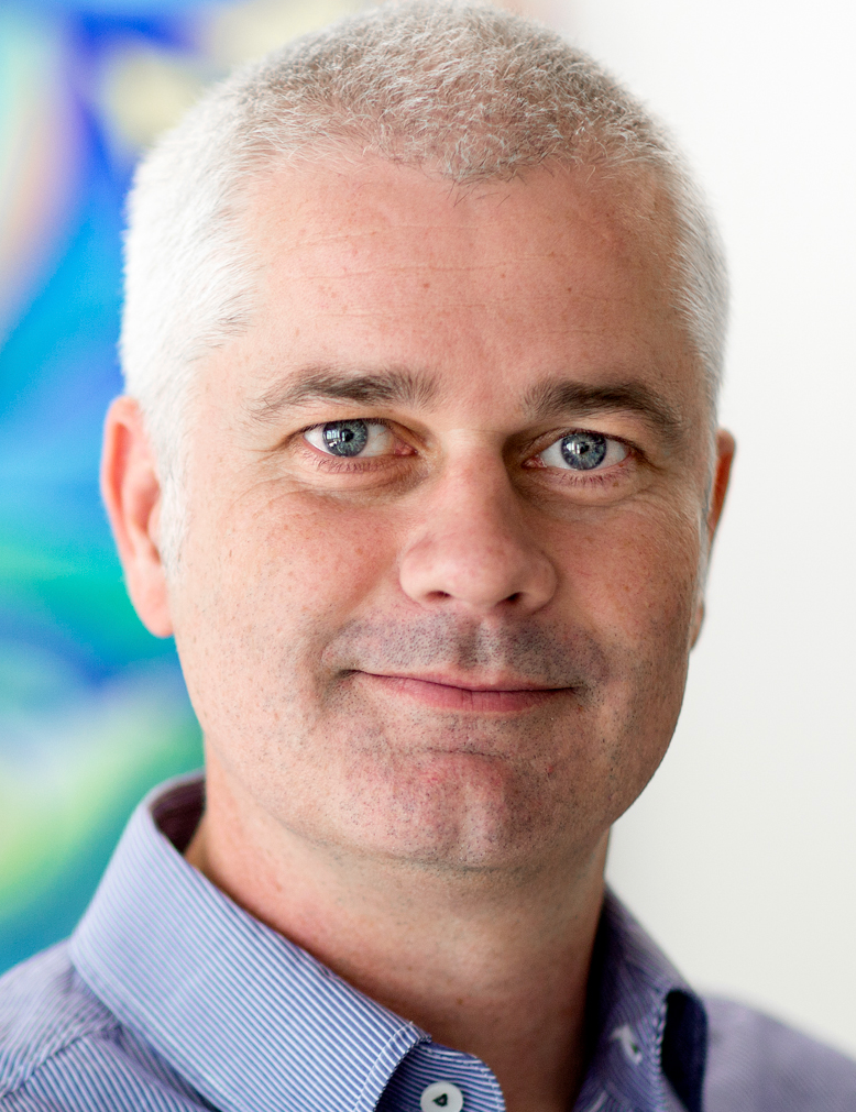 | Keynote Presentation Microfluidic Flow Control for LOAC: Bio-Inspired Engineering
Jaap den Toonder, Professor and Chair of Microsystems, Eindhoven University of Technology, Netherlands
Control of fluid flow at small length scales (typically < 1 mm) i.e. “microfluidics”, is important for many applications. Examples are devices for healthcare diagnostics, in which complex tasks of (bio-)fluid manipulation and detection need to be performed; organ-on-a-chip devices in which suitable micro-environments for multicellular tissue structures need to be created; water quality monitoring chips for continuous detection of bacteria, algae and chemicals; and manipulation of fluids droplets over surfaces. We are developing new technologies for active control of microfluidic flow in these applications. In doing this, we are inspired by nature in which a variety of microfluidic manipulation principles can be recognized. |
| 17:30 |  | Keynote Presentation Microfluidic Technologies in Liquid Biopsy: Needle-to-Needle Challenges
Victor Ugaz, Professor & Interim Department Head, Texas A&M University, United States of America
Liquid biopsy techniques are revolutionizing healthcare by enabling sophisticated bioanalysis to be performed in a minimally invasive manner. And these methods have shown particular promise in the diagnosis and treatment of cancer, where screening of cellular and extracellular material makes it possible to monitor disease progression and develop personalized therapies. But existing approaches to isolate, detect, and analyze liquid biopsy samples are complex and deliver limited throughput, making them challenging to deploy as routine diagnostic tools. Therefore, we are developing a microfluidic platform that combines high-throughput isolation and label-free imaging to rapidly screen rare cell populations. I will describe these efforts and provide a broader perspective of challenges and opportunities for microfluidic technologies in the areas of liquid biopsy and personalized medicine. |
| 18:00 | Networking Reception in the Exhibit Hall with Beer and Wine. Engage with the Exhibitors, View Posters in a Relaxed Setting at the De Doelen Rotterdam | 19:00 | Close of Day 1 of the Conference |
Wednesday, 6 June 201807:00 | Morning Coffee, Tea and Networking | |
Session Title: Methodologies and Approaches in Lab-on-a-Chip Device Development and Commercialization |
| | 07:30 |  | Keynote Presentation Integrated Optofluidic Devices for Manipulation and Imaging of Cells
Roberto Osellame, Professor, Politecnico di Milano; Senior Researcher, Institute for Photonics and Nanotechnologies (IFN) – National Research Council (CNR), Milan, Italy
Single cell analysis aims at unravelling the biological complexity due to the well-recognized diversity in cell populations. The integration of optical forces with microfluidic networks, in so-called optofluidic chips, allows advanced cell manipulation and characterization. In addition, the development of microscopy on a chip for the 3D tomography of single to few cells agglomerates paves the way to rapid analysis of a large quantity of samples for drug screening and personalized medicine. |
| 08:00 | Modelling and Characterisation of Droplet Generation, Trapping and Detection in Impedance-Based Cell Analytical Microfluidic System
Peter Furjes, Principal Scientist, Hungarian Academy of Sciences, Hungary
Present work focuses on the design, modelling and characterisation of a two-phase microfluidic device with integrated electrode system for impedance based cell analysis. The developed system is capable of creating, manipulating, trapping and detection droplets with cell-size fitted diameter. | 08:15 | SERS Active Periodic 3D Structure for Trapping and High Sensitive Raman-Spectroscopy of Molecular Surface Analysis of Particles or Cells
Peter Furjes, Principal Scientist, Hungarian Academy of Sciences, Hungary
In this work the applicability of special periodic 3D structure was demonstrated for simultaneous size selective trapping, fluorescent identification and extremely sensitive Surface-Enhanced Raman Spectroscopy based detection of characteristic molecules immobilized on the surfaces of the particles or cells.
| 08:30 | Tissue Microprocessing: Shaping Sub-nanoliter Volumes of Liquids on Tissue Sections for Multimodal Analysis
Govind V Kaigala, Research Staff Member, IBM Research Laboratory-Zurich, Switzerland
In contrast to standard microfluidics, which are typically closed, we are developing a scanning, non-contact microfluidic technology that can shape liquids in the "open space" over surfaces. This technology utilizes a microfluidic probe (MFP) having microfabricated structures for localizing a liquid of interest on a surface using hydrodynamic flow confinement. MFP permits patterning surfaces with proteins and other biomolecules in an additive and subtractive manner, forming complex gradients on surfaces, and interacting with cells on surfaces. With flow confinement operating at volumes smaller than 1 nanoliter, a few cells can be targeted in a human tissue section for the specific staining of disease markers. This confinement concept has also been scaled for targeting 1000’s of cells. Flow confinement and efficient use of chemicals can be further optimized using a concept called "hierarchical" hydrodynamic flow confinement. I will show how this family of liquid scanning probe devices is evolving as a bioanalytical tool to alter the physics and chemistry of biological interfaces at the micrometer to centimeter-length scales. I will also propose concepts pertaining to tissue microprocessing encompassing local phenotyping for interrogating tumor heterogeneity and spatially resolved molecular profiling which may contribute to the multi-modal analysis of critical biopsy samples in the context of next-generation pathology. | 09:00 |  | Keynote Presentation Microtubular NEMS for On- and Off-Chip Microfluidic Applications
Oliver Schmidt, Professor & Director, Leibniz-Institut für Festkörper- und Werkstoffforschung, Germany
Microtubular MEMS with outstanding properties are self-assembled into fully functional and integrative microtubular architectures. This makes them attractive for a broad range of applications and scientific research fields ranging from novel hybrid heterostructure devices to 3D microsystems both on and off the chip. Microtubular MEMS are exploited to construct ultra-compact and ultra-sensitive advanced electronic circuitry, sensors and optofluidic components around fluidic channels towards the implementation of a lab-in-a-tube system. They are also useful to study basic mechanisms of single cancer and stem cell migration, growth and mitosis in realistic 3D confined environments. Off-chip applications include biomimetic microelectronics for regenerative cuff implants and the development of hybrid microbiorobotic motors for paradigm shifting reproduction technologies. |
| 09:30 |  | Keynote Presentation Whole Blood Microfluidics: Fractionation and Isolation of Cells
Ian Papautsky, Richard and Loan Hill Professor of Bioengineering, Co-Director, NSF Center for Advanced Design & Manufacturing of Integrated Microfluidics, University of Illinois at Chicago, United States of America
Inertial microfluidics is receiving considerable attention for applications in liquid biopsy. However, while these devices have been widely explored for cell separation, sample dilution is necessary. Here, we report on a novel approaches to achieve cell separation directly from unprocessed whole blood based solely on cell size. The separation is achieved through coupling of inertial effects with shear-induced diffusion. Our results from high-speed imaging reveal that focusing of larger cells near the channel centerline is possible, leading to easy separation. Whole blood spiked with fluorescently labeled beads and cells was used to demonstrate the separation principle and its performance without any sample pretreatment. Results confirm the high quality of performance in terms of efficiency (>90%) and RBC rejection rate (> 96%). Ultimately, we successfully demonstrate the use of an inertial microfluidic device as a laboratory tool for sorting target cells from undiluted whole blood.
|
| 10:00 | Morning Coffee Break and Networking in the Exhibit Hall | 10:30 |  Challenges and Solutions of Lab-on-a-Chip and In Vitro Diagnostic Devices: From Test Development to High Throughput Production Challenges and Solutions of Lab-on-a-Chip and In Vitro Diagnostic Devices: From Test Development to High Throughput Production
Hans Dijk, Senior Account Manager, Scienion AG
Diagnostic test production might currently face several challenges. Among them, the high price of the capturing molecules and their precise immobilisation within a given surface. Therefore, saving precious samples and reagents are key to minimize costs. Handling small amounts of liquids with tiny dead volumes is a key part to achieve this. Advances in technology have allowed over the years to dispense less and less reagent per test in a very precise location and to dispense multiple other analytes onto the same test area. In other words, ultra-low volume handling technology has enable a significant decrease in production costs by miniaturising and multiplexing these tests. Scienion provides a single picoliter liquid handling technology to address all miniaturization aspects of rapid tests from early research to full scale production technologies.
| 11:00 |  Multimaterial Integration for Lab-on-a-Chip Devices Multimaterial Integration for Lab-on-a-Chip Devices
Holger Becker, Chief Scientific Officer, Microfluidic ChipShop GmbH
Highly integrated microfluidic devices usually consist out of components made out of different materials such as thermoplastic polymers for the cartridge body, membranes and filters, blister pouches for liquid reagents and/or silicon dies for sensors. The presentation will discuss challenges and solutions for developing and manufacturing of such multimaterial cartridges.
| 11:30 | High Resolution 3D Printing Inside a Chip
Aleksandr Ovsianikov, Professor, Head of Research Group 3D Printing and Biofabrication, Technische Universität Wien (TU Wien), Austria
3D printing opens exciting perspectives towards rapid engineering of complex 3D structures for microfluidic applications. In this context, multiphoton lithography (MPL) is an outstanding approach, since photosensitive material formulations and cells are delivered by perfusing the channels, thus enabling 3D printing within already assembled microfluidic chips. The latter aspects implies that different materials, construct geometries and cell types can be tested with the same set of chips without changing their initial design or fabrication process. In addition MPL offers high spatial resolution, unmatched by other 3D printing approaches, and can produce features down to sub-100 nm level directly in the volume of the material, without the necessity to deposit it layer by layer. An increasing portfolio of available materials enables utilization of the versatile capabilities of MPL, from producing complex volumetric 3D structures by means of cross-linking, to creating void patters within hydrogels already containing living cells. In this contribution, our recent progress on MPL for microfluidic applications and the development of according materials will be presented. | 12:00 | 3D-Printed Microfluidic Systems Built From Functional Modular Elements
Noah Malmstadt, Professor, Mork Family Dept. of Chemical Engineering & Materials Science, University of Southern California, United States of America
The integration of functional components into microfluidic systems is necessary for building compact analytical devices. Optical detection and measurement, electromechanical fluid switching, active mixing, and magneto- and electrophoretic separations all require the integration of components on top of the underlying fluidic architecture. There has been significant work towards integrating such functional components by including them directly in the microfabrication process. This approach, however, leads to expensive fabrication workflows that are often limited to a narrow set of applications. To overcome these limits, we are designing a microfluidic architecture that is easily adaptable to a wide variety of applications. This architecture is based on 3D-printed elements that can be assembled into analytical devices. Functional components are integrated into these elements; the structures are printed to directly accommodate off-the-shelf components including photodiodes, heaters, sensors, and fiber optic fittings. We have demonstrated the utility of this approach by assembling a system capable of performing automated ELISA assays from 3D-printed blocks with integrated functional components. | 12:30 | Networking Lunch -- Meet the Exhibitors and View Posters | 13:10 |  Enhanced Flow Control & Microfluidics Enhanced Flow Control & Microfluidics
François Mazuel, Head of R&D & Production, Elvesys
Introduction to enhanced flow control solutions and applications in microfluidics.
| 13:15 |  Lunch Technology Spotlight Presentation: Precise Contactless Spotting for Lab-on-Chip Applications Lunch Technology Spotlight Presentation: Precise Contactless Spotting for Lab-on-Chip Applications
Wilhelm Meyer, Managing Director, Microdrop Technologies GmbH
Lab-on-chip devices are used for a wide variety of health care applications, especially in the area of point-of-care diagnostics. These small microfluidic devices equipped with microchannels, chambers and other features carry out diagnostic tests by enabling reactions between patient samples and reagents. The devices deliver test results with very small sample volumes and in a short period of time. For production of lab-on-chip devices a precise and fast material deposition method is needed. Typically, the devices are produced at high numbers under high throughput conditions. The substrates may be made of different materials e.g. polystyrene or silicone, but in common is that these substrates typically have a structured surface which may consist of channels, mixing chambers or even wells which are used for precise detection. Also, different coating materials may be used as pretreatment for better adhesion of reagents or confinement of fluids. Deposition or Spotting of reagents onto the chip is a demanding task and requires the filling of small cavities and microchannels with a high accuracy. This can efficiently be done with a microspotting platform based on in inkjet technology. The flexibility of this technology allows for custom-made solutions adapted to the specific customer needs. Especially how high accuracy concerning volume of spotted reagents and also precision of placement is achieved under high throughput production conditions is demonstrated by examples.
| 13:30 | Frontier in Direct Observation and Visualization of Molecular Interactions
Gijs Wuite, Professor, Vrije Universiteit Amsterdam, Netherlands
The genetic information of an organism is encoded in the base pair sequence of its DNA. Many specialized proteins are involved in organizing, preserving and processing the vast amounts of information on the DNA. In order to do this swiftly and correctly these proteins have to move quickly and accurately along and/or around the DNA constantly rearranging it. In order to elucidate these kind of processes we perform single-molecule experiments on model systems such as restriction enzymes, DNA polymerases and repair proteins. The data we use to extract forces, energies and mechanochemistry driving these dynamic transactions. The results obtained from these model systems are then generalized and thought to be applicable to many DNA-protein interactions. In particular, I will report on a new single molecule methods. I’ll introduce Acoustic Force Spectroscopy (AFS). With AFS we extend the force-spectroscopy toolbox with an acoustic manipulation device that allows exerting acoustic forces on tethered molecules. AFS is a Lab-on-a-chip device consisting of a flow cell of two glass plates with a fluid chamber in between and a piezo element glued on top. While applying an alternating voltage to the piezo element, forces from sub-pN to hundreds of pNs are exerted to thousands of biomolecules in parallel, with sub-millisecond response time and inherent stability. AFS distinguishes itself by its relative simplicity, low cost and compactness, which allow straightforward implementation in lab-on-a-chip devices. I’ll discuss the application of this technique to DNA-protein interaction as well as for antibody-protein interaction detection and for its use to quantify cell adhesion. | 14:00 | Gut on-a-Chip: Towards a More Predictive and Physiological Human Intestinal Barrier Model
Evita Steeg, Researcher, TNO Quality Of Life, Netherlands
A majority of the preclinical intestinal models do not properly reflect the complex physiology of the human intestinal tract, resulting in low translational value. We have developed the InTESTine model, using fresh ex vivo (human) intestinal tissue mounted into a 2-compartmental microfluidic model with luminal and basolateral flow. Using InTESTine on-a-chip model the human intestinal tissue remains functional for 24 hours during incubations, as presented by proper transport and metabolic functionality and remained barrier integrity, and the model shows additional value in predicting human intestinal absorption of pharmaceuticals and nutrients. Besides ex vivo intestinal tissue, (human) intestinal stem-cell derived organoids possess promising features, especially when cultured as an epithelial monolayer. By applying microfluidic flow we stimulated growth of a confluent epithelial monolayer by showing an increase in TEER, limited vectorial leakage of FD4, while remaining phenotypic properties like cell proliferation and differentiation into the distinct cell types of the intestinal epithelium. In conclusion, we now can successfully use the human intestinal tissue and intestinal organoid monolayers for longer-term incubations, which can be applied as a reliable tool for studying processes that determine human intestinal permeability as well as (anaerobic) host-microbe-immune responses when mounted in the microfluidic device. | 14:30 | Scale-up and Sustainability for Large and Small Microfluidic Laboratories
Maiwenn Kersaudy-Kerhoas, Professor of Microfluidic Engineering, Heriot-Watt University, United Kingdom
The rapid translation of research from bench to bedside, as well as the generation of commercial impact, has never been more important for both academic and industrial researchers. We propose low-capital investment and sustainable rapid-prototyping solutions for almost all LOAC applications, with potential to accelerate microfluidic research and technology acceptance dramatically. | 15:00 | Thermoplastic Elastomer Microfluidic Platform For Methylation-specific Digital Droplet PCR
Lidija Malic, Research Officer, Industrial Materials Institute / National Research Council Canada, Canada
A thermoplastic polymer based microfluidic droplet generator platform is presented that enables digital droplet PCR (ddPCR) for determining degree of methylation as an epigenetic measure of WBC differential count. | 15:30 | Rapid Prototyping in Microfluidics: New Perspective by New Materials
Bastian Rapp, Principal Investigator and Head of NeptunLab, Karlsruhe Institute of Technology, Germany
Microfluidics is a very important discipline for many disciplines including, among others, synthetic and analytical chemistry, biochemistry and biology. However, the design of most microfluidic systems has remained largely unchanged since the introduction of soft lithography. In academia, microfluidic systems made from polydimethylsiloxane (PDMS) have become the de-facto standard. These are usually created in a replication process from a photolithographically structured layer of photoresist. However, in many applications fast concept-to-chip intervals are required for efficient experimental throughput and optimization. This is where 3D printing and additive manufacturing (AM) can make a significant contribution. We recently introduced “Computer Aided Microfluidics (CAMF)” a process that allows the conversion of digital chip layout to a testable physical structure in less than a day [1]. Using digital models allows a streamlined manufacturing process with digital models first being evaluated by numerical simulation before being transferred to a physical structure in a one-step manufacturing process. We recently expanded the material choice for CAMF to include glass [2] (which is created by sintering from photocurable glass prepolymers) and thermoplasts, most notably polymethylmethacrylate (PMMA) for which we created photostructurable precursors. This allows the rapid manufacturing of microfluidic systems in materials which have a long history in microfluidics and have been thoroughly investigated. We will demonstrate the advantages of this prototyping method and its potential use in many aspects of life sciences and analytics. | 16:00 |  Single Cell Manipulation and Detection Single Cell Manipulation and Detection
Marine Verhulsel, Life Sciences Product Manager, Fluigent SA
Microfluidics has emerged as a powerful tool to manipulate cells at single cell level. These manipulations have been extensively used to evaluate the degree of heterogeneity of a population. Cell to cell variability can be quantified in terms of genetics, transcriptomics or biomechanics. In this context, Fluigent has developed highly sensitive and stable instruments which allow for the measurement of single cell tension within a tissue through pipette micro-aspiration. This technique is economical, easy to use and non-invasive compared to traditional AFM, cytoindenter and optical tweezers. Regarding proteomic and genetic analysis, Fluigent has also designed a droplet chip for single cell encapsulation. Decreased volumes (down to picoliters), reduced costs, higher sensitivity and throughput, faster sample processing and readout are valuable benefits of this technology. Finally, identifying specific single cells out of a patient sample can also be determinant for diagnostic applications. High surface to volume ratio related to microfluidics enables detection of rare biomarkers. Increased sensitivity and ability to fully automate protocols using Fluigent products will be presented in the context of circulating tumor cell (CTC) detection.
| 16:30 | Key Enabling Functionalities for Point-of-Care Diagnostics
Aliki Tsopela, R&D Scientist, Micronit Microtechnologies, Netherlands
Rui Carvalho, R&D Scientist, Surfix BV, Netherlands
This presentation will focus on the work performed during the Coat PoCKET project, where Micronit Microtechnologies and Surfix join forces to bring the technology for fabricating advanced point-of-care diagnostic tests to the next level. The project aims at the development of a polymer-based microfluidic chip enabling sequential flow of liquids without using external pumps. This is achieved through an innovative combination of local surface modification and electrostatically actuated capillary valves. | 17:00 | LSC-PM: A Biomimetic Approach to Increase Solar Harvesting for Synthetic Chemistry
Timothy Noël, Professor, University of Amsterdam, Netherlands
In this lecture, we will discuss a novel device integrating the luminescent solar concentrator (LSC) concept with photomicroreactors (LSC-PM), allowing the direct use of solar light in photochemistry without the need for any intermediate energy conversion. This device is capable of capturing direct and diffuse sunlight, converting it into a narrow wavelength and delivering it to the embedded microchannels. | 17:30 | Single-cell Imaging in Novel 3D Hydrodynamic Focusing Microfluidic Devices
Iordania Constantinou, Postdoctoral Fellow, Zentrum für Molekulare Biologie der Universität Heidelberg, Germany
This presentation will discuss the development of a novel microfluidic device and its use in continuous-flow, high-resolution, single-cell imaging. | 17:45 | Single Molecule Microfluidic Methods to Characterise Vesicle-based Cell Mimetics
Ali Salehi-Reyhani, Fellow, Imperial College London, United Kingdom
We have developed single molecule microfluidic methods to absolutely quantify the biomolecular constituents of an artificial cell and measure their encapsulation efficiency. We show how the heterogeneity in their synthesis must be accounted for when used as simplified biological models. | 18:00 | Low Cost Thermoplastic Microfluidic Devices to Investigate the Ribosome Biogenesis and DNA Synthesis in Yeast
Elif Gencturk, Researcher, Bogazici University, Turkey
The aim is to fabricate a thermoplastic made microbioreactor that is capable of cell trapping. Yeasts are treated with RNA and DNA syntheses inhibitors to investigate ribosome biogenesis and DNA damage response. The device can be used for therapeutic applications. | 18:15 | Bone Marrow Scaffolds For Hematopoietic Stem Cell Utilizing the BioLithoMorphie®-Approach
Joerg Hampl, Researcher, Ilmenau University Of Technology, Germany
| 18:30 | BioLithoMorphie® Approaches to Gain 3D-Structures Including Fluidic Entities
Andreas Schober, Head of Department Nano-biosystem Technology, University of Technology Ilmenau, Technische Universitat Ilmenau, Germany
| 18:45 | Close of Day 2 of the Conference. |
|


 Add to Calendar ▼2018-06-05 00:00:002018-06-06 00:00:00Europe/LondonLab-on-a-Chip and Microfluidics Europe 2018Lab-on-a-Chip and Microfluidics Europe 2018 in Rotterdam, The NetherlandsRotterdam, The NetherlandsSELECTBIOenquiries@selectbiosciences.com