Co-Located Conference AgendasNGS, SCA, SMA & Mass Spec: Research to Diagnostics 2016 | Point-of-Care Diagnostics & Global Health World Congress 2016 | 

Monday, 26 September 201609:00-12:00 Microfluidics and Lab-on-a-Chip (LOAC) for
Point-of-Care (POC) Diagnostics Applications: Technologies,
Applications, Research Trends [Pre-Conference Training Course]
Presented by Dr. Holger Becker, microfluidic ChipShop GmbH.
Separate Registration Required for this Pre-Conference Training Course. | 12:00-14:30 Luncheon Training Course: Basic Principles in
Lab-on-a-Chip (LOAC) Technologies for the Study of Circulating
Biomarkers: Applications in Liquid Biopsies [Pre-Conference Training
Course]
Presented by Professor Steve Soper, Professor and Director, University of North Carolina-Chapel Hill.
Separate Registration Required for this Training Course. | 14:30 | Conference Registration, Conference Materials Pick-Up and Networking | |
Session Title: Conference Plenary Session -- Convergence of Technologies in Microfluidics, Diagnostics and Single Cell Analysis |
| | 15:30 |  | Keynote Presentation The Personalized Health Care Environment: How in vitro Diagnostics, Mobile Health and Medical Devices will Converge to Improve Health Care
Alan Wright, Chief Medical Officer, Roche Diagnostics Corporation, United States of America
|
| 16:00 |  | Keynote Presentation Digital Microfluidics: A Platform Whose Time Has Come
Aaron Wheeler, Canada Research Chair of Bioanalytical Chemistry, University of Toronto, Canada
Digital microfluidics is a fluid-handling technique in which droplets are manipulated by electrostatic forces on an array of electrodes coated with a hydrophobic insulator. In this talk, I will present recent results from my group’s work with digital microfluidics, with an emphasis on the topics covered in this unique venue. Specifically, I will demonstrate how digital microfluidics is particularly well-suited for Lab on a Chip applications, given its ability to automate diverse laboratory processes on a generic, programmable platform. Likewise, I will report on our work using digital microfluidics for Point of Care Diagnostics and Global Health, reporting on the results of a field trial for measles and rubella diagnostics at refugee sites in Kenya. Finally, I will describe how digital microfluidics is emerging as a useful tool for Single-Cell Analysis and for integration with Mass Spectrometry to answer questions about cell heterogeneity and cell-cell communication. Through these examples, I will make the case that digital microfluidics is emerging as a useful new tool for the next generation of analytical techniques, across a wide range of applications. |
| 16:30 |  | Keynote Presentation Microfluidics and Sensors: New Tools for Real-Time Clinical Monitoring
Martyn Boutelle, Professor of Biomedical Sensors Engineering, Imperial College London, United Kingdom
A goal for modern medicine is to protect vulnerable tissue by monitoring the patterns of changing physical, electrical and chemical changes taking place in tissue - ‘multimodal monitoring’. Clinicians hope such information will allows treatments to be guided and ultimately controlled based on the measured signals. Microfluidic lab-on-chip devices coupled to tissue sampling using microdialysis provide an important new way for measuring real-time chemical changes as the low volume flow rates of microdialysis probes are ideally matched to the length scales of microfluidic devices. Concentrations of key biomarker molecules can then be determined continuously using either optically or electrochemically (using amperometric, and potentiometic sensors). Wireless devices allow analysis to take place close to the patient. Droplet-based microfluidics, by digitizing the dialysis stream into discrete low volume samples, both minimizes dispersion allowing very rapid concentration changes to be measured, and allows rapid transport of samples between patient and analysis chip. This talk will overview successful design, optimization, automatic-calibration and use of both continuous flow and droplet-based microfluidic analysis systems for real-time clinical monitoring, using clinical examples from our recent work. |
| 17:00 |  | Keynote Presentation Microfluidics for Personalized Oncology
Chwee Teck Lim, NUS Society Chair Professor, Department of Biomedical Engineering, Institute for Health Innovation & Technology (iHealthtech), Mechanobiology Institute, National University of Singapore, Singapore
Cancer is a disease that can be characterised by the heterogeneity of the tumor cells. Such heterogeneity has led to a major hindrance in cancer classification, diagnosis and treatment. Recently, single cell analysis has become an important approach to detect variations in genetic, proteomic and molecular expressions in individual cancer cells. Here, we developed a suite of microfluidic biochips that makes use of cell mechanics principle to not only isolate, but also enable single cell analysis of circulating tumor cells (CTCs) derived from the peripheral blood of cancer patients. These include detecting the proteolytic capability of each CTC to obtain insights into their invasiveness as well as identify unique mutations with the aim of improving treatment. It is hope that this approach will lead to better personalized treatment of cancer patients. |
| 17:30 |  | Keynote Presentation High-Performance Rapid Diagnostic Tests
Bernhard Weigl, Director, Center for In-Vitro Diagnostics, Intellectual Ventures/Global Good-Bill Gates Venture Fund, United States of America
Lateral flow and similar rapid diagnostic assays (LFAs) are easy to use
and manufacture, low cost, rapid, require little or no equipment to
operate, and do not need to be refrigerated. However, they are generally
not considered to be very sensitive or able to provide a quantitative
result. Our group believes that this lack of sensitivity is not a
fundamental property of LFAs but rather a consequence of the way they
are developed, manufactured, and marketed. Historically, most lateral
flow tests were developed and optimized by relatively small
manufacturers with limited R&D capabilities and budgets, and were
generally used only for analytical targets prevalent at high
concentration in patient’s samples that were relatively easy to measure.
In contrast, our group’s mission is to develop LFA-based assays for use
in global health applications that are as sensitive as the best
conventional diagnostic assays (in some cases even better) while
retaining all their cost, simplicity, and usability advantages. |
| 18:00 | 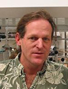 | Keynote Presentation Technologies for Personalizing Cancer Immunotherapies
James Heath, Elizabeth W. Gilloon Professor of Chemistry, California Institute of Technology (CalTech), United States of America
Cancer immunotherapy, which has taken virtually all aspects of oncology by storm over the past few years, is based upon using cellular or molecular therapies to promote tumor cell/immune cell interactions. At the heart of this therapy are the T cells that actually do the tumor cell killing, and the tumor antigens that are recognized by those T cells. Recent work has shown that neoantigens play critical roles in many immunotherapy successes. Neoantigens are tumor antigens that are fragments of mutated proteins expressed by the cancer cells, and contain those point mutations. They are presented in the clefts of major histocompatibility molecules (MHCs) by many of the cells in the tumor, where they may be recognized by neoantigen-specific T cell populations. In this presentation, I will discuss how various micro and nanotechnologies are being harnessed to identify, for a given patient which neoantigens are actively recruiting T cells into the tumor, and to carry out a deep molecular analysis of those neoantigen-specific T cells. I will further discuss how that information can then be harnessed for personalized cancer immunotherapies in the form of neoantigen-based vaccines, or engineered T cell receptor adoptive cell transfer therapies.
|
| 18:30 | Conference Opening Reception with Beer and Wine | 19:30-21:30 Dinner Training Course: Microfluidics for 3D-Printing
and Biofabrication: Technologies and Applications [Pre-Conference
Training Course]
Presented by Professor Albert Folch, University of Washington.
Separate Registration Required for this Training Course. |
Tuesday, 27 September 201607:45 | Conference Registration, Conference Materials Pick-Up, Morning Coffee and Breakfast Pastries in the Exhibit Hall | |
Session Title: Technology Trends in the Microfluidics and Lab-on-a-Chip Space, circa 2016 |
| | 09:00 | 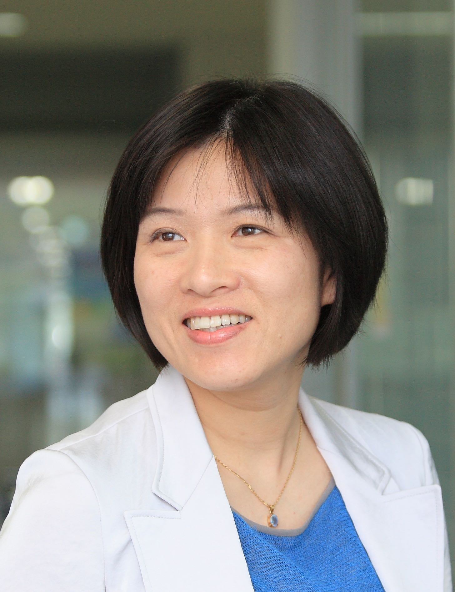 | Keynote Presentation Centrifugal Microfluidics for Biomedical Applications
Yoon-Kyoung Cho, Professor, Biomedical Engineering and Dean, UNIST (Ulsan National Institute of Science & Technology), Korea South
In this presentation, we will discuss our on-going research on “Lab-on-a-disc”, which applies centrifugal force to pump fluid for biochemical analysis. It is advantageous because of the capability to integrate and automate the analysis protocols into a disc-shaped device with simple, size-reduced, and cost-efficient instrumentation. We report various examples of fully integrated "lab-on-a-disc" for biomedical applications such as pathogen specific DNA extraction to test infectious diseases, multiplex enzyme-linked immunosorbent assay (ELISA), and isolation and analysis of circulating tumor cells starting from whole blood. Integration with centrifugal microfluidic technology allows us precise control of fluids while also reducing the expensive reagent consumption, the required analysis time and possible handling errors. We believe the presented result will not only improve the performance of the point-of-care-diagnostic devices but also potentially have great impact on global healthcare. |
| 09:30 | Centrifugal Step Emulsification Allows Miniaturized Digital Droplet-RPA, -LAMP and -PCR on the Centrifugal Microfluidic Platform
Felix von Stetten, Associate Director, Hahn-Schickard, Germany
A novel unit operation designated centrifugal step emulsification enables miniaturization of digital amplification protocols. Droplet generation, DNA-amplification and fluorescence detection was all performed within one single cavity. Results for absolute quantification of DNA by digital droplet-RPA, -LAMP and -PCR were in good agreement with those obtained by a commercial dPCR system. | 10:00 | 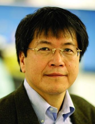 | Keynote Presentation A Billion-Droplet AC Electrospray Digital PCR Platform for Large-Dynamic Range Nucleic Acid Quantification
Hsueh-Chia Chang, Bayer Professor of Chemical and Biomolecular Engineering, University of Notre Dame, Interim Chief Technology Officer, Aopia Biosciences, United States of America
An AC-electrospray technology is shown to be able to generate 1 billion femto-liter (micron diameter) aqueous drops in immiscible silicone oil in less than 20 minutes. Unlike DC sprays, which tend to suffer from dielectric breakdown in liquid, and droplet generation technologies based on hydrodynamic shear, which can only generate pico-liter drops, we have shown that a properly tuned AC field can entrain low-mobility anions at a meniscus and the Coulombic repulsion among such entrained ions can deform the meniscus into unique 11 degree AC cones that are quite distinct from and much stable than DC cones (Phys Rev Lett, 101, 204501(2008); 109, 224301(2012); LabChip, 15, 1656(2015)). The sharper AC cones can generate 1 million femto-liter drops per second. The larger drop number and concentration (nM) allow us to get significantly better accuracy (without using Poisson statistics), dynamic range (8 decades) and reduction of inhibition and interfering effects in heterogeneous media. We have fabricated an integrated AC electrospray chip with PCR and imaging modules and have favorably compared the performance of this new droplet digital PCR platform against qPCR. The new AC droplet digital PCR platform is, however, a low-cost turn-key technology that requires no or minimum sample pretreatment. |
| 10:30 | Coffee Break and Networking in the Exhibit Hall: Visit Exhibitors and Poster Viewing | 11:15 | Sensors and Microfluidics – Challenges and Solutions for System Integration
Holger Becker, Chief Scientific Officer, Microfluidic ChipShop GmbH, Germany
| 11:45 |  | Keynote Presentation Cost-Effective Microfluidic Platform for Point-of-Care Diagnostics and Various Life-Science Applications
Alexander Govyadinov, Senior Technologist, HP Incorporated, United States of America
Recently, there is a lot of interest in microfluidic lab-on-a chip application for life science, forensic, point-of-care, molecular-diagnostic, other in-vitro-diagnostic, organs-on-a-chip, environmental and multiple others applications. Different scientific and commercial organizations explore a multitude of material sets and operational principles to forge microfluidic devices. Simultaneously, inkjet industry utilizes well established materials and principles of operation for complicated microfluidic systems developed for precision dispensing and manipulation of droplets with pico-liter accuracy on a massively parallel scale. The presentation describes our recent progress of low cost microfluidic platform development utilizing materials and processes developed for low-cost thermal inkjet business. The concept repurposes well established inkjet processes, microfluidic components and jetting elements for pumping, mixing, valving, fluid transport, sensing and other critical functions of complex integrated microfluidic systems. This presentation describes operating principles of microfluidic elements, examples of their integration in functional devices and discusses inkjet technology potential for broad range of microfluidic applications. |
| 12:15 | Networking Lunch in the Exhibit Hall: Visit Exhibitors and Poster Viewing | |
Session Title: 3D-Printing in the Microfluidics Space |
| | 14:00 | Bioanalytical Applications of Modular 3D Microfluidic Systems
Noah Malmstadt, Professor, Mork Family Dept. of Chemical Engineering & Materials Science, University of Southern California, United States of America
Assembly of microfluidic systems from modular 3D-printed components enables an innovative and powerful design workflow. While traditional fabrication approaches require design and fabrication of monolithic integrated devices, a modular approach allows for design and optimization of individual system elements. Final system design then becomes a simple iterative process based on assembling these elements by hand. An additional strength of a 3D-printed modular approach is the capacity to seamlessly integrate off-the-shelf electromechanical components into the modules. We have recently demonstrated integration of thermal sensors, optical sensors, and electromagnets into 3D-printed fluidic modules. These integrated components facilitate an array of tasks including flow rate detection, calorimetry, droplet counting, bioassay readout, and bead-based separations. Together with strategies for controlling the surface chemistry of 3D-printed parts and implementing efficient in-line mixing, these active modules form the foundation for designing and building complex integrated bioanalytical systems. | 14:30 |  | Keynote Presentation The Art of 3D-Printing Biocompatible Microfluidics
Albert Folch, Professor of Bioengineering, University of Washington, United States of America
The vast majority of microfluidic systems are presently built by replica-molding and bonding in elastomers (such as poly(dimethyl siloxane) (PDMS)) or in thermoplastics (such as poly(methyl methacrylate) (PMMA) or poly-styrene (PS)). However, biologists and clinicians typically do not have access to microfluidic technology because they do not have the engineering expertise or equipment required to fabricate and/or operate microfluidic devices. Furthermore, the present commercialization path for microfluidic devices is usually restricted to high-volume applications in order to recover the large investment needed to develop the plastic molding processes. We are developing microfluidic devices through stereolithography, a form of 3D printing, in order to make microfluidic technology readily available via the web to biomedical scientists. Most available SL resins do not have all the favorable physicochemical properties of the above-named plastics (e.g., biocompatibility, transparency, elasticity, and gas permeability), so the performance of SL-printed devices is still inferior to that of equivalent PDMS devices. Inspired by the success of hydrogel PEG-DA biocompatibility, we have developed microfluidic devices by SL in resins that share all the advantageous attributes of PDMS and thermoplastics so that we can 3D-print designs with comparable performance and biocompatibility to those that are presently molded. |
| 15:00 | 3D Printing Hydrogel-based Microfluidic Devices and Vascularized Tissue Constructs
Luiz Bertassoni, Associate Professor, Biomaterials and Biomechanics, School of Dentistry, Cancer Early Detection Advanced Research, Knight Cancer Institute, Oregon Health & Sciences University, United States of America
Fabrication of three-dimensional tissues with controlled micro-architectures has been shown to enhance tissue functionality. 3D printing can be used to precisely position cells and cell-laden materials to generate controlled tissue architectures. Therefore, it represents an exciting alternative for organ fabrication. Our group has been interested in developing innovative printing-based technologies to improve our ability to regenerate tissues with improved function, as well as to engineer hydrogel based microfluidic devices. In this seminar, we will present novel printing-based methods to fabricate vascularized tissue constructs, and 3D printed valved hydrogel based microfluidic devices that can be gated to multiple configurations for various microfluidics applications. The use of these technologies in various regenerative applications will be discussed. | 15:30 | Coffee Break and Networking: Visit Exhibitors and Poster Viewing | |
SONY DADC Symposium on Innovations in Microfluidics and Lab-on-a-Chip and their Impact on Life Sciences and Diagnostics | Session Sponsors |
| | 16:00 | Introduction to the 4th Annual SONY DADC Symposium and Topics Addressed in 2016 | 16:15 |  | Keynote Presentation Microfluidic Printing: From Combinatorial Drug Screening to Artificial Cell Assaying
Tingrui Pan, Professor, Department of Biomedical Engineering, University of California-Davis, United States of America
Microfluidic impact printing has been recently introduced, benefiting from the nature of simple device architecture, low cost, non-contamination, scalable multiplexability and high throughput. In this talk, we will review this novel microfluidic-based droplet generation platform, utilizing modular microfluidic cartridges and expandable combinatorial printing capacity controlled by plug-and-play multiplexed actuators. Such a customizable microfluidic printing system allows for ultrafine control of the droplet volume from picoliters (~10pL) to nanoliters (~100nL), a 10,000 fold variation. The high flexibility of droplet manipulations can be simply achieved by controlling the magnitude of actuation (e.g., driving voltage) and the waveform shape of actuation pulses, in addition to nozzle size restrictions. Detailed printing characterizations on these parameters have been conducted consecutively. A multichannel impact printing system has been prototyped and demonstrated to provide the functions of single-droplet jetting and droplet multiplexing as well as concentration gradient generation. Moreover, several enabling chemical and biological assays have been implemented and validated on this highly automated and flexible printing platform. In brief, the microfluidic impact printing system could be of potential value to establishing multiplexed droplet assays for high-throughput life science researchers. |
| 16:35 |  | Keynote Presentation Nanopore Sequencing for Real-Time Pathogen Identification
Kamlesh Patel, R&D Advanced System Engineering and Deployment Manager, Sandia National Laboratories, United States of America
As recent outbreaks have shown, effective global health response to
emergent infectious disease requires a rapidly deployable, universal
diagnostic capability. We will present our ongoing work to develop a
fieldable device for universal bacterial pathogen characterization based
on nanopore DNA sequencing. Our approach leverages synthetic
biofunctionalized nanopore structures to sense each nucleotide. We aim
to create a man-portable platform by combining nanopore sequencing with
advance microfluidic-based sample preparation methods for an
amplification-free, universal sample prep to accomplish multiplexed,
broad-spectrum pathogen and gene identification. |
| 16:55 |  | Keynote Presentation Polymer-based Nanosensors using Flight-Time Identification of Mononucleotides for Single-Molecule Sequencing
Steve Soper, Foundation Distinguished Professor, Director, Center of BioModular Multi-Scale System for Precision Medicine, The University of Kansas, United States of America
We are generating a single-molecule DNA sequencing platform that can acquire sequencing information with high accuracy. The technology employs high density arrays of nanosensors that read the identity of individual mononucleotides from their characteristic flight-time through a 2-dimensional (2D) nanochannel (~20 nm in width and depth; >100 µm in length) fabricated in a thermoplastic via nano-imprinting (NIL). The mononucleotides are generated from an intact DNA fragment using a highly processive exonuclease, which is covalently anchored to a plastic solid support contained within a bioreactor that sequentially feeds mononucleotides into the 2D nanochannel. The identity of the mononucleotides is deduced from a molecular-dependent flight-time through the 2D nanochannel. The flight time is read in a label-less fashion by measuring current transients induced a single mononucleotide when it travels through a constriction with molecular dimensions (<10 nm in diameter) that are poised at the input/output ends of the flight tube. In this presentation, our efforts on building these polymer nanosensors using NIL in thermoplastics will be discussed and the detection of single molecules using electrical transduction with their identity deduced from the associated flight time provided. Finally, information on the manipulation of single DNA molecules using nanofluidic circuits will be discussed that takes advantage of forming unique nano-scale features to shape electric fields for DNA manipulation and serves as the functional basis of the nanosensing platform. |
| 17:15 |  | Keynote Presentation Rapid and Ultra-sensitive Diagnostics Using Digital Detection
Weian Zhao, Associate Professor, Department of Biomedical Engineering, University of California-Irvine, United States of America
We will present our most recent droplet based digital detection
platforms for rapid and sensitive detections, which could find potential
applications at the point-of-care (POC). |
| 17:35 |  | Keynote Presentation Fractionation and Analysis of Nuclear versus Cytoplasmic Nucleic Acids from Single Cells
Juan Santiago, Charles Lee Powell Foundation Professor, Stanford University, United States of America
Single cell analyses (SCA) have become powerful tools in the study heterogeneous cell populations such as tumors and developing embryos. However, fractionating and analyzing nuclear versus cytoplasmic fractions of nucleic acids remains a challenge as these fractions easily cross-contaminate. We present a novel microfluidic system that can fractionate and deliver nucleic acid (NA) fractions from the nucleus (nNA) versus the cytoplasm (cNA) from single cells to independent downstream analyses. Our technique leverages a selective electrical lysis which disrupts the cell’s (outer) cytoplasmic membrane, while leaving the nucleus relatively intact. We selectively extract, purify, and preconcentrate cNA using isotachophoresis (ITP). The ITP-focused cNA and nNA-containing nucleus are separated by ITP and fractionated at a bifurcation downstream and then extracted for off chip analyses. We will present example applications of this fractionation including qPCR and next generation sequencing (NGS) analyses of cNA vs. nNA. This will include preliminary NGS analyses of nuclear vs. cytoplasmic RNA fractions to analyze gene expression and splicing. We hypothesize that the robust and precise nature of our electric field control is amenable to further automation to increase throughput while removing manuals steps. |
| 17:55 |  | Keynote Presentation Novel Strategies in Single Molecule Sensing
Joshua Edel, Professor, Imperial College London, United Kingdom
Analytical Sensors plays a crucial role in today’s highly demanding exploration and development of new detection strategies. Whether it be medicine, biochemistry, bioengineering, or analytical chemistry the goals are essentially the same: [1.] improve sensitivity, [2.] maximize throughput, [3.] and reduce the instrumental footprint. In order to address these key challenges, the analytical community has borrowed technologies and design philosophies which has been used by the semiconductor industry over the past 20 years. By doing so, key technological advances have been made which include the miniaturization of sensors and signal processing components which allows for the efficient detection of nanoscale object. One can imagine that by decreasing the dimensions of a sensor to a scale similar to that of a nanoscale object, the ultimate in sensitivity can potentially be achieved - the detection of single molecules. This talk highlights novel strategies for the detection of single molecules. |
| 18:15 | Close of SONY DADC Symposium | 18:30 | Cocktail Reception for All Conference Attendees: Enjoy Beer, Wine, Appetizers and Network with Fellow Delegates, Speakers, Exhibitors in the Exhibit Hall and View Posters Sponsored by Veryst Engineering, LLC | 20:00 | Close of Day 2 of the Conference. Continue Networking in Downtown San Diego (Trolleys to the City are Available Right Behind the Conference Venue). |
Wednesday, 28 September 201607:00 | Morning Coffee, Breakfast Pastries and Networking in the Exhibit Hall | |
Session Title: From Technologies to Utility -- Applications of Microfluidics/LOAC in Life Sciences and Beyond |
| | 07:30 | Micro-encapsulation of Carbon Dioxide Capture Solvents through Microfluidic Processing
Congwang Ye, Research Staff, Lawrence Livermore National Laboratory, United States of America
Microfluidics have been developed for many applications such as life science research and lab-on-a-chip. At LLNL, our work focuses on using microfluidics to produce microcapsules for energy applications, particularly the capture of CO2. Recently, new designer solvents have been developed to reduce energy cost for carbon capture. However, they suffer from many drawbacks such as high viscosity, solid precipitates and corrosivity. Separating the capture solvent with a gas permeable shell provides a practical pathway to use these solvents. In our lab, double emulsion drops were generated within a micro capillary device and are composed of liquid capture solvents as the core and different polymer precursor solutions as the shell layer. After crosslinking the polymer shell, the resulting microcapsules are mechanically robust and can be handled in fixed-bed, moving-bed or fluidized-bed reactors for different applications. While the mass transfer of gas through the capsule shell is slightly lower when compared to neat liquid sorbents, the increase in surface area enhances the overall CO2 absorption rate by 3.5-10x based on experimental data. The absorbed CO2 can then be released by heat or diffusion for reuse applications or underground storage. | 08:00 | Step by Step Fabrication of Biomaterials based on Cell Adhesion Control
Kennedy Okeyo, Senior Lecturer, Institute for Frontier Life and Medical Sciences, Kyoto University, Japan
On-chip fabrication of biomaterials such as cell sheets based on self-assembly organization of cells into tissues continues to attract attention due to potential applications in regenerative medicine as well as in the development of in vitro models for drug screening. We recently developed a technique for step-by-step fabrication of cell sheets based on adhesion control. It involves minimization of cell-substrate interaction to initiate self-assembly of cells, not into spheroids, but into planar cell sheets which can be then overlaid or stacked into 3D tissue models. For cell adhesion minimization, we developed a mesh culture system where cells are seeded and grown on suspended ultra-thin (~2µm thick) mesh scaffolds consisting of considerably large apertures (exceeding 100 µm in size) and thin mesh lines (3-5 µm in width).
| 08:30 | 3D Microfluidic Mixer for Rapid Smartphone-based Diagnosis of Anemia
Mei He, Assistant Professor, University of Kansas and Chief Science Officer, Clara Biotech, United States of America
Clinical diagnosis requiring a central facility and site visits can be burdensome for patients in resource-limited or rural areas. Therefore, development of a low-cost microfluidic chip that utilizes smartphone data collection and transmission would beneficially enable disease self-management and point-of-care diagnosis. We demonstrated 3D simulation-guided printing for fabricating micro-, millifluidic mixers, which allows rapid study and testing of various geometries in lab-on-a-chip devices. Combined with smartphone, a 3D printed microfluidic mixer for auto-mixing of reagents via capillary force has been implemented, for measuring hemoglobin levels in finger-prick blood. Self-diagnosis of anemia has been successfully demonstrated using smartphone detection, showing consistent results with clinical measurements. Capable of 3D fabrication flexibility and smartphone compatibility, this work presents a novel diagnostic strategy for advancing personalized medicine and disease management. | 09:00 |  Technology Spotlight: Technology Spotlight:
High-Sensitivity, Cost-Effective Nanophotonic Biosensor Substrates
Arash Farhang, Optics Scientist, MOXTEK, Inc.
Numerous publications demonstrate that nanophotonic substrates offer the extraordinary surface sensitivity that enables many biosensing applications. Thus far colloidal-nanoparticle-based arrays and thin metallic films are the only such truly large scale substrates that have been available commercially. Moxtek has developed nanophotonic technologies and cost-effective volume production capabilities that enable and improve numerous biosensor applications. This technology greatly enhances fluorescence and label-free assay sensitivity through the utilization of surface plasmon, guided-mode, and gap-mode resonances. Such heightened sensitivity enables significantly-lower detection limits in molecular/chemical detection, microarrays, and other bioassay technologies, applications. These in turn open the doors to additional markets such as medical/point-of-care diagnostics, forensic testing, environmental monitoring, and food safety. Theoretical and measured data on the performance of these nanophotonic technologies, possible applications, and an overview of the available developmental and commercial manufacturing capabilities will be presented.
| 09:30 |  | Keynote Presentation Biological Imaging Using Microfluidics and Electrochemistry
Charles Henry, Professor and Chair, Colorado State University, United States of America
Chemical gradients drive many processes in biology, ranging from nerve signal transduction to ovulation to cancer metastasis. At present, microscopy is the primary tool used to understand these gradients by imaging both the gradients and the resulting cell motion. Microscopy has provided many important breakthroughs in our understanding of fundamental biology, but is limited due to the need to incorporate fluorescent molecules into biological systems through either labeling or genetic manipulations. To better understand some processes, there is a need to develop tools that can measure chemical gradient formation in biological systems that do not require fluorescent modification of the targets, can be multiplexed to measure more than one molecule, and are compatible with a variety of biological sample types, including in vitro cell cultures and ex vivo tissue slices. In response to this need, we have developed a high-density electrode array containing 8,192 individual electrodes to image release of electrochemically active metabolites like nitric oxide and norepinephrine from live tissue slices. The electrode array has a resolution of 30 µm and covers and area of 2 mm by 2 mm, easily large enough to image release of neurotransmitters across an ex vivo tissue slice from a mouse model. In this talk, electrochemical characterization of this system will be discussed first using simple chemical models. Then use of the system in combination with microfluidics, for imaging spatiotemporal resolution of neurotransmitter release at the organ scale will be presented. Use of microfluidics to deliver stimulants that induce changes in release profile will be shown. |
| 10:00 | Negotiating Your Way into an Assay: Specificity has Tolerances
David Wright, CEO, Wi, Inc., United States of America
| 10:30 | Coffee Break and Networking in the Exhibit Hall: Visit Exhibitors and Poster Viewing | 11:15 | Applications of Fully Integrated Active and Passive Flow Control in the Organ-on-Chip, Analytical and Diagnostics Field
Marko Blom, Chief Technical Officer, Micronit Microtechnologies, Netherlands
We will present various applications and examples in the organ-on-chip, analytical and diagnostics field relying on fully integrated active and passive, capillary driven flow control. Active valving fabrication strategies will be shown which do not require adhesives for the integration of materials relevant to the above mentioned fields.
The applications will range from accurate dosing, distribution and dispensing for cell culturing and Organ-On-Chip (OOC) applications to examples of sequential capillary flow in polymeric devices controlled by low-power electrical triggers realized using integrated electrodes. Specifically for the OOC field we have developed scalable Design-For-Manufacturability (DFM) concepts for such OOC applications, varying from resealable devices to fully integrated microtiter plate format culturing tools. The concepts are tested and validated in the field by end-users. Here, we demonstrate their use and applicability as versatile, flexible and enabling R&D tools for prototyping and (further) development of OOC applications. | 11:45 | Modeling and Simulation of Microfluidic Organ-on-Chip Devices
Matthew Hancock, Managing Engineer, Veryst Engineering, LLC, United States of America
Modeling and simulation are key components of the engineering development process, providing a rational, systematic method to engineer and optimize products and dramatically accelerate the development cycle over a pure intuition-driven, empirical testing approach. Modeling and simulation help to identify key parameters related to product performance (“what to try”) as well as insignificant parameters or conditions related to poor outcomes (“what not to try”). For microfluidic organ-on-chip devices, modeling and simulation can inform the design and integration of common components such as micropumps, manifolds, and channel networks. Modeling and simulation may also be used to estimate a range of processes occurring within the fluid bulk and near cells, including shear stresses, transport of nutrients and waste, chemical reactions, heat transfer, and surface tension & wetting effects. I will discuss how an array of modeling tools such as scaling arguments, analytical formulas, and finite element simulations may be leveraged to address these microfluidic organ-on-chip device development issues.
| 12:15 | Networking Lunch in the Exhibit Hall: Visit Exhibitors and Poster Viewing | |
Session Title: Technology Advances in Microfluidics Drives Development of New Utility |
| | 13:30 |  | Keynote Presentation Human “Body-on-a-Chip” Devices as Tool to Improve Drug Development
Michael Shuler, Samuel B. Eckert Professor of Engineering, Cornell University, President Hesperos, Inc., United States of America
Alternatives or supplements to the use of animals in preclinical drug development that better mimic human response should reduce costs and increase the number of FDA approved drugs at the end of clinical trials. We have constructed micro-physiological (or “Body-on-a-Chip”) devices constructed from a combination of human tissue engineered constructs, micro-fabricated devices and physiologically based pharmacokinetic (PBPK) models. These human surrogates are constructed on a low cost, robust “pumpless” platform. In addition to measuring viability and metabolic responses, we can measure functional outputs such as electrical activity and force generation using integrated sensors (in collaboration with J. Hickman, University of Central Florida). We will focus our discussion on development of key organ modules and their integration with each other to form a model of the human body. |
| 14:00 |  | Keynote Presentation Microraft Array Platform for the Selection of Lymphocytes Based on Target-Cell Killing
Nancy Allbritton, Frank and Julie Jungers Dean of the College of Engineering and Professor of Bioengineering, University of Washington in Seattle, United States of America
Adoptive cellular therapy (ACT) is an emerging therapeutic in which cytotoxic T lymphocytes (CTLs) that recognize tumor cell epitopes are introduced into patients providing immunity against the cancer cells. For ACT to succeed, CTLs with high tumor-killing efficiency must be identified, isolated, and characterized. Current technologies do not enable simultaneous assay of cell behavior or killing followed by recovery of the most efficient killer cells. A microraft array technology that measures the ability of individual T cells to lyse a population of target cells followed by sorting of living cells into a multi-well plate for expansion and characterization was developed. The microraft array platform was combined with image processing and analysis algorithms to track and monitor killing assays over many hours. Automated cell collection was incorporated into the platform for facile cell collection from the array. As a proof of principle, human T cells directed against an influenza antigen were co-cultured with antigen presenting target cells on the microraft arrays. Target cell killing was measured by tracking the appearance of dead cells on each microraft over time. Microrafts with a single CTL demonstrating the greatest rate of target cell death were identified, cloned, and influenza-antigen reactivity confirmed. The platform is readily modified to measure the antigen-specific activity of individual cells within a bulk CTL culture or the cell heterogeneity within a population of gene-engineered T cells. |
| 14:30 | Free-Surface Microfluidics and SERS for High Performance Sample Capture and Analysis
Carl Meinhart, Professor, University of California-Santa Barbara, United States of America
Nearly all microfluidic devices to date consist of some type of fully-enclosed microfluidic channel. The concept of ‘free-surface’ microfluidics has been pioneered at UCSB during the past several years, where at least one surface of the microchannel is exposed to the surrounding air. Surface tension is a dominating force at the micron scale, which can be used to control effectively fluid motion. There are a number of distinct advantages to the free surface microfluidic architecture. For example, the free surface provides a highly effective mechanism for capturing certain low-density vapor molecules. This mechanism is a key component (in combination with surface-enhanced Raman spectroscopy, i.e. SERS) of a novel explosives vapor detection platform, which is capable of sub part-per-billion sensitivity with high specificity. | 15:00 | Bipolar Electrode Coupling of Nanoscale Electron Transfer Reactions to Remote Chromogenic and Luminogenic Reporter Reactions
Paul Bohn, Arthur J. Schmitt Professor of Chemical and Biomolecular Engineering and Professor of Chemistry and Biochemistry, University of Notre Dame, United States of America
The combination of fluorescence and absorption spectroscopy with electrochemistry presents new avenues for the study of redox reactions, with potential for enhanced throughput, sensitivity, and spatial resolution. Here we present a novel configuration for coupling high sensitivity voltammetric measurements implemented in nanoscale architectures - such as nanopore-confined recessed ring-disk electrode arrays - with remote electrochemically-triggered chromogenic and fluorigenic reporter reactions. Coupling is mediated by a mm-scale bipolar electrode which communicates the local solution potential in the analyte-measuring portion of the device to an opposing chromogenic or fluorigenic reporter reaction in a remote location. Oxidation (reduction) of reversible analytes at the disk working electrode is accompanied by reduction (oxidation) on the nanopore portion of the bipolar electrode and then monitored by the accompanying oxidation of the reporter to produce a change in color or luminescence. The remote end of the bipolar electrode is placed in a cell far from the nanopore ring-disk array so that highly efficient reporter measurements can be carried out conveniently against low intrinsic backgrounds. The combination of bipolar eletrodes with luminescence in the dihydroresorufin/resorufin system has been used to study in situ generation of H2O2 in electrokinetic flow and for analytical determinations down to pM limits of detection. Applications of chromogenic reporter reactions for point-of-care (POC) use will also be described. | 15:30 | Inertial Focusing in Triangular Microchannels for Flow Cytometry
Ian Papautsky, Professor, University of Cincinnati, United States of America
This work presents a microfluidic device that takes advantage of asymmetric velocity profile in a low aspect ratio triangular microchannel to focus cells and microbeads into a single position with high efficiency, and without the need for secondary flow, sheath flow or external forces. | 16:00 | Plasmonic-Enhanced Single-Molecule Detection
Steve Blair, Professor, University of Utah, United States of America
The next generation of molecular diagnostics tools are targeted to have single molecule sensitivity. Plasmonic-enhanced fluorescence can be a key enabling factor in achieving this goal. Large-scale arrays of plasmonic structures meet the requirements of enhanced signal-to-background in fluorescence detection, along with compatibility with existing instrumentation and surface chemistry. Fluorescence enhancement results from a combination of plasmonic mediated excitation and emission enhancement. Even though molecules are confined within a plasmonic structure, the spectral region of enhancement depends strongly on the metal. As such, have also been working with structures in Al, which is mass-production friendly and provides balanced enhancement throughout the visible spectrum, opening up a wider range of applications. However, new chemical passivation strategies need to be devised due to the native oxide of Al. Tuning of the relative enhancements can be accomplished by adjusting the shape of the plasmonic structures, opening up the UV spectral range where the native fluorescence of biomolecules can be accessed. | 16:30 | Cation Dependent Transport in Gated Nanofluidic Systems
Shaurya Prakash, Associate Professor, Department of Mechanical and Aerospace Engineering, The Ohio State University, United States of America
A nanofluidic device with an embedded and fluidically isolated gate electrode, analogous to solid state semiconductor field effect transistors was developed for exquisite ionic transport control. Specifically, the gated electric field allows for tunable control of both the magnitude and direction of the net ionic current through the nanochannels as a function of electrolyte concentration and gate electrode location permitting device operation as a multi-state switch. Additionally, we show that ion transport is cation dependent for negatively charged walls demonstrating that engineered surface charge can potentially lead to significant advances in separations and nanoscale selective ion transport. | 17:00 | Surface Functionalization Strategies for the Design of Thermoplastic Microfluidic Devices for New Analytical Diagnostics
Fanny d'Orlyé, Associate Professor, Chimie ParisTech, Unité de Technologies Chimiques et Biologiques pour la Santé, France
Cyclic olefin copolymer (COC) and fluoropolymer (Dyneon THV) are emerging materials attractive for the conception of microfluidic chips thanks to their UV-visible transparency and high resistance to aggressive solvents. We propose to develop new analytical microsystems using these polymers for trace quantitation in complex matrices. This involves the immobilization of selective ligands in a confined zone of the microchannel for target extraction and preconcentration. This integrated pretreatment step will be followed inside the microdevice by electrokinetic separation and on-line detection. This requires new surface treatments for chemically inert COC and Dyneon to modify the microfluidic system at two scales: (1) on the entire surface to control surface properties and fluid flows during electrokinetic separation, or (2) locally to immobilize selective ligands (aptamers) on restricted areas for target extraction. For global modification, a plasma-induced immobilization of brominated derivatives allowed further ligand immobilization through alkyne-azide “click” chemistry reaction. For local ligand immobilization, we developed an original electrochemical strategy on Dyneon THV microchannel. Local electrochemical carbonization followed by covalent linkage of ligands (aptamers) through “click” reaction leads to immobilization zones in the 50 micrometer range. | 17:30 | Design of a Lab-on-a-Chip for the Quantitation of S-Nitrosothiols: From their Separation to their Decomposition and Electrochemical Detection
Anne Varenne, Professor, Chimie ParisTech, Unité de Technologies Chimiques et Biologiques pour la Santé, France
Abdulghani Ismail, Researcher, Chimie ParisTech, Unité de Technologies Chimiques et Biologiques pour la Santé, France
NO is a diatomic free radical that has in biological fluids extremely short half-life (<1 s). The addition of NO to functional proteins is as important as phosphorylation in its consequences on cellular activities. In order to be transported and stocked in biological fluids, NO binds to peptides and proteins forming S-nitrosothiols (RSNOs). The variation of their proportion has been recognized in many diseases. RSNO are sensitive to decomposition by light, heat, and metal ions. RSNOs can exchange the NO between one other by transnitrosation reaction. Recently we studied the separation of different RSNOs and their transnitrosation reaction using capillary electrophoresis with capacitively coupled contactless conductivity detection. Also, we developed a decomposition process of RSNO using copper to quantify RSNO by electrochemical techniques (ultra-micro electrodes). We are currently down scaling these approaches within microchip devices. This opens the way for an integrated lab-on-a-chip for S-nitrosothiols quantitation.
| 18:00 | Close of Day 3 of the Conference. |
|


 Add to Calendar ▼2016-09-26 00:00:002016-09-28 00:00:00Europe/LondonLab-on-a-Chip, Microfluidics and Microarrays World Congress 2016Lab-on-a-Chip, Microfluidics and Microarrays World Congress 2016 in San Diego, California, USASan Diego, California, USASELECTBIOenquiries@selectbiosciences.com