Co-Located Conference AgendasExtracellular Vesicles & Cell-Free RNAs 2023 | Lab-on-a-Chip & Microfluidics World Congress 2023 | Materials & Tools for Developing POC & Rapid Dx 2023 | 

Tuesday, 28 November 202313:00 | Conference Registration, Materials Pick-Up and Networking | 13:50 | Welcome and Introduction to the Mehmet Toner Microfluidics Symposium 2023 by Chairs: Dino Di Carlo and Albert Folch -- Plenary Ballroom | 14:00 |  | Plenary Presentation Microfluidics in the Clinic: Applications from Infectious Diseases to Cancer Treatment Monitoring
Shannon Stott, Assistant Professor, Massachusetts General Hospital & Harvard Medical School, United States of America
|
| 14:20 |  | Plenary Presentation Early Dissemination of CTCs: Detection, Molecular Characterization and Clinical Relevance
Sunitha Nagrath, Professor of Chemical Engineering and Biomedical Engineering, University of Michigan-Ann Arbor, United States of America
My journey into the world of circulating biomarkers started under the mentorship of Mehmet Toner. I am fortunate to experience the power of microfluidics when it rendered the ability to discover more CTCs than conventional approaches. This was the foundation to further ask the challenging questions on how early these cells disseminate. With high throughput and sensitive microfluidic technologies for the isolation of CTCs, we were able to detect the CTCs not only in the setting of MRD, but also in earlier stages of cancer. We have developed microfluidic approaches for the detection and molecular characterization of these early disseminated cells. Finding CTCs in peripheral blood without any evidence of overt metastasis is further investigated for the clinical relevance. |
| 14:40 |  | Plenary Presentation Understanding Tissue Microenvironments with Miniaturization Technologies
Sangeeta Bhatia, John J. and Dorothy Wilson Professor, Massachusetts Institute of Technology, United States of America
|
| 15:00 |  | Conference Chair Interrogation of Intact Tumor Biopsies Using Microfluidics, Robotics, and 3D Printing
Albert Folch, Professor of Bioengineering, University of Washington, United States of America
There is a lack of confidence in present in vitro drug efficacy tests, as they do not properly recapitulate the dynamic physiology and pathophysiology of the human organism. This challenge is particularly acute in oncology: present tools to study drug responses fail to faithfully mimic the patient’s tumor microenvironment (TME) and thus have not kept up with drug testing needs. As a measure of this problem, on average less than 4% of oncology drugs in clinical trials end up being FDA-approved, a dismal approval rate that has dire social repercussions such as high cancer drug prices and difficult accessibility. We have developed a suite of microfluidic platforms that address this problem by multiplexing the delivery of drugs to intact-TME human biopsies. In addition, a precision slicing methodology allows for producing large numbers of cuboidal micro-tissues (“cuboids”) from a single tumor biopsy. We trap cuboids in arrays of microfluidic traps in a 96-well platform and we also use robotics for very high-throughput automated placement of mouse cuboids in 384-well plates. We believe that with these approaches it will soon be possible to bypass animal testing and perform direct testing of drugs using only human tumors. Since these new-generation tests preserve the TME intact, we envision that they will minimize FDA failure rates and could contribute to alleviate the cost of cancer drugs. |
| 15:20 | Mid-Afternoon Coffee Break and Networking in the Exhibit Hall | 16:00 |  | Plenary Presentation Ultra-High Sensitivity Diagnostics with Droplet Microfluidics
David Weitz, Mallinckrodt Professor of Physics and Applied Physics, Director of the Materials Research Science and Engineering Center, Harvard University, United States of America
This talk will describe new uses of droplet microfluidics for very high sensitivity detection of biomolecules, including nucleic acids and proteins. The talk will be dedicated to Mehmet Toner and his innumerable accomplishments in applying microfluidics for medical applications. |
| 16:20 |  | Plenary Presentation Replicating In Vivo Micro Scale Biology In Vitro: Cancer Biology and Neutrophil Migration
David Beebe, Claude Bernard Professor of Biomedical Engineering, John D. MacArthur Professor, University of Wisconsin-Madison, United States of America
Cell biology occurs at the micro scale. I will describe two projects where we are using micro scale technology to replicate in vivo micro scale biology. In the first, we build patient-specific models of cancer with cells from that patient to replicate the tumor (and it’s microenvironment) structure and function. The model includes eight different cell types including both lymph and blood vasculature and the tumor cells in a spheroid format. We than treat these models to predict which treatment (e.g. drug, radiation) will be best for that patient. In the second, we are applying a recently developed under oil microfluidic technology to replicate the in vivo single cell scale mechanics and kinetics during neutrophil migration. |
| 16:40 |  | Plenary Presentation Quantitative Microfluidic Technologies to Ensure Reproducibility of Microbial Measurements
Rustem Ismagilov, Ethel Wilson Bowles and Robert Bowles Professor of Chemistry and Chemical Engineering, California Institute of Technology, United States of America
Reproducibility and interpretability of biomedical research results is increasing in importance. In particular, sequencing microbial measurements in diagnostics and microbiome studies are of particular attention. This talk will discuss how quantitative microfluidic technologies can provide the missing piece to ensure reproducibility of microbial sequencing. |
| 17:00 |  | Plenary Presentation Identification of Different Subpopulations of Circulating Tumor Cells and Extracellular Vesicles in the Blood of Cancer Patients using Microfluidics
Steve Soper, Foundation Distinguished Professor, Director, Center of BioModular Multi-Scale System for Precision Medicine, The University of Kansas, United States of America
Liquid biopsies are an attractive source of biomarkers that can be used to manage a variety of cancer-related diseases. Two liquid biopsy markers found in blood are circulating tumor cells (CTCs) and extracellular vesicles (EVs). The challenge associated with using CTCs or EVs as biomarkers has been that affinity selection using anti-EpCAM antibodies only does not recapitulate the tumor microenvironment entirely providing limited predictive power of the assay. For example, CTCs expressing invasive phenotypes down-regulate epithelial antigens, such as the epithelial cell adhesion molecule – EpCAM. We developed a liquid biopsy selection strategy that employs plastic microfluidic chips modified with antibodies to select two different liquid biopsy marker subpopulations. In addition to the common marker used for positive selection (EpCAM), Fibroblast Activation Protein alpha (FAPa) expressing liquid biopsy markers were also selected. Using the dual selection strategy, both subpopulations are detected from patients with results showing better predictive value compared to selection in which only EpCAM is used. In this presentation, we will show two examples of EpCAM/FAPa selection to monitor response to therapy using CTCs in pancreatic cancer, and determining the best treatment option for breast cancer patients using EVs. The CTCs and EVs were selected using specially designed microfluidic chips that could be operated by a fluid handling robot to support large-scale clinical trials. In the case of CTCs, response to a PARPi therapy was serially monitored to determine treatment efficacy. Results indicated that CTCs were better in monitoring response to the PARPi compared to the serum marker, CA19-9, but only when using both CTC subpopulations. In addition, the CTCs could be sequenced using NGS without the need for single-cell picking. Both EpCAM and FAPa expressing EVs and their mRNA cargo were used to perform molecular subtyping of breast cancer patients. We found the four molecular subtypes (luminal A/B, HER2 enriched, and basal-like) could be successfully identified in patients only when using both EV subpopulations. The EV mRNA copy numbers were analyzed using the nCounter assay without requiring the need for an amplification step. |
| 17:20 |  | Plenary Presentation Microfluidics in Space
Leanna Levine, Founder & CEO, ALine, Inc., United States of America
From NASA's first successful nanosatellite technology demonstration of engineered protein expression on GeneSat to the current exponential growth of research and biomanufacturing in near earth orbit, a new frontier in microgravity-based research and manufacturing is being realized. An overview of the programs we have supported and the goals of their missions will be discussed. |
| 17:40 |  | Conference Chair Navigating the Winding Road of Inertial Focusing to the Clinic
Dino Di Carlo, Armond and Elena Hairapetian Chair in Engineering and Medicine, Professor and Vice Chair of Bioengineering, University of California-Los Angeles, United States of America
I will share a historical perspective exploring inertial microfluidic concepts, from the beginning in Mehmet Toner's Center for Engineering in Medicine, to its application for cell profiling using deformability cytometry at UCLA, to the recent FDA-clearance and clinical use of deformability cytometry as part of the IntelliSep test for early sepsis diagnosis. IntelliSep is the first FDA-cleared test for early sepsis diagnosis and the first test that employs biomechanical measurements of cells as a key marker of disease. The talk will highlight key aspects of matching microfluidic technology capabilities with clinical need, lessons learned in the journey from research lab to the clinic, and the importance of exploiting serendipity. |
| 18:00 |  | Plenary Presentation Extreme Microfluidics: Large-Volumes and Complex Fluids
Mehmet Toner, Helen Andrus Benedict Professor of Biomedical Engineering, Massachusetts General Hospital (MGH), Harvard Medical School, and Harvard-MIT Division of Health Sciences and Technology, United States of America
Microfluidics gained prominence with the application of microelectromechanical systems (MEMS) to biology in an attempt to benefit from the miniaturization of devices for handling of minute samples of fluids under precisely controlled conditions. Microfluidics exploits the differences between micro- and macro-scale flows, for example, the absence of turbulence, electro-osmotic flow, surface and interfacial effects, capillary forces in order to develop scaled-down biochemical analytical processes. The field also takes advantage of MEMS and silicon micromachining by integrating micro-sensors, micro-valves, and micro-pumps as well as physical, electrical, and optical detection schemes into microfluidics to develop the so-called “micro-total analysis systems” or “lab-on-a-chip” devices. However, the ability to process ‘real world-sized’ volumes efficiently has been a major challenge since the beginning of the field of microfluidics. This begs the question whether it is possible to take advantage of microfluidic precision without the limitation on throughput required for large-volume processing? The challenge is further compounded by the fact that physiological fluids are non-Newtonian, heterogeneous, and contain viscoelastic living cells that continuously responds to the smallest changes in their microenvironment. Our efforts towards moving the field of microfluidics to process large volumes of fluids was counterintuitive and not anticipated by the conventional wisdom at the inception of the field. We metaphorically called this “hooking garden hose to microfluidic chips.” We are motivated by a broad range of applications enabled by precise manipulation of extremely large volumes of complex fluids, especially those containing living cells or bioparticles. The use of high-throughput microfluidics to process large-volumes of complex fluids (e.g., whole blood, bone marrow, bronchoalveolar fluid) has found broad interest in both academia and industry due to its broad range of utility in medical applications. |
| 18:30 | Mehmet Toner Birthday Celebration + Networking Reception with Beer, Wine and Dinner in the Exhibit Hall - Network with Colleagues and Engage with Exhibitors | 20:30 | Close of Day 1 Main Conference Programming |
Wednesday, 29 November 202307:30 | Introduction to Microfluidics Training Course. **Separate Registration Required to Attend**
Shuichi Takayama, Professor, Georgia Research Alliance Eminent Scholar, and Price Gilbert, Jr. Chair in Regenerative Engineering and Medicine Wallace H. Coulter Department of Biomedical Engineering, Georgia Institute of Technology & Emory University School of Medicine, United States of America
Introduction to Microfluidics Training Course **A Training Course for Beginners and New Entrants into the Microfluidics Field* Date: Wednesday, 29 November 2023 Time: 07:30am-09:00am Location: Ballroom A Coffee and Pastries will be Served This presentation will introduce basics of microfluidics. - Topics include size scales of microfluidic devices and how that affect microscale fluid flows.
- The evolution and different methods of microfluidic device fabrication.
- Select biological applications including cellular, molecular, and exosome applications.
- Some of the challenges and opportunities and future perspectives will also be discussed.
- Time will also be reserved for questions and discussions.
**This is an excellent course for new entrants seeking an immersion into the microfluidics field -- the course is taught by Professor Shu Takayama, a World Leader in the Lab-on-a-Chip and Microfluidics Field.**
**Separate Registration Required to Attend this Course**
| |
Exhibit Hall Opens at 08:00 - Coffee, Tea and Pastries Served in the Exhibit Hall |
| | |
Main Conference Programming Starts at 09:00 |
| | |
Session Title: Deep Dive into Microfluidics and Lab-on-a-Chip Spaces -- Ballroom A |
| | |
Session Chairperson: Dr. Claudia Gärtner, CEO microfluidic ChipShop GmbH Germany |
| | 09:00 | | Keynote Presentation Sequencing Single RNA Molecules using a Nanofluidic Device with Dual In-Plane Nanopore Sensors and Immobilized Exoribonuclease Enzymes
Steve Soper, Foundation Distinguished Professor, Director, Center of BioModular Multi-Scale System for Precision Medicine, The University of Kansas, United States of America
With the development of next generation sequencing (NGS), the field of transcriptomics has seen tremendous advancements opening up opportunities for improved diagnostics of diseases such as cancers and infectious diseases. RNA sequencing enables measurement of single nucleotide variants (SNVs), insertions and deletions, detection of different transcript isoforms, splice variants, and chimeric gene fusions. Although NGS has been useful for unraveling RNA structure and function, several technical difficulties remain including the need for reverse transcription and PCR amplification, which can mask epitranscriptomic modifications. To address NGS issues, we are developing an exciting new sequencing technology called Exonuclease Time-of-Flight (X-ToF) for the label-free detection and identification of single molecules. The hypothesis behind X-ToF is, “individual molecules moving electrokinetically through a 2D nanotube will experience a time-of-flight (ToF) that is dependent upon its molecular identity.” X-ToF is a nanofluidic device comprised of input/output channels, a nanoscale solid-phase enzymatic bioreactor, and a flight tube equipped with a pair of in-plane nanopores to measure the ToF of a single ribonucleotide monophosphate (rNMP). Each rNMP is produced from an unamplified RNA molecule being clipped with a processive exoribonuclease, XRN1. In this presentation we will discuss the high rate manufacturing of the X-ToF chip with sub-5 nm in-plane nanopores using nano-injection molding from a cyclic olefin polymer (COP) plastic. The in-plane pores are situated at either end of a nanochannel (50 x 50 nm; 10 µm long) that generates current transient signals to detect and deduce the identity of rNMPs. The ToF is dependent on the apparent electrophoretic mobility of the molecule. The identity is determined from the ToF, the current transient amplitudes, and dwell times using multi-parameter Principle Component Analysis (PCA) or machine learning. We will show the ability to detect (detection efficiency ~100%) single rNMPs with identification accuracies >99% in a single read. Finally, we will discuss the generation of solid-phase nano-bioreactors using XRN1, which can cleave ssRNA in the 5' to 3’ direction releasing single rNMPs that are detected and identified using the in-plane nanopore sensor. Unique properties of the immobilized enzyme will be presented in terms of its processivity and kinetic rate of cleaving single RNA molecules. |
| 09:30 |  | Keynote Presentation Quantitative Biology with Droplet Microfluidics
Adam Abate, Professor of Bioengineering and Therapeutic Sciences, University of California-San Francisco, United States of America
Many questions at the forefront of biology depend on the individual properties and interactions of millions of single cells. My lab develops methods for analyzing, sorting, and engineering single cells using droplet-based microfluidics. I will describe methods in which we are using this to detect rare cells in a population and evolve new cells and enzymes with enhanced properties. |
| 10:00 |  | Keynote Presentation CRISPR-based Diagnostics: Gross Errors, Useful Specificity, and Microfluidic Assays
Juan Santiago, Charles Lee Powell Foundation Professor, Stanford University, United States of America
Molecular diagnostics based on clustered regularly interspaced short palindromic repeats (CRISPR) enzyme systems have been the subject of intense research and development. CRISPR-associated (Cas) enzyme assays are easily reconfigurable to different nucleic acid targets, highly specific, and compatible with simple kits and microfluidic components. We are conducting studies of the basic CRISPR enzyme kinetics. We discovered that the great majority of all CRISPR enzyme kinetics studies show data that are inconsistent and which grossly violate basic rules of mass conservation and rate laws. This widespread inconsistency makes it difficult to assess the potential of CRISPR as a diagnostics tool. Following up on this, we quantified the kinetics of a range of CRISPR-Cas systems and demonstrated how these kinetics fundamentally limit detection sensitivity. We also performed a study of CRISPR specificity to small mutations, including single-nucleotide polymorphisms. We are currently developing assays that leverage CRISPR specificity for cancer detection. We will also present a review of microfluidic CRISPR assays and report on our CRISPR assays using on-chip electric field control with a method called isotachophoresis (ITP). |
| 10:30 | Mid-Morning Coffee Break and Networking in the Exhibit Hall | 11:00 |  Integration of Mixed Materials Components for Microfluidic Design and Manufacturing Integration of Mixed Materials Components for Microfluidic Design and Manufacturing
Stefano Begolo, Director of Microfluidic Engineering, ALine, Inc.
In order to achieve the desired functionalities, microfluidic products typically require integration of multiple components produced with different materials. Selecting the right integration strategies and number of components is critical to enable scale up manufacturing at target cost. In this presentation we will review examples of components and integration strategies from commercial products, and showcase hybrid glass/plastic devices developed in collaboration by ALine and IMT.
| 11:30 |  Consumables for Life Sciences and Diagnostics Made of Glass Consumables for Life Sciences and Diagnostics Made of Glass
Tobias Bauert, Business Development Manager, IMT Microtechnologies
Material choice for microfluidic flow cells or biochips is not so much a choice in many cases but a given specification. The application defines the material. Microfluidic consumables made of glass dominate the applications where photonics meet microfluidics. Scaling up from proof-of-concept prototypes to the lab-on-a-chip application in large quantities is full of challenges. One of these challenges is integration of glass microfluidic chips in a system or cartridge. Glass microfluidic chips and sensors can be integrated into plastic cartridges to make use of the best of both worlds: get the high quality and material properties of glass and combine it with an easy to use plastic cartridge.
| 12:00 |  Elveflow, Microfluidics One-Stop-Shop: PDMS Microfabrication and Flow Control Elveflow, Microfluidics One-Stop-Shop: PDMS Microfabrication and Flow Control
Théo Champetier, Technical Sales Engineer, Elvesys
Elveflow develops state-of-the-art microfluidics equipment so scientists can focus on the science while we take care of the instruments. We specialize in chip microfabrication in PDMS and high-performance automated flow control, with solid expertise in system design for countless applications. Our plug-and-play microfluidics packs provide easy access to microfluidics for non-specialists.
Joint presentation with Anatole Héliot.
| 12:30 | Networking Buffet Lunch in the Exhibit Hall -- Networking with Colleagues, Engage with Exhibitors, View Posters | |
Session Title: 3D-Printing of Microfluidics |
| | |
Session Chairperson: Professor Albert Folch, University of Washington |
| | 14:00 |  | Keynote Presentation Progress in Advanced 3D Printing for Microfluidics
Gregory Nordin, Professor, Brigham Young University, United States of America
While there is great interest in 3D printing for microfluidic device fabrication, a main challenge has been to achieve feature sizes that are in the truly microfluidic regime (<100 µm). A key issue is that microfluidic devices are comprised primarily of negative space features, which therefore dominate 3D printing resolution requirements, as compared to positive space features that are typical for many other 3D printing applications. Consequently, we have developed our own stereolithographic 3D printers and materials that are specifically tailored to meet these needs. We have shown 3D printed channels as small as 18 µm x 20 µm, and have recently reduced this to 2 µm x 2 µm. We have also developed active elements such as valves and pumps. With these capabilities, we demonstrate highly integrated 3D printed microfluidic devices such as a 10-stage 2-fold serial dilutor that simultaneously creates a 3 order of magnitude range of concentrations, high density chip-to-chip interconnects (53 interconnects per square mm) that are directly 3D printed as part of a device chip, and droplet-on-demand structures to efficiently entrain individual bacteria in relatively few droplets despite initial large sample volume. These advances open the door to 3D printing as a replacement for expensive cleanroom fabrication processes, with the additional advantage of fast (~5-15 minute), parallel fabrication of many devices in a single print run due to their small size. |
| 14:30 |  DLP 3D Printing: Modern Technology for In-House Manufacturing DLP 3D Printing: Modern Technology for In-House Manufacturing
Ken Wang, Technical Manager, ASIGA
Learn how DLP 3D printing produces accurate, fast, and economical microfluidic devices with Asiga’s proprietary technologies and open material platform.
| 15:00 |  | Keynote Presentation Design Principles For 3D-Printed Microfluidics
Noah Malmstadt, Professor, Mork Family Dept. of Chemical Engineering & Materials Science, University of Southern California, United States of America
As 3D printing replaces traditional clean room manufacturing for microfluidic engineering applications, it’s becoming clear that this transition offers not only lower cost and faster design iterations, but also new opportunities for fluidic routing and control that are only possible due to the inherent three-dimensional nature of these systems. Over the past several years, we have developed design principles that take advantage of this three-dimensionality, as well as demonstrating several applications that benefit from this approach.
Designing fluidic paths in three dimensions can be facilitated using standard finite element modeling tools for fluid simulations. For instance, we used a FEM fluid mechanics simulation to design and optimize a 3D flow-focusing junction for manufacturing lipid vaccine nanoparticles. In addition, modular 3D printed devices allow for the application of rules from circuit design. And the nature of 3D printing as an easily characterized manufacturing technique facilitates the statistical analysis of tolerances to predict operational ranges of microfluidic systems.
3D printing also presents materials opportunities and challenges for microfluidic applications. For instance, stereolithographic resins can often poison enzymatic reactions, limiting applications to biochemical processing. We have explored surface modification techniques to passivate 3D-printed channel surfaces and enable on-chip enzymatic reactions. |
| 15:30 |  Microfluidics and Direct Laser Write Technology: How to Make the Right Choice? Microfluidics and Direct Laser Write Technology: How to Make the Right Choice?
Nicolas Brillouet, CTO, Kloé
Direct laser write technology is really appreciated when considering the fabrication of microchips, and particularly in microfluidics, since it first offers the flexibility of fast prototyping, meaning fabricating at minimum time and development costs, by preventing, in particular, from the need to fabricate some several photomasks before achieving the goals in terms of performance and functionality. Direct laser write technology also enables producing microchips in small and medium series with a very satisfying yield, thanks to the high repeatability of laser processing technology. However, all direct laser writers are not all as suitable as some others to particularly address microfluidics challenges and making the choice of the right equipment/supplier remains very important to ensure that the performance of the equipment is fully compatible with the expected performance/rendering, in particular in terms of edge roughness and verticality, resolution, depth of focus, writing strategy and maximum versatility. This presentation will highlight the most important specifications/parameters that must be considered and thus how to ensure making the right choice of equipment.
| 16:00 | Mid-Afternoon Coffee Break and Networking in the Exhibit Hall | 16:30 |  Standardization Meets Customization Standardization Meets Customization
Claudia Gärtner, CEO, microfluidic ChipShop GmbH
The Shortcut to your Microfluidic Device.
| 17:00 |  The Smaller Dimension – The Relevance of Nano-Scale Precision in Clinical Diagnostics The Smaller Dimension – The Relevance of Nano-Scale Precision in Clinical Diagnostics
Magdalena Schimke, Business Development and Sales, Single Cell Diagnostics, STRATEC Consumables GmbH
Microfluidics (10^-6) is nowadays well established in the world of clinical diagnostics and single cell analysis. Many novel applications however strongly benefit from nano-scaled (10^-9) structural features and precision to allow analysis of rare biomarkers or phenotypical single-cell characteristics. We will discuss examples of both aspects and display the mitigation of challenges in upscaling such high-accuracy consumables.
| 17:30 |  Scaling Up PDMS Microfluidic Chips: Prototyping, Production Challenges, and Transition to Injection Molding Scaling Up PDMS Microfluidic Chips: Prototyping, Production Challenges, and Transition to Injection Molding
Jing Chen, Founder & CEO, Hicomp Microtech
This talk covers the spectrum of PDMS microfluidic chip manufacturing, spanning from initial prototyping to mass production and the transition to plastic injection molding. It begins with an exploration of various prototyping methods and their practical applications, followed by an overview of the demand for volume PDMS chip production and its applications in life science. Challenges in scaling up from prototyping to mass production are examined, encompassing mold issues, consistency, and scalability, along with strategies for overcoming these obstacles. Furthermore, the talk delves into emerging trends in PDMS manufacturing and the potential of PDMS injection molding. A detailed case study illustrates a successful transition from PDMS to injection-molded microfluidic chips, showcasing the benefits of enhanced production efficiency and reduced costs.
| 18:00 |  | Keynote Presentation Microfluidic T Cell Engineering For Immunotherapies
Abraham Lee, Chancellor’s Professor, Biomedical Engineering & Director, Center for Advanced Design & Manufacturing of Integrated Microfluidics, University of California-Irvine, United States of America
Adoptive cell therapy (ACT) is a type of immunotherapy that involves the processing of blood from a donor to isolate immune cells (e.g. T cells) for genetic manipulation followed by reinfusion of the cells into patients. Specifically for CAR T cell therapy, genetic coding material (e.g. DNA, mRNA) is inserted into the T cells to express chimeric antigen receptors to target biomarkers of cancer cells and trigger an activated immune response towards the tumor of interest. This process that starts from blood drawn from one person and ends with specialized engineered cells delivered to the same patient includes multiple tedious and costly steps, and can require a long time that the patient may not have. Microfluidics techniques are being developed that can address all steps of this cell manufacturing process, including cell harvesting, cell isolation, cell activation and expansion, and cell transfection. In this talk I will introduce two microfluidic platforms in my lab, one is the lateral cavity acoustic transducer (LCAT) and the other is droplet microfluidics. LCAT was used for processing blood samples, isolating T cells, transfecting T cells, and finally expanding T cells to scale up for treatment. Based on LCAT, we developed the acoustic electric shear orbiting poration (AESOP) device to uniformly deliver genetic cargo dosage into a large population of cells simultaneously. Based on droplet microfluidics we constructed a single cell artificial antigen presenting cells (aAPCs) for T cell activation. By trapping single cells in microfluidic compartments, we are able to study the cell morphology and cell-cell communications to further understand immune cell activation and immune cell synapses. |
| 18:30 | Networking Reception with Beer and Wine in the Exhibit Hall - Engage with Exhibitors and View Posters | 19:30 | Close of Day 2 Conference Programming |
Thursday, 30 November 202308:00 | Morning Coffee, Pastries and Networking in the Exhibit Hall | 08:30 |  | Keynote Presentation Engineering Organotypic Disease On-a-Chip Models; Harnessing Innovations in Microfluidics, Biomaterials and Single-Cell Resolution Analysis
Mehdi Nikkhah, Associate Professor of Bioengineering, Arizona State University, United States of America
Three-dimensional (3D) ex vivo organotypic tissue models have garnered significant attention for a wide range of applications in the landscape of biomedical and pre-clinical research. Tissue-on-a-chip technologies have effectively addressed the limitations associated with animal models, providing a better understanding of the intricate biological mechanisms underlying complex human diseases. These innovative technologies have also greatly streamlined the drug development and discovery process by establishing scalable and high-throughput miniaturized platforms for efficiently assessing the effectiveness of multiple drugs and compounds. In this seminar, Dr. Nikkhah will introduce his laboratory's multidisciplinary research focus, which centers on the integration of microfluidics technologies, advanced biomaterials, and single-cell level analysis to create the next generation of physiologically relevant organotypic tissue-on-chip platforms for disease modeling and drug testing applications. The seminar will particularly highlight their work in engineering tumor microenvironment (TME) models, aimed at studying the earliest stages of cancer progression in the metastatic cascade. Additionally, he will briefly touch upon the development of a 3D vascularized human stem cell-derived tissue-on-a-chip model designed for investigating cardiovascular and cerebrovascular diseases. |
| 09:00 |  | Keynote Presentation It Took 20 Years for Our Overnight Success!
Ravi Kapur, CEO, AutoIVF Inc., United States of America
A 20 year journey to make Microfluidics mainstream in Clinical Medicine- the science, the scale, the people, the heartbreaks, and the breakthrough moments. |
| 09:30 |  | Conference Chair How Does ChipShop See the Evolution of the Lab-on-a-Chip and Organ-on-a-Chip Fields
Claudia Gärtner, CEO, microfluidic ChipShop GmbH, Germany
|
| 10:00 |  | Keynote Presentation Microfluidics for Assessing Breast Cancer Susceptibility
Lydia Sohn, Almy C. Maynard and Agnes Offield Maynard Chair in Mechanical Engineering, University of California-Berkeley, United States of America
More than 75% of women with newly diagnosed breast cancer are over the age of 50. Women who carry germline mutations have a lifetime risk as high 80% for developing breast cancer. Microfluidic applications in breast cancer have focused on diagnosis and therapeutic screening, and more recently, on recapitulating tumor microenvironments to study breast cancer biology. In this talk, I will describe how my lab has taken a different path: using microfluidics to assess a woman’s susceptibility for developing breast cancer.
|
| 10:30 |  | Keynote Presentation Wet Parylene-based Sensors
Ellis Meng, Shelly and Ofer Nemirovsky Chair of Convergent Biosciences and Professor of Biomedical Engineering , University of Southern California, United States of America
The prevailing paradigm for transduction of physical parameters in wet environments is to encapsulate and protect sensing elements originally designed for use in dry environments. Examples include capacitive and piezoresistive sensors which may require additional protection when intended for use as implantable monitoring devices. This talk will describe an alternative strategy for sensing in which the underlying principle leverages the presence of the local liquid environment. Impedimetric sensing is particularly versatile and, when combined with microfabrication, can be adapted for a wide range of applications from force and pressure sensing to flow sensing. |
| 11:00 | Mid-Morning Coffee Break and Networking in the Exhibit Hall | 11:30 |  | Keynote Presentation Prototyping of Microphysiologic Cell Culture Systems That Mimic Key Aspects of the Human Body
Mandy Esch, Project Leader, National Institute of Standards and Technology (NIST), United States of America
Single and multi-organ microphysiologic systems (MPS) can be used to detect secondary drug toxicities stemming from drug metabolites. Here we describe how to design and prototype such systems to replicate key aspects of the human body that influence the concentration of drug metabolites within the system. Using 3D printing we have prototyped and tested several microfluidic MPS that can recirculate near-physiological amounts of cell culture medium. For example, the body cube represents a part of the human body that is scaled down by a factor of 73000. It can recirculate 80 µL of medium (the equivalent of 5L to 6L of blood scaled down by a factor of 73000). We have also developed several devices that recirculate small amounts of cell culture medium in a way that makes it feasible to culture mechanosensitive cells such as HUVEC or GI tract epithelial cells within the system. The talk given here is a summary of our efforts in this area. |
| 12:00 |  | Keynote Presentation Building Bespoke Artificial Cells and Tissues For Drug Discovery
Katherine Elvira, Associate Professor, Canada Research Chair, Michael Smith Health Research BC Scholar, University of Victoria, Canada
Cells are complex and it is not always possible to know what effect each component of the cell has on drug transport. The goal of my research is to use microfluidic technologies to build bespoke artificial cells and tissues from the bottom up, starting with the cell membrane and then adding other cellular components such as transporter proteins and the cell microenvironment. This allows us to quantify how each component of a cell affects the uptake of drugs. We use these artificial cells to mimic how an orally administered drug moves from the intestine into an intestinal cell, and then from the cell into the blood stream, to mimic how the cell membrane changes during cancer and to build artificial tissues such as the blood brain barrier on a chip. We want our new in vitro models to help us predict the in vivo drug behaviour. |
| 12:30 |  | Keynote Presentation A Novel Microfluidic Platform for Biomedical Applications –Thin-film Direct Coating (TDC) Technology
An-Bang Wang, Distinguished Professor, Institute of Applied Mechanics, National Taiwan University, Taiwan
Microfluidics can bring significant advantages in various applications, e.g., chemical reaction, process control and bio-medical detection etc. In the past twenty-five years, we shifted the research topics from different microfluidic components, including micropump, microvalve, and micromixer etc., to various integration systems for industrial applications, e.g., microreactor / multi-emulsion generators, high-throughput fluid / flow properties analyzer, and the maskless pattern coating system by multi-phase microfluidic technology, etc. Special efforts were made for microfluidic applications in the life science studies and clinical diagnoses with the advantages of fast, easy use, and excellent cost performance, e.g., a sequential lab-on-a-chip control for biomedical point-of-care testing. Recently, an extremely material- and time-saving TDC (Thin-film Direct Coating) microfluidic platform for fast immunoassay and detection, e.g., the fast TDC WB (western blotting) strip for fast evaluation of neutralizing response to SARS-CoV-2 and fast TDC IHC (Immunohistochemistry) was developed and tested for the future practical use. |
| 13:00 | Networking Buffet Lunch in the Exhibit Hall -- Networking with Colleagues, Engage with Exhibitors, View Posters | 14:00 | Neutrophil Extracellular Traps (NETs) Measurements In a Drop of Blood
Daniel Irimia, Associate Professor, Surgery Department, Massachusetts General Hospital (MGH), Shriners Burns Hospital, and Harvard Medical School, United States of America
The pathology of several conditions, from acute respiratory distress syndrome (ARDS) and COVID-19 complications, acute kidney injury (AKI), liver failure, and stroke, to rheumatic diseases, atherosclerosis, and cancer is mediated by the release of neutrophil extracellular traps (NETs) in the circulation. Monitoring the levels of NETs in the blood of patients with these conditions is important for monitoring to progression of disease and for evaluating the efficacy of treatments that block the release of NETs or accelerate NETs degradation. However, the assays available today for measuring NETs are imprecise because they lose NETs during separation from blood, cannot distinguish between intact and degraded NETs, and are laborious and difficult to integrate in clinical studies. To circumvent the shortcomings of current assays, we developed a class of microfluidic devices that capture and measure intact NETs directly from blood samples. These devices rely on the physical properties of NETs rather than their biochemistry, which is the basis of current assays. Moreover, our devices can measure NETs in samples as small as a drop (10 microliters), measure the degradation rate of NETs in plasma, and study the trapping of microbes on NETs, processes that are important in pathology and cannot be evaluated using current assays. | 14:30 | Tracking Tumor Heterogeneity and Plasticity using Microfluidics and Biomaterials
Ian Wong, Associate Professor of Engineering and of Pathology, Brown University, Brown University, United States of America
Microfluidic and biomaterial approaches to track collective and individual cancer cell migration. | 15:00 | Microfluidics For High-Resolution Analysis of Genomic Adaptation, Rapid Immunodiagnostics and Beyond
Yuguang Liu, Assistant Professor and Associate Consultant, Microbiome Program, Mayo Clinic, United States of America
Microfluidic technologies are revolutionizing our scientific research and healthcare practices in many aspects. This talk is focused on how we develop and use microfluidic tools to investigate long-standing unanswered questions in fundamental processes in biology and disease mechanisms, as well as to contribute to unmet clinical needs. Briefly, we present 1) how microfluidic tools have advanced our understanding in single microbial cells’ ability to genomically adapt to extreme stresses in unusual environments (e.g.,high UV radiation, extreme temperature cycles) by analyzing their point mutation; 2) how we use microdroplets to solicit and study the interaction between microbes and immune cells on a single cell level, and 3) the integration of microsensors (impedance and modified electrochemical microsensors) with a digital microfluidic device to detect key indicators of immune responsiveness to immune checkpoint inhibitors (e.g., immune cell subpopulations, extracellular vesicles and soluble molecules), which can eventually provide a tool to closely monitor the therapeutic responsiveness to cancer immunotherapies in an automated manner.
| 15:30 | Mid-Afternoon Coffee Break and Networking in the Exhibit Hall | 16:15 | An Automated Digital Microfluidic Platform Integrated with Electrochemical Biosensor to Detect Circulating PD-L1 in Extracellular Vesicle and Soluble Forms
Yuqian Zhang, Postdoctoral Research Fellow, Mayo Clinic, United States of America
PD1/PD-L1 immune checkpoint inhibitors are at the forefront of cancer immunotherapy. However, the overall response remains low; among many initial responders, drug resistance develops. Closely monitoring patients’ responsiveness is vital to prompt personalized therapies. Circulating PD-L1 molecules are increasingly recognized as a non-invasive key indicator of therapeutic responsiveness, whether in extracellular vesicle (EV) or soluble forms. Conventional methods to measure PD-L1 molecules involve ultracentrifugation to separate EVs and soluble PD-L1 (sPD-L1) and detect them with ELISA. These are tedious processes that require trained technologists in centralized facilities. Therefore, we develop a proof-of-concept digital microfluidic (DMF) device integrated with electrochemical biosensors to separate EVs directly from biofluids (culture media at this stage), and quantify PD-L1-positive EVs and sPD-L1 automatedly. In this device, EVs are captured by immunomagnetic beads, and the PD-L1 expression level are quantified with the electrochemical sensor < 2 hours. Meanwhile, sPD-L1 is detected by functionalizing a reduced graphene oxide-based 3D matrix structure on the sensor surface to increase binding sites. We dynamically incubate the sample by continuously moving the droplets, enabling detection sensitivity as low as 1 pg/mL. The integrated system is portable and automated, which makes it suitable for monitoring their dynamic changes regularly. | 16:45 | NanoToxChip: A Novel Microfluidic Chip for In-vivo Assessment of Nanoplastics Toxicity
Preyojon Dey, PhD Candidate, Florida International University, United States of America
Nanoplastics (NPs) can affect marine ecosystems and travel up the food chain. NP aggregation, fouling, limited oxygen, non-uniform environment, and animal relocation limit nanoparticle toxicity study. These challenges are addressed by a novel microfluidic chip for in-vivo NP toxicity evaluation. | 17:15 | Microfluidic Multi-well Plate for Biaxial Intestinal Cell Stretching and Compression
Liyuan Gong, Researcher, University of Rhode Island, United States of America
Unleash the secret health conditions of intestine mucosal layer to mechanical cues with our affordable microfluidic multiwell cell stretching plate. Study intestinal cells that mimic in vivo intestine motion, unravel mechanotransduction pathways, and revolutionize gastrointestinal and mucosal layer research | 17:30 | Rheotaxis of Mammalian Sperm in Micron-sized Apertures Determines Embryonic Development
Mohammad Yaghoobi, GRA Cornell University, Cornell University, United States of America
Female reproductive tract (FRT) imposes physical or chemical barriers in
sperm path to fertilization that naturally selects superior sperm.
These barriers include a fluid flow from ovaries to uterus and the
narrow lumen of uterotubal junction (UTJ). We have shown that the
spermatozoa from high fertility bulls exhibit higher capability to swim
against the flow in a microfluidic device that mimics the structure of
UTJ containing narrow apertures. Bulls with higher rheotaxis index also
possessed lower levels of DNA fragmentation index (DFI). By
parallelizing the apertures in another microfluidic device, we have
separated more than 2 million spermatozoa of various rheotaxis
capabilities and performed conventional in vitro fertilization. Our
results indicated that at higher flow rates there are a greater number
of embryos turning into blastocysts. Compared with centrifugation-based
sperm sorting, the rheotaxis based sperm selection resulted in 23% more
blastocysts resulting from spermatozoa selected at shear rate of 7 s-1.
The cleavage rate of embryos, however, did not change significantly. We
attributed the higher blastocyst yield to lower DFI of spermatozoa. For
human sperm we showed the same pattern of low DFI with increasing the
flow rate where minimum occurred at shear rate of 5 s-1. |
|


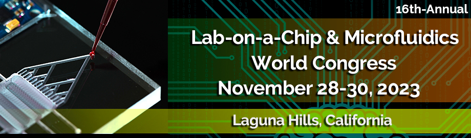
 Add to Calendar ▼2023-11-28 00:00:002023-11-30 00:00:00Europe/LondonLab-on-a-Chip and Microfluidics World Congress 2023Lab-on-a-Chip and Microfluidics World Congress 2023 in Laguna Hills, CaliforniaLaguna Hills, CaliforniaSELECTBIOenquiries@selectbiosciences.com