Co-Located Conference AgendasExtracellular Vesicles (EVs): Technologies & Biological Investigations | Lab-on-a-Chip and Microfluidics 2021 | Organoids & Organs-on-Chips 2021 | Point-of-Care Diagnostics and Biosensors 2021 | 

Monday, 13 December 202113:00 | Conference Registration and Materials Pick-Up | |
Session Title: Conference Plenary Session |
| | |
Venue: Marriott Coronado Island Ballrooms C & D |
| | 14:00 |  | Conference Chair Welcome and Introduction by Conference Co-Chair -- Lab-on-a-Chip and Microfluidics -- Inputs and Outputs in Product Engineering
Leanna Levine, Founder & CEO, ALine, Inc., United States of America
A unique aspect to point-of-care product development is to determine if the biological or chemical test will perform in a microfluidic system often before the rest of the product concept is thoroughly fleshed out. This means the programmatic priorities are focused on translational research before developing design inputs for a regulated product with an instrument. ALine’s new partnership with Nectar Product Development will enable us to support not just the assay implementation, but also create the entire product development roadmap, including the path to regulatory approval. |
| 14:10 |  | Conference Chair Welcome introduction by Conference Co-Chair Lab-on-a-Chip, Microfluidics & Associated Fields – Current Status: The Two Pathways – Simple Standard Devices Versus Highly Individualized Integrated Devices
Claudia Gärtner, CEO, microfluidic ChipShop GmbH, Germany
This presentation will pinpoint two interesting completely different ways for the use and the style of Lab-on-a-Chip devices: namely the simpler standard devices and the highly specific multifunctional integrated lab-on-a-chip devices. Point-of-care diagnostic has been one of the most prominent fields for lab-on-a-chip devices in particular since the last years during the pandemic. These devices are rather specific and highly individualized from the different companies commercializing them. A different pathway are easier standard devices, less application specific that are provided off-the-shelf from various companies and are frequently used with existing non-specific-lab-on-a-chip laboratory equipment. This “Lab-on-a-Chip-Catalogue” of an ever-growing number of different devices from various suppliers round the globe is worth to be highlighted and the use of existing standards and the creation of new ones should be shown as “the second way” for microfluidics. |
| 14:20 |  | Conference Chair Welcome and Introduction by Conference Co-Chair -- Extracellular Vesicles and Cellular Communication
Lucia Languino, Professor of Cancer Biology, Thomas Jefferson University, United States of America
This conference is focused on current Technologies and Biological Investigations on Extracellular Vesicles. Extracellular vesicles are nano-sized membranous structures released by cells into the extracellular space, and easily detectable in body fluids. Extracellular vesicles are highly heterogenous; large extracellular vesicles are plasma membrane-derived extracellular vesicles, while small extracellular vesicles are of endosomal or non-endosomal origin and are secreted upon fusion with the plasma membrane. Extracellular vesicle biological functions contribute to cell-cell communications in physiological and pathological conditions by carrying unique cargo (proteins, lipids, mRNAs and miRNAs) that modifies the functional state of the recipient cells. Recent investigations have clearly demonstrated the therapeutic and diagnostic potential of extracellular vesicles. The therapeutic potential of extracellular vesicles is of great interest especially because these vesicles can be engineered to achieve specific targeting. Currently, more than 200 trials that utilize extracellular vesicles are already registered in clinicaltrials.gov in the US. In this conference, emerging biological topics and novel technologies will be presented to stimulate discussion between areas of research of microfluidics, organoids and extracellular vesicles. |
| 14:30 |  | Keynote Presentation Development of Novel Intestine-on-Chip Models
Nancy Allbritton, Frank and Julie Jungers Dean of the College of Engineering and Professor of Bioengineering, University of Washington in Seattle, United States of America
Organ-on-chips are miniaturized devices that arrange living cells to
simulate functional subunits of tissues and organs. These microdevices
provide exquisite control of the biochemical and biophysical
microenvironment for the investigation of organ-level physiology and
disease. 2D and 3D models displaying a polarized human colonic
epithelium were developed to recapitulate gastrointestinal physiology.
The 2D crypt mimic displays a spatially patterned monolayer of
epithelium displaying a stem-cell niche and differentiated cell zone
with cells migration between the two regions. This planar 2D format
enables efficient image cytometry for high-throughput screening
applications as well physiologic measurements difficult to perform in a
3D format e.g., calcium signaling measurements. The 3D model builds on
the 2D model by providing the full architecture of the in vivo human
crypt. These models support formation of gradients of growth factors,
microbial metabolites, and gases. A thick impenetrable layer of mucus
with biophysical parameters similar to that of a living human can be
formed for epithelial cell-microbe studies. These bioanalytical
platforms are envisioned as next-generation systems for assay of
microbiome-behavior, drug-delivery, and toxin-interactions with normal
and diseased intestinal epithelia. |
| 15:00 |  | Keynote Presentation Affinity Selection of Extracellular Vesicles using Plastic-based Microfluidic Devices for the Management of Different Diseases
Steve Soper, Foundation Distinguished Professor, Director, Center of BioModular Multi-Scale System for Precision Medicine, The University of Kansas, United States of America
We have been developing tools for the diagnosis of a variety of
diseases. The commonality in these tools is that they consist of
microfluidic devices made from plastics via injection molding. Thus, our
tools can be mass produced at low-cost to facilitate bench-to-bed side
transition and point-of-care testing (PoCT). We have also been
generating novel assays focused on using liquid biopsy samples that are
enabled using microfluidics. In this presentation, I will talk about the
evolution of our fabrication efforts of plastic-based microfluidic and
nanofluidic devices as well their surface modification to make the
devices biocompatible for in vitro diagnostics. One tool that we have
generated is a plastic device (38 × 42 mm) that consists of 1.5M
pillars, which are surface decorated with affinity agents targeting
certain disease-associated extracellular vesicles (EVs). The affinity
agents are covalently attached to the surface of the microfluidic device
using a bifunctional linker, which consists of a coumarin moiety to
allow for the photolytic release of the captured EVs using a blue-light
LED to minimize photodamage to the EVs’ molecular cargo. We have also
developed a high-throughput nano-Coulter counter (nCC) made from a
plastic via injection molding for the counting of captured EVs from
clinical samples to allow their enumeration. The nCC consists of
multiple pores that are ~350 nm to allow for high throughput counting
with exquisite LODs (500 EVs/mL). In this presentation, I will discuss
the utility of these microfluidic and nanofluidic devices in several
diseases, for example, using EVs as a source of mRNAs for molecular
sub-typing of breast cancer patients. EVs were affinity selected from
breast-cancer patients’ plasma by searching for both epithelial and
mesenchymal expressing EVs to allow for highly efficient sub-typing
using the PAM50 gene panel. In an addition, the microfluidic and
nanofluidic devices were integrated into a single platform
(modular-based system) for PoCT to screen for early stage ovarian
cancer. Affinity probes were used to target EVs specifically generated
from tumor cells that signal early-stage ovarian cancer disease with the
nCC used for enumerating the number of EVs captured. Finally, the
modular system was used for the detection of COVID-19 at the PoC by
affinity selecting SARS-CoV-2 viral particles. The integrated system
could process saliva samples to search for the viral particles and count
them in <20 min. |
| 15:30 | Coffee Break and Networking | 16:00 |  | Keynote Presentation From Droplet-based Microfluidics Developments to Droplet-based Digital PCR
Valérie Taly, CNRS Research Director, Professor and Group leader Translational Research and Microfluidics, Université Paris Cité, France
Droplet-based microfluidics has led to the development of highly powerful
systems that represent a new paradigm in High-Throughput Screening
where individual assays are compartmentalized within microdroplet
microreactors. The integration of such systems for series of complex
individual operations on droplets has offered a solution to the
necessary miniaturization and automation of individual biological
assays. By combining a decrease of assay volume and an increase of
throughput, this technology goes beyond the capacities of conventional
screening systems. We will show how by combining droplet-based
microfluidic systems and clinical advances in molecular diagnostic we
have developed an original method to perform millions of single molecule
PCR in parallel to detect and quantify a minority of mutant sequences
within a high quantity of non-mutated sequences in complex mixture of
DNA with a sensitivity and precision that was unreachable by
conventional procedures. |
| 16:30 |  | Keynote Presentation High-Purity and High-Sensitivity Rare-Cell Isolation with Aliquot Sorting
Daniel Chiu, A. Bruce Montgomery Professor of Chemistry, University of Washington, United States of America
This presentation describes the high-sensitivity isolation of rare
cells, including circulating tumor cells and fetal cells, using
fluorescence activated aliquot sorting. Here, a blood sample is divided
into nanoliter volumes or aliquots, then analyzed and sorted based on
the presence or absence of a rare cell. By performing two rapid, on the
millisecond timescale, back-to-back sorting, we also demonstrate
isolation of rare cells with high purity. Finally, we drastically
increased the number of nucleated cells sorted by first concentrating
peripheral blood mononuclear cells from whole blood, which translated
into an equivalent blood volume analyzed by over an order of magnitude,
from 8mL to over 100mL per experiment. |
| 17:00 |  | Keynote Presentation Extracellular Vesicles: Liquid Biopsies and Biological Nanocarriers For Therapy
Dominique PV de Kleijn, Professor Experimental Vascular Surgery, Professor Netherlands Heart Institute, University Medical Center Utrecht, The Netherlands, Netherlands
|
| 17:30 |  | Keynote Presentation Over-Engineering in the Life Sciences: Microfluidics and Microphysiological Systems Meet Artificial Intelligence
John Wikswo, Gordon A. Cain University Professor, A.B. Learned Professor of Living State Physics; Founding Director, Vanderbilt Institute for Integrative Biosystems, Vanderbilt University, United States of America
It is a worthwhile exercise to examine whether the addition of microfluidic pumps and valves to organ-chip systems represents either “over-engineering” or the means by which microphysiological systems can be made massively parallel. While several cited examples of over-engineering represent overly complicated or frivolous solutions to a problem, such as the $400 Juicero juice-pouch-squeezer or a $1,390 Porsche roof box rated at 200 km/hour, others are prescient and ultimately yield great value, for example the NASA Opportunity rover whose mission was planned to last 90 Mars sols (~93 Earth days) but in fact lasted 14 Earth years, and Mercedes Benz’s introduction in the 1970s of antilock braking systems and in 1980 the driver’s airbag and seat-belt tensioner. From the perspective of a biologist who uses an agar-filled Petri dish for microbial colony picking, a 1536 well plate and its associated high-throughput screening infrastructure are overkill; for big Pharma, it is a welcome tool that took 15 years to be refined and put into practical use. As we approach the 15th anniversary of Mike Shuler’s landmark 2007 patent “Devices and Methods for Pharmacokinetic-based Cell Culture System,” we realize that microfluidic tissue and organ chips have been perfused using gravity, pressurized reservoirs, external syringe or peristaltic pumps, or on-chip peristaltic pumps. Although the smallest devices abound with Quake-style pneumatic valves that also serve as pumps, very few organ-chip systems utilize valves. For over a decade, our group has been developing microfluidic pumps and valves, and we have long argued that multi-organ microphysiological systems will ultimately need automation of pumps and valves to control each chip, their closed-loop sensors, and organ-organ interconnections. Our Missing Organ MicroFormulator concept spawned an award-winning 96-channel MicroFormulator, which served as the foundation for CN Bio Innovations’ recently introduced PhysioMimixTM pharmacokinetic (PK) system that supports PK studies of oncological drugs. As our group develops its fourth generation of rotary microfluidic pumps and valves to enable our construction and Ross King’s coding of the machine-learning algorithms for the 1000-channel Genesis Self-Driving Chemostat for yeast systems biology, we realize that this hardware can be readily adapted to perfuse and control a dozen of Steve George’s and Scott Simon’s bone marrow and skin chips in an in vitro infection model, maintain and analyze multiple, coupled organ chips for six months or longer, support remote studies of select agents in a BSL-3 or BSL-4 facility, apply circadian rhythms to thousands of wells in dozens of well plates or hundreds of zebrafish or perfused organ chips, and accelerate and optimize the microbial production of antibodies, industrial feed stocks, food protein, and sequestered carbon dioxide. Is this over-engineering or the future of artificial intelligence in biology? |
| 18:00 |  | Keynote Presentation Development and Deployment of ‘Smart Diagnostics’: Next Generation Point of Care Sensors with Capacity to Learn
John T McDevitt, Professor, Division of Biomaterials, New York University College of Dentistry Bioengineering Institute, United States of America
While COVID-19 has yielded devastating consequences over the past few years, the global pandemic also has opened the door for acceleration of development of core diagnostic capabilities that have the potential to lead to lasting impact for our society. With this vantage point in mind, in the recent past the McDevitt laboratory has launched a series of efforts that target the development and deployment of ‘smart diagnostics’ that serve as distributed point of care sensor nodes with capacity to learn. These mini-sensor ensembles with embedded artificial intelligence integrate programmable chip-based diagnostic systems capable of multiplexed measurements alongside clinical decision support tools that utilize strategically chosen nonclinical data elements that elicit signatures that can be used to capture diseases before they spiral out of control. As such, these efforts link for the first-time the following five key disciplines: i) lab-on-a-chip technologies, ii) in vitro diagnostics, iii) -omics research, iv) artificial intelligence, and v) digital healthcare delivery systems.
Importantly, the combination of point-of-care medical microdevices and machine learning has the potential transform the practice of medicine. In this area, scalable lab-on-a-chip devices have many advantages over standard laboratory methods including portability, faster analysis, reduced cost, lower power consumption, and higher levels of integration and automation. Despite significant advances in medical microdevice technologies over the years, several remaining obstacles are preventing clinical implementation and market penetration of these novel medical microdevices. Similarly, while machine learning has seen explosive growth in recent years and promises to shift the practice of medicine toward data-intensive and evidence-based decision making, its uptake has been hindered due to the lack of integration between clinical measurements and disease determinations.
In this talk, our recent advances in ‘smart diagnostics’ will be highlighted. These smart diagnostics include single-use microfluidic cartridges that serve as fully integrated, self-contained devices that contain aqueous buffers suitable for automated completion of all assay steps within nontraditional healthcare settings. Further, a portable analyzer instrument is fashioned to integrate fluid delivery, optical detection, image analysis, and user interface, representing a universal system for acquiring, processing, and managing clinical data while overcoming many of the challenges facing the widespread clinical adoption of lab-on-a-chip technologies. Intimate linkages between these medical microdevices and cloud connected databases allows for early disease detection algorithms to be used to impact clinical progress. |
| 18:30 | Networking Reception with Beer, Wine and a Light Dinner in the Exhibit Hall -- Meet Colleagues and Network with Sponsors and Exhibitors | 20:00 | Close of Day 1 of the Conference |
Tuesday, 14 December 202108:00 | Conference Registration and Continental Breakfast Served in the Exhibit Hall | |
Session Title: Opening Session -- Extracellular Vesicles, circa 2021 |
| | |
Venue: Marriott Coronado Island Ballroom D |
| | 08:30 | Conference Welcome and Opening Remarks by Conference Chairperson
Lucia Languino, Professor of Cancer Biology, Thomas Jefferson University, United States of America
| 08:45 |  | Conference Chair Welcome and Introduction by Conference Co-Chair
Michael Graner, Professor, Dept of Neurosurgery, University of Colorado Anschutz School of Medicine, United States of America
|
| 09:00 | CANCER CELL TARGETING VIA Small EXTRACELLULAR VESICLES
Lucia Languino, Professor of Cancer Biology, Thomas Jefferson University, United States of America
Cancer cells crosstalk with the tumor microenvironment by releasing small extracellular vesicles (sEVs). sEVs, isolated from cancer cell culture media, express the epithelial-specific alphaVbeta6 integrin. After confirming the fidelity of the sEV preparations by electron microscopy, density gradient, and immunoblotting, we determined that the alphaVbeta6 integrin is actively packaged into sEVs isolated from cancer cells. sEVs mediate protein transfer of alphaVbeta6 integrin to microvascular endothelial cells and increase the number of their junctions and tubules. We also show that alphaVbeta6 is transferred from cancer cells to monocytes by sEVs and that sEVs, purified via density gradients, promote M2 polarization. In addition, as evaluated by our proteomic analysis, alphaVbeta6 down-regulation causes a significant increase in donor cancer cells, and their sEVs, of two molecules that have a tumor suppressive role, STAT1 and MX1/2. De novo alphaVbeta6 expression in an alphaVbeta6-negative recipient cell is not a result of a change in mRNA levels but is a consequence of sEV-mediated transfer of this integrin.
We have now selected alphaVbeta6, which is expressed on the cell surface in many cancers but absent in normal tissues, as candidate for therapeutic targeting via sEVs. The benefits of sEV-mediated cancer therapy are: low immunogenicity, ability to infiltrate biological barriers and ‘targetability’. We demonstrate an efficient strategy to therapeutically target alphaVbeta6 by using short interfering RNA (siRNA) loaded into sEVs. We first demonstrate that fluorescently labeled siRNAs can be efficiently loaded into sEVs by electroporation. By confocal microscopy, we show efficient internalization of these siRNA-loaded sEVs into recipient cells. We then show that sEV-mediated delivery of alphaVbeta6 -targeting siRNA into cancer cells specifically downregulates expression and function of alphaVbeta6. Overall, this study shows that sEVs from cancer cells may contribute to a horizontal propagation of integrin-associated phenotypes from cancer cells to the tumor microenvironment. | 09:30 | Biogenesis of RNA-containing Extracellular Vesicles
Alissa Weaver, Cornelius Vanderbilt Professor of Cell and Developmental Biology, Vanderbilt University Medical Center, United States of America
Extracellular RNAs (exRNAs) carried by extracellular vesicles (EVs) can affect gene expression, function, and phenotypes of recipient cells. While a number of RNA binding proteins (RBPs) are known to carry RNAs into extracellular vesicles (EVs), where and how in the cell this occurs is unclear. As many RBP-RNA complexes are associated with the endoplasmic reticulum, we investigated whether membrane contact sites (ER MCS) with endosomes may drive the biogenesis of RNA-containing EVs. To carry out this study, we used RNA-sequencing, lipidomic, confocal and transmission electron microscopy, tumor xenograft and various biochemical techniques to analyze EV biogenesis and cargo content in colon cancer cell lines molecularly engineered for molecules that control ER MCS. We uncovered a novel pathway of EV biogenesis that takes place at ER MCS. Our data suggest a model in which lipid transfer at ER MCS drives biogenesis of a select subpopulation of EVs containing RNA-RBP complexes. Beyond improving our understanding of EV biogenesis, we anticipate that these findings may be useful for future engineering of therapeutic EVs as well as exploring the functions of RNA-containing EVs. | 10:00 | Morning Coffee, Pastries and Networking in the Exhibit Hall | 10:30 |  Insights into EV Characterization Technologies Insights into EV Characterization Technologies
Jean-Luc Fraikin, CEO, Spectradyne
Methods for characterizing EVs are evolving, and new technologies are being developed that deliver size, concentration, and fluorescent phenotyping, each to a varying degree of success. Learn about how some of these new and cutting-edge technologies work, their strengths and weaknesses, and where we at Spectradyne see the technology landscape moving next.
| 11:00 |  Improvements in Fluorescent Nanoparticle Tracking Analysis: Reliable Characterization of Extracellular Vesicles Improvements in Fluorescent Nanoparticle Tracking Analysis: Reliable Characterization of Extracellular Vesicles
Sven Rudolf Kreutel, Chief Executive Officer, Particle Metrix GmbH and CEO, Particle Metrix Inc., USA
| 11:30 | Developing Technologies for Single Vesicle Isolation and for Regenerative Medicine
Danilo Tagle, Director, Office of Special Initiatives, National Center for Advancing Translational Sciences at the NIH (NCATS), United States of America
Extracellular vesicles (EVs) are lipid membranous vesicles released from almost all cell types, and they provide a tremendous opportunity as sources of novel biomarkers from liquid biopsies, as well as agents for tissue repair and wound healing in regenerative medicine. EVs carry complex molecular cargoes, such as proteins, RNAs [e.g., mRNA and noncoding RNAs (microRNA, transfer RNA, circular RNA and long noncoding RNA)], and DNA fragments; these cargoes are delivered to recipient cells and serve as a cell-to-cell communication system. The molecular contents of EVs largely reflect the cell of origin and thus show cell-type specificity. Exosomes are endogenous nanoparticles that constitute a fraction of extracellular vesicles that are secreted by all cell types into the extracellular environment, and play an important role in intercellular signalling. Exosomes are being utilized in a variety of biomedical applications, including targeted drug delivery, gene therapy, diagnosis, and tissue regeneration. Despite significant efforts made in this relatively new field of research, progress has been held back by challenges such as inefficient separation methods, difficulties in characterization, and lack of specific biomarkers. This presentation will elaborate on the NIH support for exosome isolation, as well as its potential use in regenerative medicine. | 12:00 | Plasma Extracellular Vesicles for Cardiovascular Disease
Dominique PV de Kleijn, Professor Experimental Vascular Surgery, Professor Netherlands Heart Institute, University Medical Center Utrecht, The Netherlands, Netherlands
Cardiovascular Disease (CVD) is with the cardiovascular events of Ischemic Heart Disease and Stroke, the number 1 and 2 cause of death in the world and expect to increase especially in Asia. We are focussing on prognosis of secondary cardiovascular events after stroke and diagnosis of Ischemic Heart Disease. Ischemic heart disease (IHD) comprises 2 entities: Chronic Coronary Syndrome (CCS) and Acute Coronary Syndrome (ACS). Because CCS is associated with 6-8 times increased risk of adverse cadviovascular events like myocardial infarction and death, early recognition of CCS. Blood markers for CCS do not exist, resulting in that 80-90% of chest pain patients undergoing costly imaging do not have CCS. We use plasma extracellular vesicle protein content of vesicles from plasma subfractions as an accurate source for early diagnosis of CCS better then a clinical risk model. The same EV subfraction technology is used on plasma of stroke patients that after carotid atherectomy are at high risk of a second event (myocardial infarction, stroke or cardiovascular death). Protein as well as lipid content can identify patients at high risk for a secondary event better then a clinical risk prediction model. | 12:30 | Buffet Lunch and Networking in the Exhibit Hall with Exhibitors and Conference Sponsors | |
15-Minute Oral Presentations of Selected Posters in the EVs Track -- Venue: Coronado Ballroom D |
| | 13:30 | Efficient Production of Extracellular Vesicles Using Cell Spheroids
KEUNSUN AHN, CEO, SPHEBIO, Korea South
Cell-derived extracellular vesicles(EVs) contain miRNA, mRNA, and various proteins, and many studies have been conducted on the therapeutic potential due to their intracellular communication capabilities. However, for commercial use, it is necessary to improve the production efficiency and quality control standards of existing EV production technology. Here, we have developed EV production technology using cell aggregates/spheroids to improve EV production efficiency. Since the EV production environment changes according to the size of the cell spheroid used for EV production, we developed cell spheroid production automation technology to produce uniform cell spheroids. The production of EVs derived from cell spheroids significantly increased the production index of number of EVs per cell(# of EVs/cell) compared to the conventional 2D cell culture, and a change in productivity was confirmed depending on the size. The results of this study could suggest mass production technology for EV commercialization. | 13:45 | Circulating microRNAs in Extracellular Vesicles as Prognostic and Predictive Biomarker for Metastatic Colorectal Cancer
Klara Cervena, Ph.D. Student, Institute of Experimental Medicine, Czech Republic
Systematic therapy, whose gold standard is a combination of chemotherapy and targeted therapy, remains a mainstay of metastatic CRC (mCRC) treatment. The choice of the treatment strategy is affected by various clinical factors and biomarkers. Although several predictive biomarkers have previously been suggested, only a few of them are being used in routine clinical practice. The importance of these markers is highlighted by the fact that approximately 20% of CRC patients have metastatic disease at the time of diagnosis. Recently, we have identified that circulating microRNAs (miRNAs) - miR-122-5p and miR-142-5p - show high potential for early detection of CRC as well as for the assessment of patient´s outcome and response to therapy. These miRNAs, isolated from plasma extracellular vesicles of repeated samplings from 20 mCRC patients, were further studied for their potential to predict the therapy response. The first sampling was performed at the time of post-therapy developed liver metastasis and other samplings were collected approximately every 3 months over one year. Detailed results of the study will be presented during the meeting. We believe that our results may add to new prognostic and predictive biomarkers associated with therapy response in mCRC patients. | |
Session Title: Emerging Technologies in EV Research |
| | |
Venue: Marriott Coronado Island Ballroom D |
| | 14:00 |  The Next Generation of Extracellular Vesicle Isolation and Characterization The Next Generation of Extracellular Vesicle Isolation and Characterization
Jared Lynch, Director of Business Development, IZON Science
In this talk, I will discuss Izon’s new Exoid TRPS instrument and how it fits into Izon’s complete EV characterization ecosystem of new and existing product pipelines. I will also introduce Izon’s new qEV1.0 column with Izon’s next generation SEC gel.
| 14:30 |  Single-Molecule Imaging of Extracellular Vesicles Below the Diffraction Limit Single-Molecule Imaging of Extracellular Vesicles Below the Diffraction Limit
Dina Sirypangno, Sales Director, USA West Coast, ONI
Extracellular Vesicles (EVs) are membranous particles that enable cell-cell communication via their surface components and cargo which includes proteins, lipids, and genetic material such as DNA, mRNA and miRNA. Understanding the unique molecular signatures of sub-populations of EVs and their association with specific cargo can enhance our understanding of how EVs function in cell signalling pathways and give deeper insights into the phenotypic consequences they enact at their target sites. Single-molecule localization microscopy (SMLM) through the Nanoimager platform offers a means of characterizing heterogeneous populations of EVs with single-molecule sensitivity, overcoming the resolution limits associated with conventional fluorescence microscopy techniques. Furthermore, ONI has developed a novel analysis software termed CODI, a platform which hosts a wide range of advanced image analysis tools that have been designed specifically to aid the characterization of EVs through clustering-based methods. Through this platform users can count and quantify thousands of EVs from a single super-resolution image, assess their morphology with 20 nm resolution and visualize multiple biomarkers with single-molecule sensitivity. This allows EV researchers to unveil previously invisible insights from their sample through this simple, reliable, and easy to use platform.
| 15:00 | Afternoon Coffee Break and Networking in the Exhibit Hall | 15:30 | Utilizing Flow Cytometry For Multi-Parametric Analysis
Joshua Welsh, Research Fellow, National Cancer Institute (NCI), NCI, United States of America
Extracellular vesicles (EVs) are sub-micron lipid spheres derived from cells. Interest in EVs is growing due to their potential applications in translational medicine, therapeutics, as well as their role in basic biology. Progress in the understanding of EVs has been hampered by the lack of equipment and standardization in the EV field. In recent years, the development of highly sensitive commercial flow cytometry platforms, along with societal initiatives, has led to flow cytometry being one of the most informative EV characterization methods. Our work at NIH has developed flow cytometry assays, standardization methods, and software for study of EVs at a single particle level and as part of multiplex analysis techniques. Our methods have allowed EV data to be reported in standard units allowing cross-platform data integration, with the ultimate aim of developing an EV atlas. | 16:00 | Multi-Parametric High-Throughput Flow-Based Analysis of Single Extracellular Vesicles
Daniel Chiu, A. Bruce Montgomery Professor of Chemistry, University of Washington, United States of America
We have developed a multi-parametric high-throughput flow-based method for the analysis and sorting of individual extracelluar vesicles (EVs), which are highly heterogeneous and comprise a diverse set of surface protein markers as well as intra-vesicular cargoes. Yet, current approaches to the study of EVs lack the necessary sensitivity and precision to fully characterize and understand the make-up and the distribution of various EV subpopulations that may be present. In fact, most current EV isolation methodologies, including differential centrifugation, affinity/immuno-magnetic isolation, polymer-based precipitation, size-based exclusion, can be prone to contaminations and cannot resolve single EVs. Our new technology platform is able to immune-phenotype EVs and quantify proteins associated with EVs, at the single-EV level, with high precision and throughput. | 16:30 |  Enrichment of microRNA Using a Novel Avalanche Photodiode-based Benchtop Cell Sorter Enrichment of microRNA Using a Novel Avalanche Photodiode-based Benchtop Cell Sorter
John Tigges, Flow Cytometry Science Center Director, Beth Israel Deaconess Medical Center
In the past few years, extracellular vesicles (EVs) have rapidly become one of the most studied entities. EVs are characterized by their outer lipid layer and internalized cargo such as microRNA (miR). EVs and their cargo have been shown to regulate gene expression and alter cell function in various cell types (Kreimer et al., 2015, Mantel et al., 2013). Isolation and molecular profiling of subsets of EVs (i.e., RNAs, proteins, lipids, metabolites) are critical for understanding the biogenesis of EVs and their potential utility as biomarkers (Pereira et al., 2020). To this end, flow cytometry has been employed for the analysis, characterization, and phenotyping of EV populations and their cargo in a technique called nano-flow cytometry. While nano-flow cytometry has become more prevalent as an analysis tool and undergone standardization formats (Welsh et al., 2020), nano-sorting is neither well defined nor a commonly used technique. Historically, this is due to the complexity of the technique and instrumentation necessary for sorting of EVs (Morales-Kastresana et al., 2019, Kormelink et al., 2015). In this study, the CytoFLEX SRT, a semiconductor based benchtop cell sorter equipped with avalanche photodiodes and optimized signal detection, (Brittain et al., 2019) is utilized for the detection and nano-sorting of miRs in EV populations. Platelet EVs, red blood cell EVs, and neutrophil EVs are mixed with molecular beacons (MBs) targeted to miR495, miR451, and miR148 respectively. MBs are nucleic acid sequences locked in a hairpin conformation with a quencher signal and fluorophore attached to the end (Tyagi, 1996). Upon binding with a specific target RNA sequence, the quencher is spatially moved from the fluorophore and a fluorescent signal is produced. Upon analysis of the EV populations with MBs via VSSC and fluorescent thresholding, fluorescent positive signals corresponding to the particular miR target were sorted. Sorted samples were verified via microscopy and PCR analysis and compared to fluorescent negative miR EVs The presence of EV populations and subsequent increased target miRs, verified via dark field and fluorescent microscopy, confirm sorting efficiency compared to the negative population. Additionally, CT values from the PCR analysis show 8x difference in miR detection between positive and negative sorted samples. These results show the CytoFLEX SRT to be a viable instrument for nano-sorting.
| 17:00 |  Multiparameter Analysis of EVs by Vesicle Flow Cytometry (vFC) Multiparameter Analysis of EVs by Vesicle Flow Cytometry (vFC)
John Nolan, CEO, Cellarcus Biosciences, Inc.
Progress in understanding the basic biology and applied uses of extracellular vesicles (EVs) depends on transition from bulk biochemical and physical analysis to single EV analysis. Single vesicle flow cytometry (vFC) has emerged as a highly sensitive and specific approach to counting and sizing EVs as well as measuring molecular cargo of individual EVs. Quantitative immunofluorescence.
| 17:30 |  The ExoView R200 Platform for Single EV and Virus Phenotyping and Sizing The ExoView R200 Platform for Single EV and Virus Phenotyping and Sizing
Clayton Deighan, North American Sales and Applications Manager, NanoFCM
NanoView recently launched the ExoView R200, our latest analytical platform for purification-free analysis of extracellular vesicles and viruses. ExoView works by affinity capturing exosomes onto a protein microarray (ExoView chip) via common or custom exosomal markers. Once bound to the surface of the ExoView chip each individual exosome can be sized, counted and characterized in terms of protein expression. The latest R200 platform adds an additional fluorescent channel and EVs can now imaged in 4 fluorescent channels (blue, green, red and far-red) meaning that individual exosomes can be phenotyped by up to 5 surface or cargo proteins (4 fluorescent and 1 capture antibody) with single-molecule sensitivities. To complement the new platform, NanoView will also be discussing existing and new assays designed to enable EV and virus research. Standard assays as designed to measure common EV markers from cell culture or plasma samples (with or without purification) and can be extended to detect EV cargo which will be demonstrated. These assays have been optimized to promote biomarker colocalization on even the smallest EVs and have been extended to include fully user customizable assays that allow customers to capture and detect EVs via any custom protein of their choice. We will review newly developed assays for detecting lenti-viruses and their capsids.
| 18:00 |  EV Analysis Using Imaging Flow Cytometry EV Analysis Using Imaging Flow Cytometry
Stephanie Brunelle, Senior Product Manager, Flow Cytometry, Luminex Corp.
Amnis flow cytometers leverage the superior photonic sensitivity provided by time delay integration charge coupled device (TDI CCD) cameras. High gain mode on the ImageStream® and small particle mode on the CellStream® systems further enhance that sensitivity making these systems ideal for measuring extracellular vesicles (EVs) and other small particles. With multiple excitation lasers and up to 20 fluorescent channels, EV experiments can include probes for cargo proteins, RNA, and other molecules when needed. In this presentation, we will review the life cycle of EVs, talk about the critical controls needed for EV experiments, and show data on particle detection sensitivity, cargo protein and RNA, and uptake of EVs by other cells.
| 18:30 | Networking Reception with Beer and Wine: Network with Colleagues and Engage with Exhibitors and Conference Sponsors | 19:30 | Close of Main Conference Day 2 | |
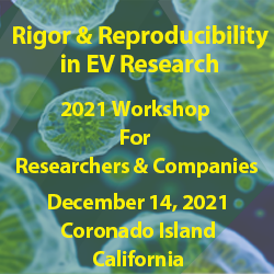
Session: Standardization & Single Extracellular Vesicles (sEV) Analysis Workshop |
| | 19:40 | Rigor & Reproducibility in EV Research and Single EV Analysis: Workshop Chaired by Dr. John Nolan, The Scintillion Institute and Cellarcus
John Nolan, CEO, Cellarcus Biosciences, Inc., United States of America
Topics Covered and Presenters:
- Guidelines for measurement of EV number, size, and cargo
- Calibration of single EV measurement instruments and assays
- Reference materials for standardization of single EV measurements
- Flow cytometry instrumentation: Current status and pathway to single molecules on single vesicles
- John Nolan -- Chair
- Josh Welsh -- Speaker
- Daniel Chiu -- Speaker
 | 21:30 | Close of Day 2 Programming |
Wednesday, 15 December 202108:00 | Morning Coffee and Continental Breakfast in the Exhibit Hall | |
Venue: Marriott Coronado Island Ballroom D |
| | 08:30 | Engineered Synthetic Bacterial Vesicles as Cancer Immunotherapy and as Vaccines against Bacteria and SARS-CoV-2
Jan Lötvall, Professor, University of Gothenburg; Founding President of ISEV; Editor-in-Chief, Journal of Extracellular Vesicles, Sweden
We are developing a novel platform of “exosome-like” synthetic bacterial vesicles (SyBV) that can be utilized as cancer immunotherapy, and as preventive vaccines against viral or bacterial infection. Briefly, the SyBV are produced from isolated bacterial membranes, and exhibit many of the features of natural bacterial veicles (OMV), but have been detoxified efficiently by removal of multiple bacterial components that activate pattern recognition receptors (PRR), including Toll Like Receptors (TLR). This strongly reduces the dose-limiting side effects of the SyBVs, but retains their stimulatory effects on adaptive immunity. The SyBV efficiently induce specific immunity via T-cell and humoral responses in both cancer models, and against bacterial components. SyBV combined with tumor exosomes induce immune responses in models of malignant melanoma and colon cancer, and synergize with checkpoint inhibitors to reduce or stop tumor growth. Further, SyBV can function as adjuvant in virus vaccine candidates, including SARS-CoV-2. SyBV is a safe and highly efficient platform with potentially very wide clinical applications in both cancer immunotherapy, as well as preventive vaccines against both bacterial and viral infections. | 09:00 | Defining the Role of Small Extracellular Vesicles in Lymph Node Pre-metastatic Niche Formation
Hector Peinado, Group Leader, Microenvironment and Metastasis Group, Spanish National Cancer Research Centre (CNIO), Spain
We have analyzed the dynamics of tumor-derived small EVs (sEVs) in the lymphatic system and their role establishing the pre-metastatic niche formation in sentinel lymph nodes. Our data shows how both tumor intrinsic and extrinsic factors such as tumor-secreted sEVs are involved in lymphangiogenesis and pre-metastatic niche formation in sentinel lymph nodes through a neurotrophin receptor-dependent mechanism favoring metastatic spread in melanoma. | 09:30 | EV Genomic and Proteomic Characterization Using Izon’s Next Generation qEV1 SEC Column and qEV Kits
Henry Scheffer, Research Associate, IZON
Izon Science is the world leading manufacturer of nano-biological separation and characterization tools. Izon’s next generation qEV1 SEC column has superior EV isolation and purification performance allowing for high signal to noise EV downstream analysis using the qEV RNA Extraction and qEV Concentration kits, leading to easily identified commonly found EV proteomic and genomic markers. In this talk, we will show the efficient use of Izon consumables and reagents in generating commonly recognized EV biomarkers. | 10:00 | Mid-Morning Coffee Break and Networking in the Exhibit Hall | 10:30 | NCI-Supported Extracellular Vesicle Research: Perspectives and Opportunities
Elisa Woodhouse, Program Director, Tumor Biology and Microenvironment Branch, NIH/NCI, United States of America
Extracellular vesicles (EVs) have emerged in the recent past as important mediators of communication between cells or with the micro- and macro-environments (TME). Nevertheless, NCI portfolio analysis of the past 5 years indicates that while exosome research remains stable it has not expanded in step with its biological significance. Numerous NCI funding mechanisms may be leveraged towards directly or indirectly achieving short- and long-term growth in this research field. Direct measures include a wide range of training programs or administrative supplement initiatives that can further support current funded EV projects and the next generation of researchers. Furthermore, EV-focused or technology-development NCI funding announcements and ongoing NCI/NIH Programs also provide opportunities for specific aspects of EV research. Indirectly, parallel development and implementation of new NCI initiatives are also being strategized, including a new series of cooperative networks that – broadly speaking – place greater emphasis on the micro-/macro-environments (and numerous TME components such as EVs) as modulators of disease progression. Several programs have been established to comprehensively study and differentiate various stages of disease progression as distinct entities governed by nuanced biological rules and dynamics across the tumor-TME continuum, including the role of the TME as a co-organizer – from early lesions to frank tumors and advanced disease/therapeutic resistance. This recent recalibration in TME emphasis – away from the classical tumor-centric/autonomous dogma will likely further stimulate EV research in different stages of disease progression and as an integral component of tumor-stromal dynamics. Central to this set of objectives are three NCI programs: Translational and Basic Research in Early Lesions (TBEL) focused on risk stratification and understanding the determinants of early lesion evolution; PDAC Stromal Reprogramming Consortium (PSRC) tackling the role of the stroma as a co-organizer of early lesion fate in a disease-specific manner; and Acquired Resistance to Therapy Network (ArtNet) examining the TME response in driving therapy resistance. | 11:00 | Biomarkers of an effective Antitumor Response to Immune Checkpoint Inhibitor in Head and Neck Cancer
My Mahoney, Professor and Vice Chair, Thomas Jefferson University, United States of America
Head and neck squamous cell carcinoma (HNSCC) is the 6th leading cancer worldwide with common risk factors including chronic alcohol and tobacco use as well as human papillomavirus (HPV) infection. Despite the prevalence, there are no clear clinical biomarkers that could delineate normal homeostasis versus tumorigenic processes, HPV positivity, and response to immune checkpoint inhibitors (ICI). In fact, profiling and validation of any cancer biomarkers have proven so complex that HE4 was the last biomarker approved by the FDA for breast cancer in 2009. Emerging as a potentially reliable source for cancer biomarkers are exosomes or small extracellular vesicles (sEVs), which have been shown to play critical roles in cell-to-cell communication and cancer progression. Here, sEVs were isolated from circulating plasma of HNSCC patients and control volunteers by the immunoaffinity ExoRelease method (Clara Biotech) and subjected to bead-based Multiplex profiling of cancer-specific antigens (Luminex). Results were compared to respective total plasma showing substantial differences between the profiles. Interestingly, higher levels of several cancer biomarkers were detected in HNSCC sEVs, particularly in HPV(-) as compared to HPV(+) patients. Most importantly, HE4 associates with HPV(-). In addition to protein biomarkers, we performed small RNAseq for miRNAs in HNSCC sEVs. Here, in a 4-week window of opportunity trial, HNSCC patients received 2 doses of the ICI anti-PD1 mAb, nivolumab. Patients were stratified according to pathologic treatment response, as demonstrated by reduction in tumor size. sEVs isolated from post-treatment tumor cultured supernatant were analyzed by FACS analysis, which confirmed the downregulation of immune checkpoint receptors in responders. Profiling by miRNAseq and analysis by Weka Explorer revealed that 10 miRNAs were strongly correlated with HPV status and 18 miRNAs with response to ICI treatment. KEGG analysis revealed that their targets were associated with pathways in cancer. While the levels of miRNAs did not change in response to treatment, there was a statistically higher level of anti-inflammatory miRNAs, including tumor suppressor miR-146a, in responders versus non-responders. The results from these studies suggest there are new cancer biomarkers that could contribute to better diagnosis and treatment of HNSCC cancer patients. | 11:30 | Targeting Cancer-Stromal Cell Communication by Novel Drugs that Disrupt the Nuclear Entry of Extracellular Vesicles
Aurelio Lorico, Professor of Pathology, Touro University Nevada School of Medicine, United States of America
Extracellular vesicles (EVs) are mediators of intercellular communication under both healthy and pathological conditions, including the induction of pro-metastatic traits, but it is not yet known how and in which host cell compartment(s) EVs deliver their functional cargo. We recently described a novel intracellular pathway whereby EV-containing late endosomes enter in the nucleoplasmic reticulum through type II nuclear envelope invaginations (NEI) and transfer EV cargo into the nucleoplasm of recipient cells. A tripartite protein complex named VOR, containing nuclear membrane-associated VAP-A, ORP3 and late endosome-associated Rab7 orchestrates entry and/or docking of EV-containing late endosomes into NEI. In search for drugs that could inhibit ORP3 function as component of the VOR complex, we came across ORPphilin molecules, such as anti-proliferative natural product OSW-1 and the FDA-approved antifungal azole itraconazole (ICZ). In addition to its antifungal activity, due to the inhibition of lanosterol 14a-demethylase, an inhibitory activity of ICZ on enterovirus and hepatitis C virus replication has been reported and attributed to OSBP and ORP4 inhibition. The five ring-backbone structure and the sec-butyl chain of ICZ are important for antiviral activity, whereas the triazole moiety is critical for the antifungal activity. We recently found that ICZ, but not its main metabolite hydroxy-ICZ or ketoconazole, disrupts the VOR complex and inhibits EV-mediated pro-metastatic morphological transformation and migratory properties of colon cancer cells. By computer modelling, we have synthesized novel, smaller chemical drugs, that maintain the property of inhibiting the VOR complex without the ICZ moieties responsible for the antifungal activity and interference with intracellular cholesterol distribution. Knowing that cancer cells hijack their microenvironment and that EVs derived from them determine the pre-metastatic niche, small-sized inhibitors of nuclear transfer of EV cargo into host cells could find cancer therapeutic applications, particularly in combination with direct targeting of cancer cells. | 12:00 | Diagnostic Potentials of Circulating EV/EXO-related Biomarker Signatures for the Discrimination of Pneumonia, Sepsis, ARDS and Covid-19
Michael Pfaffl, Professor, Technical University of Munich, Germany
Extracellular vesicles (EVs) circulate in body liquids and are involved in the intercellular communication. They have important regulative functions in almost any physiological or pathological process. In recent time, especially the exosomes (EXO) or exosome-like small EV have gained huge scientific interest because of their molecular diagnostic potentials, mainly based on their microRNA signature. The past decade has brought about the development and commercialization of a multitude of extraction methods to isolate EV/EXO, primarily from blood compartments, plasma, serum or full blood. The EV/EXO purity and which EV subpopulations are captured strongly depend on the applied isolation method, which in turn determines how suitable resulting samples are for potential downstream applications and biomarker discovery. Herein we compared the overall performance of various optimized isolation principles for serum derived EV/EXO in healthy individuals with various manifestations of critically ill patients. Furthermore, we applied combined EV/EXO isolation methods and compared cell-free and vesicular microRNAs from matched arterial and venous sera. Applied isolation methods were benchmarked regarding their suitability for microRNA biomarker discovery as well as biological characteristics of captured vesicles, according to the latest MISEV 2018 guidelines. To analyze the small-RNA deep sequencing results various self-established bioinformatic tools were used: microRNA analysis pipeline (based on R), analysis of microRNA isoforms (via isomiRROR), identification of stable references (via miREV). Differential expressed microRNAs candidates were identified by multivariate statistics (HCA, PCA, PLS-DA) to find reliable biomarkers. Final goal was the development of microRNA biomarker signature for an early diagnosis and a valid classification of critical ill patients. Various independent patient cohorts were investigated: healthy volunteers, mild- or severe pneumonia, acute pulmonary failure (ARDS), septic shock, and recently Covid-19 patients. Distinct miRNA signatures could be identified, which are applicable to indicate disease progression from limited inflammation present in pneumonia, to severe inflammatory changes as seen in ARDS, septic shock or Covid-19. The study results indicate that EV/EXO miRNA biomarkers have high potential for early diagnosis of pneumonia and to indicate disease progression towards severe inflammation events. Furthermore, the methodological findings provides guidance for navigating the multitude of EV/EXO isolation methods available, and helps researchers and clinicians in the field of molecular diagnostics to make the right choice about the optimal isolation strategy to get the most valid EV/EXO biomarker signature. | 12:30 | Buffet Lunch and Networking in the Exhibit Hall with Exhibitors and Conference Sponsors | |
Venue: Marriott Coronado Island Ballroom D |
| | 13:30 | Oligodendrocyte-derived Extracellular Vesicles as Antigen-specific Therapy in Experimental Models of Multiple Sclerosis
Abdolmohamad Rostami, Nicholas Maiale Distinguished Professor and Chair of the Department of Neurology, Thomas Jefferson University, United States of America
Autoimmune diseases such as multiple sclerosis (MS) develop because of failed peripheral immune tolerance for a specific self-antigen (Ag). Numerous approaches for Ag-specific suppression of autoimmune neuro-inflammation have been proven effective in experimental autoimmune encephalomyelitis (EAE), an animal model of MS. One such approach is intravenous tolerance induction by injecting a myelin Ag used for triggering EAE. However, the translation of this and similar experimental strategies into therapy for MS has been hampered by uncertainty regarding relevant myelin Ags in MS patients. To address this issue, we developed a therapeutic strategy that relies on oligodendrocyte (Ol) derived extracellular vesicles (Ol-EVs), which naturally contain multiple myelin Ags. Intravenous Ol-EV injection reduced disease pathophysiology in a myelin Ag–dependent manner, both prophylactically and therapeutically, in several EAE models. The treatment was safe and restored immune tolerance by inducing immunosuppressive monocytes and apoptosis of autoreactive CD4+ T cells. Furthermore, we showed that human Ols also released EVs containing most relevant myelin Ags, providing a basis for their use in MS therapy. These findings introduce an approach for suppressing central nervous system (CNS) autoimmunity in a myelin Ag–specific manner, without the need to identify the target Ag and no systemic immunosuppression. | 14:00 | Features of IgG Antibodies and Extracellular Vesicles in Patients with Brain Tumors
Xiaoli Yu, Assistant Professor, Department of Neurosurgery, University of Colorado Anschutz Medical Campus, United States of America
Glioblastoma (GBM) is the most common malignant primary brain tumor in humans. Meningioma (MMA) is a generally benign brain tumor formed from the meningeal layers of the brain. Previous findings suggest that tumor antibodies have decreased function from subtle proteolytic cleavage. We hypothesized that immunoglobulin G (IgG) antibodies in the plasma of brain tumor patients are abnormal and may play an important role in tumor pathogenesis. Using multiple immunoassays, we characterized IgG antibodies in plasma and in plasma extracellular vesicles (EVs) from patients with GBM, MMA, and controls. Using capture ELISA, we showed that significantly higher levels of Fc heavy chain IgG antibodies and IgG1 subclass are present in GBM plasma compared to MMA. Similarly, EVs purified from GBM plasma contained higher levels of IgG Fc heavy chain compared to that of MMA. In addition, there are no relationship between size and concentration of brain tumor EVs, in contract to a strong correlation found in EVs from healthy plasma. Proteomics analysis revealed the unique protein profiles between brain tumor EVs and control EVs. We further demonstrated that both GBM plasma and cell line EVs produce complement-dependent cytotoxicity in human neurons and mouse brain tissues in dose- and time-dependent manner. The higher levels of IgG in the plasma and EVs in GBM patients, and the high capacity of cell killing suggest that GBM IgG antibodies and EVs may play an important role in tumor pathogenesis. | 14:30 | Reversing EV Induced Tumor Immune Suppression at the Sentinel Lymph Node: Role of Secretory Autophagy/Mitophagy
Lance Liotta, Co Director, George Mason University, United States of America
The goal is to reverse cancer immune evasion at the level of the sentinel lymph node. Reduced expansion of CD8+Tcells and other innate immune effector cells in the cancer draining SLN is associated with progression and resistance to checkpoint inhibitors. Cancer derived extracellular vesicles (EVs) that are PD-L1 positive suppress immune recognition at the level of the SNL. Experimental goal: To remodel the SNL to overcome cancer EV-associated immune suppression and induce immune rejection of the tumor. We developed three methodologies for this project a) Collection of draining lymph fluid to characterize extracellular vesicles (EVs) shed by 4T1 syngeneic breast tumors growing in the mammary fat pad. b) Chromatographic separation and characterization of the repertoire of EVs shed by tumors into the tumor microenvironment interstitial space using western blotting, mass spectrometry, and electron microscopy. c) Nanoparticle delivery of purified classes of EVs to the tumor draining SNL in combination with cytokine chemoattractants for innate immune cells. Results: Murine and human breast cancer cell lines shed classic exosomes and autophagosomes. The latter are shed extracellularly by the activation of the secretory autophagy pathway. The classic exosomes(exosome marker positive) contained PD-L1. In contrast the LC3 I/II P62 positive double membrane extracellular autophagosomes contained mitochondrial components (mitophagy). SLN immune cell populations could be massively remodeled by introducing hydrogel nanoparticles that affinity release chemoattractants locally in the subcapsular sinus. Nanoparticles could also be used to deliver concentrated packages of EVs of different classes to the SLN. Introduction of the large CD81 negative VEGF positive EV population (amphisome characteristics) to the SLN augmented tumor growth, angiogenesis, and metastasis, even when cytokine induction was used to remodel the SLN. In marked contrast, introduction of the PD-L1 positive exosomes to the SLN in combination with nanoparticle release of T-cell and dendritic cell chemoattractants, induced immune rejection of the syngeneic breast cancer, reducing tumor growth and blocking metastasis. The findings demonstrate that different classes of EVs have opposite effects on cancer immune evasion at the level of the SLN and that EV mediated immune suppression can be reversed by SLN remodeling to augment dendritic and CD8 T cells. | 15:00 | Afternoon Coffee Break and Networking in the Exhibit Hall | 15:30 | Small Extracellular Vesicles Modulated by the alphaVbeta3 Integrin Reprogram Recipient Prostate Cancer Cells Towards an Aggressive Neuroendocrine Phenotype
Christopher Shields, Research Assistant, Thomas Jefferson University, United States of America
Neuroendocrine prostate cancer (NEPrCa) is a highly aggressive subtype of prostate cancer (PrCa) that can arise as a result of androgen deprivation therapies or it may arise de novo. The alphaVbeta3 integrin is shown to be upregulated in NEPrCa primary tumor samples and treatment of PrCa cells with small extracellular vesicles (sEVs) containing alphaVbeta3 has been observed to play role in tumor formation, proliferation, and survival in vitro and in vivo. Moreover, upon treatment with alphaVbeta3 vesicles, neuroendocrine differentiation is observed, implying a functional role of sEV integrins in reprogramming of recipient cells. | 15:45 | IFIT3 Modulates STAT1 Expression in sEVs Derived From Prostate Cancer Cells
Nicole Naranjo, PhD Candidate, Thomas Jefferson University, United States of America
Maisha Harris, Masters Student, Thomas Jefferson University, United States of America
We have previously shown that the alpha 5 beta 6 integrin plays a key role in promoting prostate cancer (PrCa) as it can be transferred to recipient cells via small extracellular vesicles (sEVs). Furthermore, we have reported in a proteomic analysis that alpha 5 beta 6 integrin down-regulation increases the expression of IFIT3 (interferon induced protein with tetratricopeptide repeats 3) in PrCa cells and their derived sEVs. IFIT3 is a protein well known for being an antiviral effector, but recently its role in cancer has also been elucidated. To study the relationship between IFIT3 and STAT1 (signal transducer and activator of transcription 1), an upstream regulator of IFIT3, in PrCa cells and their released sEVs, we used CRISPR/Cas9 techniques to down-regulate the expression of the beta 6 integrin subunit, IFIT3 or STAT1. Our results show that IFIT3 and STAT1 are highly expressed in PrCa cells devoid of the beta 6 integrin subunit. However, IFIT3 but not STAT1, is present in sEVs derived from PrCa cells lacking the beta 6 integrin subunit. We demonstrate that loss of IFIT3 generates sEVs enriched in STAT1 but reduces the levels of STAT1 in the cells. As expected, IFIT3 is not detectable in STAT1 negative cells or sEVs. We thus propose that the observed STAT1 enrichment in sEVs is a compensatory mechanism for the loss of IFIT3. Overall, these results provide new insights into the intrinsic role of IFIT3 as a regulator of STAT1 expression in PrCa derived sEVs and in intercellular communication in PrCa. | |
Conference Closing Session: Viruses and Extracellular Vesicles (EVs) -- A View to the Future |
| | 16:15 |  | Keynote Presentation Future Directions of EVs and Viruses
Fatah Kashanchi, Professor and Director of Research, Lab of Molecular Virology, George Mason University, United States of America
EVs naturally are bioactive containers for delivering various bioactive molecules between cells. A growing body of evidence indicates that such EVs are released from virus-infected cells and transfer viral components between cells including DNA, RNA and proteins, as well as viral receptors mediating entry into recipient cells. The released EVs constitute a range of markers that can serve as biomarker for viral infection both in vitro and in vivo. These cells release EVs with distinct cargos, which differs from healthy cells and viral infection alters EV loading and biogenesis in donor cells. The main mechanism of viral cargo release through EVs is alteration in the autophagic pathways, including microautophagy, chaperone-mediated autophagy, macroautophagy, and secretory autophagy. Specially, secretory autophagy has been shown to be responsible for the secretion of these EVs containing parts of the virus or EVs that contain fully infectious virions. Multiple RNA and DNA virally infected cells including HIV-1; CHIKV; HBV; EBV; SV40; PV; IAV; DENV; ZIKV; HTLV-1; HSV-1; KSHV; and MHV-68 utilize alteration of autophagy for their cargo release. Examples of some of the RNA and DNA viruses that utilize degradative or secretory autophagy in relations to homeostasis or disease will be discussed. |
| 16:45 | Live Question & Answer Session and Discussion Moderated by Professors Kashanchi and Graner | 17:30 | Close of Conference |
|


 Add to Calendar ▼2021-12-13 00:00:002021-12-15 00:00:00Europe/LondonExtracellular Vesicles (EVs): Technologies and Biological InvestigationsExtracellular Vesicles (EVs): Technologies and Biological Investigations in Coronado Island, CaliforniaCoronado Island, CaliforniaSELECTBIOenquiries@selectbiosciences.com