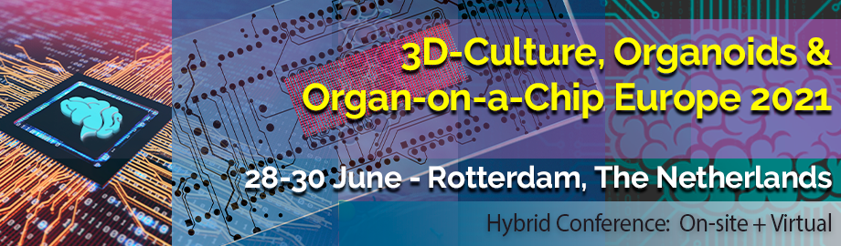
Co-Located Conference Agendas3D-Culture, Organoids & Organ-on-a-Chip Europe 2021 | Lab-on-a-Chip and Microfluidics Europe 2021 | Point-of-Care, Biosensors & Mobile Diagnostics Europe 2021 | 

Monday, 28 June 2021 |
Full Details of Day 1 Agenda -- Please Check the Lab-on-a-Chip Europe 2021 Agenda |
| |
Tuesday, 29 June 202108:00 | Morning Coffee, Pastries and Networking | 09:00 |  | Keynote Presentation Applications of Organ-on-a-Chip Systems in Precision Medicine
Peter Ertl, Professor of Lab-on-a-Chip Systems, Vienna University of Technology, Austria
Organs-on-a-chip systems contain three-dimensional human living cell cultures that are grown in a dynamic microenvironment under controlled conditions. These microphysiological systems allow biological, chemical and physical manipulation and analysis of minimal functional units of human organs and tissues. Consequently, organ-on-a-chip systems can be used to establish personalized disease models with the aim of providing clinical-relevant information from a patient’s own cells. In this presentation the current state and future of personalized medicine will discussed and two examples using primary cells and iPCS-derived organoids presented. |
| 09:30 |  | Keynote Presentation Organ-on-a-Chip Models for Cancer Research
Séverine Le Gac, Professor, Applied Microfluidics for Bioengineering Research, MESA+ Institute for Nanotechnology, University of Twente, Netherlands
Organ-on-a-chip (OoC) technology has become a game-changer by providing advanced in vitro models of human tissues and organs, allowing thereby conducting a variety of experimentation and assays, ranging from drug testing, toxicity screening, tissue engineering, to disease modeling to, e.g., elucidate mechanisms at play in disease onset and progression. Organ-on-a-chip devices are hybrid models combining cells and microfabricated structures in a microfluidic format, aiming altogether to mimic functional and/or structural features of an organ. OoCs exhibit a number of advantages compared to conventional in vitro and in vivo models: an in vivo–like and tunable microenvironment, dynamic culture, possibility to incorporate a variety of (bio)chemical and (bio)physical cues, with spatial and temporal control thereon, suitability to prepare patient-specific (disease) model, amenability to parallelized and automated studies, and compatibility with routine imaging and molecular assay. As such, OoC is currently acknowledged as a promising technology to reduce and replace experimentation on animals, which are poor mimics of human diseases and physiology. In my presentation, I will discuss recent work from our group in the field of cancer research: to develop tumor models including different features found in the tumor microenvironment in vivo; to evaluate the penetration of nanomedicines in multi-cellular tumor spheroids, in the presence of a flow or under the assistance of microbubbles and ultrasound activation; to assess the impact of (bio)mechanical cues on a tumor model; and to study cancer metastasis using a multi-organ-on-a-chip approach. |
| 10:00 |  | Keynote Presentation Blood Perfusion of Organs-on-Chips
Andries D. van der Meer, Associate Professor, Scientific lead, Organ-on-Chip Center Twente, University of Twente, Netherlands
Organs-on-chips are unique cell culture platforms in that they offer unparalleled control over fluid dynamics and flow. This dynamical nature also is precisely why it is very attractive to perfuse organs-on-chips with whole blood. Whole blood perfusion of organs-on-chips offers unique opportunities to broaden the impact of 3D cell culture models in biomedical and pharmacological science. Not only is blood perfusion interesting from a perspective of realistically modeling tissue physiology, but it also enables unique in vitro studies of aspects of disease that involve blood clotting, platelet activation and immune cell extravasation. In this talk, I will discuss the advantages of including blood as a routine component in organ-on-chip-based cell culture, and I will give examples of how blood-perfused organs-on-chips can be used to study aspects of COVID-19, drug toxicity and sepsis. |
| 10:30 | Morning Coffee Break and Networking | 11:00 | Modeling Human Fatty Liver Disease Using Stem Cell-derived Liver Tissue
David Hay, Principal Investigator/RCUK Fellow, MRC Centre for Regenerative Medicine, United Kingdom
Liver disease represents an increasing cause of global morbidity and mortality. Although liver transplant is a curative treatment for end stage liver disease, donor organs cannot meet the demand. Therefore, scalable treatments and new disease models are required to improve clinical intervention. A major contributor to liver disease worldwide is obesity. This has led to a surge in fatty liver disease. This term encompasses a spectrum of pathologies, ranging from benign hepatic steatosis to disease initiating non-alcoholic steatohepatitis. Although there are a number of in vitro approaches for the study of fatty liver disease, they do possess drawbacks. For example, cell line based models display perturbed metabolic function, arising from malignant transformation and immortalization. Alternatively, primary mouse and human hepatocytes can be used to model hepatic steatosis in vitro; however, those systems display unstable phenotype post isolation, limiting their application. Moreover, human hepatocytes are commonly isolated from transplant-rejected organs, often fatty livers, which may adversely affect model performance. In contrast to this, pluripotent stem cell derived systems represent a renewable source of human tissue to model disease and physiology. Most recently, we have used our human liver models to study hepatic steatosis, providing insight into the disease process. Those studies revealed that steatotic liver tissue displayed a compromised electron transport chain as well as a truncated TCA cycle. The steatotic hepatocytes rewired their metabolic circuitry to compensate for this, generating increased levels of fumarate through the purine nucleotide cycle, and to a lesser extent the malate-aspartate shuttle. We are currently developing those models to probe other aspects of fatty liver disease and our most recent studies will be presented at the meeting. | 11:30 | Microfabrication Technologies for Engineering a Joint on Chip
Marcel Karperien, Professor, University of Twente, Netherlands
Osteoarthritis is a degenerative joint disease affecting more than 300.000.000 patients world-wide. Despite tremendous efforts in the past, the etiology of the disease is still poorly understood and cannot be cured. While initially considered as a disease of cartilage it is now clear that the disease involves all tissues in the joint. Furthermore a disease trigger in each of these tissues can initiate the onset of disease. Despite the different origin they culminate over time in a similar disease presentation. It has also become clear that currently used animal models poorly reflect the complexity of human disease nor do allow the detailed study of the complex interactions between the different joint tissues at various stages of disease. To address these shortcomings my group has started the development of a joint-on-chip. The joint-on-chip has a modular chip design. We are engineering chips for each individual tissue, i.e. cartilage, subchondral bone, synovium, ligament and/or meniscus which together form the joint. The individual chips are connected to each other through blood vessel mimicking microfluidic channels as well as with a chip mimicking the intra-articular space. This latter chip will contain features allowing non-invasive imaging / sensing of inter tissue communication. Since movement is a critical and an essential feature in every joint, the individual chips can all be independently actuated mimicking both compression and shear stress. Prototype chips of the synovium and the cartilage including actuation have become available and we have shown that these devices can be used for studying critical biological processes in the healthy and diseased joint. Additionally we developed various strategies for introducing cell laden membranes composed of natural polymers and/or tissue constructs in the chip. Finally, we have developed SPRi based sensors for assessing the secretion of cytokines over time and dedicated sensors that can be used to locally assess matrix metalloproteinase activity, a key factor driving joint degeneration on chip. In my presentation I will discuss various engineering aspects of our microfabrication technology platforms needed for recreating a representative and functional human joint and show our efforts to functionally characterize these devices and will illustrate how we use these devices to further our understanding of a healthy and diseased human joint. | 12:00 | Networking Lunch, Meet Exhibitors and View Posters | 12:40 | Poster Presentation by Angeline Lim, Molecular Devices. Poster Title: "A streamlined approach for multiplexed phenotypic profiling based on the Cell Painting assay." Download Poster PDF by Clicking Here | 13:00 | 3D Cultures to Study Parkinson’s Disease
Silvia Bolognin, Research Scientist, University of Luxembourg, Luxembourg
Parkinson’s disease (PD) is the second most common neurodegenerative disease affecting the aging population. Despite tremendous research efforts in the course of more than a century, the cause of the disease is still unknown. Our lab focuses on the optimization of advanced human in vitro models to understand the molecular basis underlying PD. We have established the cultivation of PD patient-specific neurons, derived from induced pluripotent stem cells carrying the LRRK2-G2019S mutation, in 3D microfluidics. In contrast to conventional 2D cultures, this 3D approach reveals robust endophenotypes. High-content imaging data show decreased dopaminergic differentiation and branching complexity, altered mitochondrial morphology, and increased cell death in LRRK2-G2019S neurons compared to controls without using stressor agents. Pushing forward the optimization of a model recapitulating the human brain tissue, the cultivation of midbrain brain organoids was also established in microfluidic devices. PD organoids show fewer dopaminergic neurons compared to organoids derived from healthy subjects. These data support the use of 3D models to study complex cellular mechanisms underlying PD and allow for testing the effect of disease-modifying compounds using human-derived material. | 13:30 | Population Effect on Drug Metabolism and Absorption Using Ex Vivo Intestinal Tissue Explants in the Intestine Explant Barrier Chip
Joanne Donkers, Scientist, TNO, Netherlands
The Intestine Explant Barrier Chip integrates ex vivo intestinal tissue between two microchannels and enables drug permeability and metabolism studies under physiological conditions, including studying population effects in combination with microbiome supernatant. | 14:00 |  Automation of 3D Organoid Culture with Monitoring, Imaging and Analysis of Complex Biological Effects Automation of 3D Organoid Culture with Monitoring, Imaging and Analysis of Complex Biological Effects
Oksana Sirenko, Sr. Imaging Applications Scientist, Molecular Devices
There is an unmet need for cell-based model systems that better resemble human biology to improve pre-clinical selection of drug candidates. 3D cell models representing various tissues were successfully used for studying complex biological effects, tissue architecture, and functionality. While complexity of 3D models remains a hurdle for the wider adoption in research and drug screening, significant progress has been made in developing tools for automated cell culture, monitoring, and image analysis. We will describe an integrated cell culture and a high-content imaging process that allows automated culture, monitoring, maintenance, and characterization of growth and differentiation of organoids and stem cells, as well as testing the effects of various compounds. We will discuss the methods for automation of three complex workflows: maintenance and monitoring of iPSC cell culture, culturing and testing toxicity effects in lung organoids, and monitoring of the development of intestinal organoids. The automated integrated system included ImageXpress Confocal HT.ai High-Content Imaging System, (IXM-C HT.ai confocal imaging system), automated CO2 incubator (Wave STX44), a liquid handler (Biomek i7), as well as an automation robotic system and software. Described workflow demonstrates the useful tools for increased throughput and automation in organoid assays and compound screening, and analysis methods and descriptors for gaining more information from complex assays ?useful for studying disease phenotypes and compound effects.
| 14:30 | Tissue Organoids For Disease Modeling and Therapy Development
Shay Soker, Professor of Regenerative Medicine and Chief Science Program Officer, Wake Forest Institute for Regenerative Medicine, United States of America
3D human tissue organoids replicate native tissue structure and function and can be studied in vitro for several weeks to allow intensive investigations. Besides their advantages in drug toxicity testing and for development of new drugs, the human tissue organoid platform serves as a model system to explore human tissue development and disease. Our recent research was focused on the use of tissue organoids to study human development and congenital diseases as well as other common diseases such as tissue fibrosis and cancer. Once a disease model is established, patient-specific (personalized) therapies can be developed. | 15:00 | Afternoon Coffee Break and Networking | 15:30 |  Optical Sensors in Microfluidics Devices Optical Sensors in Microfluidics Devices
Jake Boy, Senior Application Scientist, Scientific Bioprocessing
Scientific Bioprocessing (SBI) develops leading-edge instruments that make the work of cell scientists easier and more reproducible with optical sensing. Here, we introduce SBI’s optical sensing technology and explain how it works. We will show you its use in applications using various vessel types including bioreactors and microfluidics applications with data using these optical sensors. SBI is changing the way cell scientists are able to optimize their bioprocessing applications and continues to introduce new and exciting technologies to enhance efficiency, reduce variability, and increase reproducibility with real-time monitoring.
| 16:00 | Assessing Contractility of 3D iPSC-derived Engineered Muscle Tissues for Safety and Drug Discovery Applications Using a High-throughput and Novel Label-free Method
Nicholas Geisse, Chief Science Officer, Curi Bio Inc., United States of America
Preclinical assays often fail to predict drug action, resulting in high clinical trial failure rates. Cellular models can screen for cardiotoxicity, but oftentimes utilize complex 3D systems that are challenging to deploy at sufficient scale. We designed, optimized, and validated a novel method to fabricate and measure the contractility of 24 3D Engineered Muscle Tissues (EMT) in parallel. Our approach allows for successful tissue casting rates >95% (n > 100) and produces consistently-sized constructs (confirmed across 6 laboratories). The consumable features embedded magnets. As tissues contract, the magnet’s displacement is quantitatively detected in a highly-parallel manner using specialized sensors. Displacement can be detected simultaneously across all 24 tissues with suitable acquisition rates for measuring contractility parameters such as upstroke velocity and decay time. Our platform can be used to detect changes in contractility, including those induced by structural cardiotoxicants like doxorubicin. EMTs treated with doxorubicin showed no differences over controls at acute (30 min) or chronic timepoints to Day 2 post exposure. The highest-dosed (1 µM) tissue slowed at Day 4 and Day 5 until complete cessation of twitching by Day 6. We also show phenotype stratification across EMTs from healthy and diseased individuals, demonstrating potential use in drug discovery studies. | 16:30 |  | Keynote Presentation Three Things Count for MPS and Organoids: Quality, Quality and Quality!
Thomas Hartung, Professor and Doerenkamp-Zbinden Chair for Evidence-based Toxicology, Director of the Center for Alternatives to Animal Testing (CAAT), Johns Hopkins Bloomberg School of Public Health, United States of America
The promise of the novel cell culture technologies (microphysiological systems, MPS, including organ-on-chip models ad organoids) replicating organ architecture and function is counterbalanced by the demanding technologies, efforts and costs, often limiting throughput and number of replicates. With increasing desire to use them also in the regulatory context, the quality of work comes into focus. The first quality concerns the fidelity of the model in replicating (patho-)physiology and relevant responses. This is typically addressed by validation, but the complexity of models and responses requires an adaptation of the validation process. The second quality refers to the quality of performance of the work. In the regulatory context, Good Laboratory Practice (GLP) and the associated Good In Vitro Method Practice (GIVIMP) still require adaptation to MPS. Good Cell Culture Practice (GCCP), which is more geared toward defining minimum standards also applicable in non-GLP environments, is undergoing already such adaptations and with stakeholder review on the way, GCCP 2.0 is expected to be finalized later in 2021. The third quality refers to the quality of reporting. Two initiatives, i.e., the In Vitro Critical Appraisal Tool (IV-CAT) and Good In Vitro Reporting Standards (GIVReSt) are on the way. Together with organizations like the US Food and Drug Administration (FDA) defining their performance standards, these quality assurance and control initiatives pave the way for relevant, reproducible science and maximum impact of MPS technologies. The 2022 MPS World Summit aims to foster these quality initiatives also with the planned creation of an International society. |
| 17:00 |  | Keynote Presentation Organ-specific Models of Barrier Function
Roger Kamm, Cecil and Ida Green Distinguished Professor of Biological and Mechanical Engineering, Massachusetts Institute of Technology (MIT), United States of America
Barrier function, the ability to selectively allow passage of molecules or nanoparticles based on their physicochemical properties, is critical in numerous physiological and pathological settings. Here we discuss two examples. The first is transport across the blood-brain barrier of therapeutic molecules or nanoparticles. Focusing on the exchange of proteins of varying size and functionalized nanoparticles for targeting cytotoxic agents to glioblastoma multiform the mechanisms and control of delivery will be discussed. The second model involves subcutaneous delivery of monoclonal antibodies and their bioavailability. Brain and skin models are all human-derived and utilize either primary cells or iPSC-derived cells. Studies are based on a common microfluidic platform in which vascular networks (both blood and lymphatic) are grown by self-assembly within a 3D matrix. Flows and pressures are controlled via simple onboard pumping systems capable of continuous circulation of a small volume of medium at physiological flow rates, with or without circulating immune cells. These systems are compatible with moderate throughput studies in a standard 96-well format. |
| 17:30 | Close of Day 2 of the Conference |
Wednesday, 30 June 2021 |
Full Details of Day 3 Agenda -- Please Check the Point-of-Care Diagnostics Europe 2021 Agenda |
| |
|


 Add to Calendar ▼2021-06-28 00:00:002021-06-30 00:00:00Europe/London3D-Culture, Organoids and Organ-on-a-Chip Europe 20213D-Culture, Organoids and Organ-on-a-Chip Europe 2021 in Rotterdam, The NetherlandsRotterdam, The NetherlandsSELECTBIOenquiries@selectbiosciences.com