Co-Located Conference AgendasLab-on-a-Chip and Microfluidics: Companies, Technologies and Commercialization | Lab-on-a-Chip and Microfluidics: Emerging Themes, Technologies and Applications | NGS, SCA, SMA & Mass Spec: Research to Diagnostics 2016 | Point-of-Care Diagnostics & Global Health World Congress 2016 | 

Monday, 26 September 201609:00-12:00 Microfluidics and Lab-on-a-Chip (LOAC) for Point-of-Care (POC) Diagnostics Applications: Technologies, Applications, Research Trends [Pre-Conference Training Course]
Presented by Dr. Holger Becker, microfluidic ChipShop GmbH.
Separate Registration Required for this Pre-Conference Training Course.
| 12:00-14:30 Luncheon Training Course: Basic Principles in Lab-on-a-Chip (LOAC) Technologies for the Study of Circulating Biomarkers: Applications in Liquid Biopsies [Pre-Conference Training Course]
Presented by Professor Steve Soper, Professor and Director, University of North Carolina-Chapel Hill.
Separate Registration Required for this Training Course.
| 14:30 | Conference Registration, Conference Materials Pick-Up and Networking | |
Session Title: Conference Plenary Session -- Convergence of Technologies in Microfluidics, Diagnostics and Single Cell Analysis |
| | 15:30 |  | Keynote Presentation The Personalized Health Care Environment: How in vitro Diagnostics, Mobile Health and Medical Devices will Converge to Improve Health Care
Alan Wright, Chief Medical Officer, Roche Diagnostics Corporation, United States of America
|
| 16:00 | 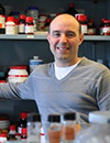 | Keynote Presentation Digital Microfluidics: A Platform Whose Time Has Come
Aaron Wheeler, Canada Research Chair of Bioanalytical Chemistry, University of Toronto, Canada
Digital microfluidics is a fluid-handling technique in which droplets are manipulated by electrostatic forces on an array of electrodes coated with a hydrophobic insulator. In this talk, I will present recent results from my group’s work with digital microfluidics, with an emphasis on the topics covered in this unique venue. Specifically, I will demonstrate how digital microfluidics is particularly well-suited for Lab on a Chip applications, given its ability to automate diverse laboratory processes on a generic, programmable platform. Likewise, I will report on our work using digital microfluidics for Point of Care Diagnostics and Global Health, reporting on the results of a field trial for measles and rubella diagnostics at refugee sites in Kenya. Finally, I will describe how digital microfluidics is emerging as a useful tool for Single-Cell Analysis and for integration with Mass Spectrometry to answer questions about cell heterogeneity and cell-cell communication. Through these examples, I will make the case that digital microfluidics is emerging as a useful new tool for the next generation of analytical techniques, across a wide range of applications. |
| 16:30 |  | Keynote Presentation Microfluidics and Sensors: New Tools for Real-Time Clinical Monitoring
Martyn Boutelle, Professor of Biomedical Sensors Engineering, Imperial College London, United Kingdom
A goal for modern medicine is to protect vulnerable tissue by monitoring the patterns of changing physical, electrical and chemical changes taking place in tissue - ‘multimodal monitoring’. Clinicians hope such information will allows treatments to be guided and ultimately controlled based on the measured signals. Microfluidic lab-on-chip devices coupled to tissue sampling using microdialysis provide an important new way for measuring real-time chemical changes as the low volume flow rates of microdialysis probes are ideally matched to the length scales of microfluidic devices. Concentrations of key biomarker molecules can then be determined continuously using either optically or electrochemically (using amperometric, and potentiometic sensors). Wireless devices allow analysis to take place close to the patient. Droplet-based microfluidics, by digitizing the dialysis stream into discrete low volume samples, both minimizes dispersion allowing very rapid concentration changes to be measured, and allows rapid transport of samples between patient and analysis chip. This talk will overview successful design, optimization, automatic-calibration and use of both continuous flow and droplet-based microfluidic analysis systems for real-time clinical monitoring, using clinical examples from our recent work. |
| 17:00 |  | Keynote Presentation High-Performance Rapid Diagnostic Tests
Bernhard Weigl, Director, Center for In-Vitro Diagnostics, Intellectual Ventures/Global Good-Bill Gates Venture Fund, United States of America
Lateral flow and similar rapid diagnostic assays (LFAs) are easy to use and manufacture, low cost, rapid, require little or no equipment to operate, and do not need to be refrigerated. However, they are generally not considered to be very sensitive or able to provide a quantitative result. Our group believes that this lack of sensitivity is not a fundamental property of LFAs but rather a consequence of the way they are developed, manufactured, and marketed. Historically, most lateral flow tests were developed and optimized by relatively small manufacturers with limited R&D capabilities and budgets, and were generally used only for analytical targets prevalent at high concentration in patient’s samples that were relatively easy to measure. In contrast, our group’s mission is to develop LFA-based assays for use in global health applications that are as sensitive as the best conventional diagnostic assays (in some cases even better) while retaining all their cost, simplicity, and usability advantages. |
| 17:30 | 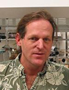 | Keynote Presentation Technologies for Personalizing Cancer Immunotherapies
James Heath, Elizabeth W. Gilloon Professor of Chemistry, California Institute of Technology (CalTech), United States of America
Cancer immunotherapy, which has taken virtually all aspects of oncology by storm over the past few years, is based upon using cellular or molecular therapies to promote tumor cell/immune cell interactions. At the heart of this therapy are the T cells that actually do the tumor cell killing, and the tumor antigens that are recognized by those T cells. Recent work has shown that neoantigens play critical roles in many immunotherapy successes. Neoantigens are tumor antigens that are fragments of mutated proteins expressed by the cancer cells, and contain those point mutations. They are presented in the clefts of major histocompatibility molecules (MHCs) by many of the cells in the tumor, where they may be recognized by neoantigen-specific T cell populations. In this presentation, I will discuss how various micro and nanotechnologies are being harnessed to identify, for a given patient which neoantigens are actively recruiting T cells into the tumor, and to carry out a deep molecular analysis of those neoantigen-specific T cells. I will further discuss how that information can then be harnessed for personalized cancer immunotherapies in the form of neoantigen-based vaccines, or engineered T cell receptor adoptive cell transfer therapies.
|
| 18:00 |  | Keynote Presentation Wearable Eccrine Sweat Biosensing: Uncovering The Real Challenges That Lie Ahead
Jason Heikenfeld, Professor and VP Operations, UC Office of Innovation, University of Cincinnati, United States of America
Despite the many ergonomic advantages of eccrine perspiration (sweat) compared to other biofluids (particularly in “wearable” devices), sweat remains an underrepresented source of biomarker analytes compared to the established biofluids blood, urine, and saliva. Upon closer comparison to other non-invasive biofluids, the advantages may even extend beyond ergonomics: sweat might provide superior analyte information. A number of challenges, however, have historically kept sweat from its place in the pantheon of clinical samples. These challenges include very low sample volumes (nL to µL), unknown concentration due to evaporation, filtration and dilution of large analytes, mixing of old and new sweat, and the potential for contamination from the skin surface. More recently, rapid progress in “wearable” sweat sampling and sensing devices has resolved several of the historical challenges. However, this recent progress has also been limited to high concentration analytes (µM to mM) sampled at high sweat rates (>1 nL/min/gland, e.g. athletics). Progress will be much more challenging as sweat biosensing moves towards use with sedentary users (low sweat rates or not sweating at all) and/or towards low concentration analytes (pM to nM). Fortunately, none of the remaining challenges appear to be fundamentally blocking, and scientific and engineering innovations have the opportunity to enable broader application of sweat biosensing technology. |
| 18:30 |  | Keynote Presentation The Challenge in Building Phenotype Body-on-a-Chip Models for Toxicological and Efficacy Evaluations in Drug Discovery as well as Precision Medicine
James Hickman, Professor, Nanoscience Technology, Chemistry, Biomolecular Science and Electrical Engineering, University of Central Florida; Chief Scientist, Hesperos, United States of America
The utilization of human-on-a-chip or body-on-a-chip systems for toxicology and efficacy that ultimately should lead to personalized, precision medicine has been a topic that has received much attention recently. Key characteristic needed for these systems are the ability for organ-to-organ communication in a serum-free recirculating medium and incorporation of induced pluripotent stem cells that allow for understanding genetic variation as well as to construct systems utilizing stem cells from diseased patients and also from individuals. Additional characteristics that have been discussed are functional readouts that would enable non-invasive monitoring of organ health and viability for chronic studies that now are only possible in animals or humans at this time. In addition, in order to achieve wide spread adoption of these technologies they should also be low cost, easy to use and reconfigurable to allow flexibility for platforms to be examined with small variation. Our group, in collaboration with Dr. Michael Shuler from Cornell University, has been constructing these systems with up to 6 organs and have demonstrated long-term (>28 days) evaluation of drugs and compounds, that have shown similar response to results seen from clinical data or reports in the literature. We have accomplished the construction of these systems utilizing mostly 2D systems in serum-free medium with functional readouts that employs a pumpless platform that enables ease of use of these assays. Our group’s ability to control the interface between the biological and non-biological components in these systems has enabled the straightforward integration of multiple cell types in the same platform. Results with the functional multi-organ systems will be presented as well as results of five workshops held at NIH to explore what is needed for validation and qualification of these systems by the FDA and EMA. |
| 19:00 | Conference Opening Reception with Beer, Wine and a Light Dinner Sponsored by Veryst Engineering, LLC | 20:00-22:00 Dinner Training Course: Microfluidics for 3D-Printing and Biofabrication: Technologies and Applications [Pre-Conference Training Course]
Presented by Professor Albert Folch, University of Washington.
Separate Registration Required for this Training Course.
|
Tuesday, 27 September 201607:45 | Conference Registration, Conference Materials Pick-Up, Morning Coffee and Breakfast Pastries in the Exhibit Hall | |
Session Title: The Translation of Next Generation Sequencing from Research to Clinical Deployment |
| | 09:00 | Opportunities and Challenges of Implementing Next Generation Sequencing (NGS) for Delivery of Genomic Medicine
Nazneen Aziz, Executive Director, Kaiser Permanente Research Bank, United States of America
Many important advances in technologies for molecular analyses have fostered the rapid growth in molecular medicine. The use of these technologies in testing of DNA, RNA, proteins for screening, diagnosing and prognosis of patient’s disease, and also in the development of new biologics and small molecule drugs have given rise to a science that 30 years ago was practically non-existent. Each of these technologies has led to significant advances, but one in particular, can be undoubtedly labeled as ‘disruptive’. Next generation sequencing (NGS), introduced in 2005, has revolutionized the field by increasing the speed at which the genome can be sequenced at an exponentially lower cost. Within 5 years of its introduction and widespread use in research, NGS is transforming molecular medicine. NGS has a higher throughput and lower cost per base and therefore has been rapidly adopted into clinical testing. What is particularly intriguing about NGS’s rapid adoption into clinical testing is that it has a number of intricacies associated with its implementation that is unfamiliar to clinical laboratory or to healthcare professionals. Importantly, the implementation of genomic analysis into routine clinical practice will take a dedicated and collaborative effort between physicians, scientists, and all healthcare professionals. There are knowledge gaps in understanding when and what tests to order, how tests are validated and made ready for clinical use and how to interpret test reports. Additionally, there is lack of data sharing to eradicate the limited understanding of genomic variants and their involvement in health and disease. Dr. Aziz will address the opportunities, complexities and challenges associated within our healthcare system and institutions, for implementing genomics into clinical practice. | 09:30 | The Challenges in Clinical Implementation of NGS Tests
Erick Lin, Director of Medical Affairs, Ambry Genetics, United States of America
The adoption and clinical implementation of NGS tests has become a benchmark in advancing precision medicine, but the realities of adopting it and obtaining reimbursement for tests presently being offered are difficult. This presentation will focus on Ambry’s experience in adopting and offering NGS tests to its clients, including the need to help clients navigate the complexities of interpreting and understanding the tremendous amounts of data generated by NGS tests. | 10:00 | Next Generation Sequencing for Clinical Applications: Challenges and Current Trends in Developing Clinical Tests
Martin Siaw, Vice President of Science and Innovation, BasePrime Genomics, United States of America
Next Generation Sequencing has evolved very rapidly and is now used for molecular testing in clinical laboratories. This powerful technology offers many exciting opportunities for use in clinical testing. However, NGS also presents many challenges, including the differences between research and clinical labs, and the rapid evolution of platforms, protocols, and reagents. There is a need to recognize the challenges and current trends that are specific to CLIA lab environments when developing and validating clinical NGS tests. Some specific clinical applications will be presented.
| 10:30 | Coffee Break and Networking in the Exhibit Hall: Visit Exhibitors and Poster Viewing | 11:15 |  | Keynote Presentation Genomics in Clinical Practice: Patient Stories from the Frontline
Jennifer Friedman, Clinical Professor of Neurosciences and Pediatrics, Rady Children's Hospital in San Diego, United States of America
Advances in genome sequencing hold tremendous promise for providing answers, tailored therapies and in some case cures for undiagnosed patients. However, how to interpret and act upon volumes of complex genomic data remains a challenge for sequencing providers, physicians and their patients and families. Uncertain and non-validated results present obstacles in attaining goals of diagnosis and cure. Off-target results may create unforeseen medical and ethical challenges. This presentation will use case-based examples to demonstrate promises and pitfalls encountered in application of genomic sequencing to diagnosis of patients with rare disease. |
| 11:45 | NGS to Assess Low Input exRNA Samples
Kendall Van Keuren-Jensen, Professor and Deputy Director, Translational Genomics Research Institute, United States of America
It is challenging to work with and achieve low-bias comprehensive profiles with low input RNA samples. We will describe various kits and data analysis strategies. | 12:15 | Networking Lunch in the Exhibit Hall: Visit Exhibitors and Poster Viewing | |
Session Title: Study of Circulating Tumor Cells (CTCs) and Vesicles as Paradigms for Single Cell Analysis (SCA) |
| | 14:00 | Enabling Microfluidics and Nanosensing Technologies for Circulating Biomarker Analysis
Yong Zeng, Associate Professor, University of Florida, United States of America
Developing circulating biomarkers and blood-based tests is extremely appealing for non-invasive cancer diagnosis and monitoring of therapy response where tissue biopsy is highly invasive and costly. However, in many case circulating biomarkers (e.g., ctDNA, proteins and exosomes) are present at very low concentration levels. In comparison with nucleic acids that can be amplified, quantitative detection of low-level proteins and extracellular vesicles remains very challenging. We have been developing new nanotechnology and microfluidics-based sensing platforms for ultrasensitive analysis of circulating biomarkers with broad dynamic range. These technologies substantially enhances the analytical performance, while reducing the sample demand and analysis time. We will demonstrate the applications of these microfluidic platforms to quantitative analysis of protein biomarkers and tumor-derived exosomes in circulation as a means for liquid biopsy based non-invasive diagnosis of cancer diagnostics. Overall, these microfluidic systems would provide enabling bioanalytical technologies to promote quantitative measurement of complex biological systems and clinical disease diagnosis. | 14:30 | Interrogation of Single Circulating Tumor Cells using Microchannel Enrichment
Lyle Arnold, Chief Scientific Officer, Biocept, United States of America
Liquid biopsies offer the opportunity to interrogate a number of different target sample types, including CTCs. At Biocept both ctDNA and CTCs are used for identifying medically actionable biomarkers. In this presentation the use of a unique patented microchannel will be high-lighted that enables the capture, enrichment, and analysis of CTCs at the single cell level. This allows for the direct visual analysis of CTCs, and has enabled the clinical validation of an array of biomarkers using FISH and protein analysis methods for HER2, FGFR1, MET, ALK, ROS1, ER, PR, AR, and PDL-1 across a range of cancer types. In addition, CTCs can be isolated from the microchannel and interrogated using other means, including NGS. | 15:00 | Next-Generation Liquid Biopsy
Eric Kaldjian, Chief Medical Officer, RareCyte, United States of America
To fully exploit the non-invasive potential of liquid biopsy, rare cells must be collected and identified without size or protein expression bias and be available for assessment of pertinent phenotypic and molecular characteristics. This presentation will describe pre-clinical and clinical applications of the recently commercialized AccuCyte-CyteFinder system, an integrated platform technology for collection, up to six-parameter visualization, and single-cell retrieval for genomic analysis of circulating tumor and fetal cells. | 15:30 | Coffee Break and Networking in the Exhibit Hall: Visit Exhibitors and Poster Viewing | |
STRATEC Consumables Symposium on Innovations in Microfluidics and Lab-on-a-Chip and their Impact on Life Sciences and Diagnostics | Session Sponsors |
| | 16:00 | Introduction to the STRATEC Consumables Symposium and Topics Addressed in 2016 | 16:15 | 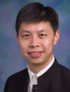 | Keynote Presentation Microfluidic Printing: From Combinatorial Drug Screening to Artificial Cell Assaying
Tingrui Pan, Professor, Department of Biomedical Engineering, University of California-Davis, United States of America
Microfluidic impact printing has been recently introduced, benefiting from the nature of simple device architecture, low cost, non-contamination, scalable multiplexability and high throughput. In this talk, we will review this novel microfluidic-based droplet generation platform, utilizing modular microfluidic cartridges and expandable combinatorial printing capacity controlled by plug-and-play multiplexed actuators. Such a customizable microfluidic printing system allows for ultrafine control of the droplet volume from picoliters (~10pL) to nanoliters (~100nL), a 10,000 fold variation. The high flexibility of droplet manipulations can be simply achieved by controlling the magnitude of actuation (e.g., driving voltage) and the waveform shape of actuation pulses, in addition to nozzle size restrictions. Detailed printing characterizations on these parameters have been conducted consecutively. A multichannel impact printing system has been prototyped and demonstrated to provide the functions of single-droplet jetting and droplet multiplexing as well as concentration gradient generation. Moreover, several enabling chemical and biological assays have been implemented and validated on this highly automated and flexible printing platform. In brief, the microfluidic impact printing system could be of potential value to establishing multiplexed droplet assays for high-throughput life science researchers.
|
| 16:35 |  | Keynote Presentation Nanopore Sequencing for Real-Time Pathogen Identification
Kamlesh Patel, R&D Advanced System Engineering and Deployment Manager, Sandia National Laboratories, United States of America
As recent outbreaks have shown, effective global health response to
emergent infectious disease requires a rapidly deployable, universal
diagnostic capability. We will present our ongoing work to develop a
fieldable device for universal bacterial pathogen characterization based
on nanopore DNA sequencing. Our approach leverages synthetic
biofunctionalized nanopore structures to sense each nucleotide. We aim
to create a man-portable platform by combining nanopore sequencing with
advance microfluidic-based sample preparation methods for an
amplification-free, universal sample prep to accomplish multiplexed,
broad-spectrum pathogen and gene identification. |
| 16:55 |  | Keynote Presentation Polymer-based Nanosensors using Flight-Time Identification of Mononucleotides for Single-Molecule Sequencing
Steve Soper, Foundation Distinguished Professor, Director, Center of BioModular Multi-Scale System for Precision Medicine, The University of Kansas, United States of America
We are generating a single-molecule DNA sequencing platform that can
acquire sequencing information with high accuracy. The technology
employs high density arrays of nanosensors that read the identity of
individual mononucleotides from their characteristic flight-time through
a 2-dimensional (2D) nanochannel (~20 nm in width and depth; >100 µm
in length) fabricated in a thermoplastic via nano-imprinting (NIL). The
mononucleotides are generated from an intact DNA fragment using a
highly processive exonuclease, which is covalently anchored to a plastic
solid support contained within a bioreactor that sequentially feeds
mononucleotides into the 2D nanochannel. The identity of the
mononucleotides is deduced from a molecular-dependent flight-time
through the 2D nanochannel. The flight time is read in a label-less
fashion by measuring current transients induced a single mononucleotide
when it travels through a constriction with molecular dimensions (<10
nm in diameter) that are poised at the input/output ends of the flight
tube. In this presentation, our efforts on building these polymer
nanosensors using NIL in thermoplastics will be discussed and the
detection of single molecules using electrical transduction with their
identity deduced from the associated flight time provided. Finally,
information on the manipulation of single DNA molecules using
nanofluidic circuits will be discussed that takes advantage of forming
unique nano-scale features to shape electric fields for DNA manipulation
and serves as the functional basis of the nanosensing platform. |
| 17:15 | 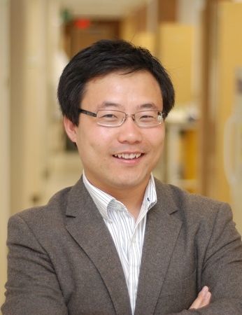 | Keynote Presentation Rapid and Ultra-sensitive Diagnostics Using Digital Detection
Weian Zhao, Associate Professor, Department of Biomedical Engineering, University of California-Irvine, United States of America
We will present our most recent droplet based digital detection
platforms for rapid and sensitive detections, which could find potential
applications at the point-of-care (POC). |
| 17:35 | 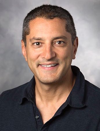 | Keynote Presentation Fractionation and Analysis of Nuclear versus Cytoplasmic Nucleic Acids from Single Cells
Juan Santiago, Charles Lee Powell Foundation Professor, Stanford University, United States of America
Single cell analyses (SCA) have become powerful tools in the study heterogeneous cell populations such as tumors and developing embryos. However, fractionating and analyzing nuclear versus cytoplasmic fractions of nucleic acids remains a challenge as these fractions easily cross-contaminate. We present a novel microfluidic system that can fractionate and deliver nucleic acid (NA) fractions from the nucleus (nNA) versus the cytoplasm (cNA) from single cells to independent downstream analyses. Our technique leverages a selective electrical lysis which disrupts the cell’s (outer) cytoplasmic membrane, while leaving the nucleus relatively intact. We selectively extract, purify, and preconcentrate cNA using isotachophoresis (ITP). The ITP-focused cNA and nNA-containing nucleus are separated by ITP and fractionated at a bifurcation downstream and then extracted for off chip analyses. We will present example applications of this fractionation including qPCR and next generation sequencing (NGS) analyses of cNA vs. nNA. This will include preliminary NGS analyses of nuclear vs. cytoplasmic RNA fractions to analyze gene expression and splicing. We hypothesize that the robust and precise nature of our electric field control is amenable to further automation to increase throughput while removing manuals steps. |
| 17:55 |  | Keynote Presentation Chip-Scale Microfluidic Physiological Circulation Systems
Abraham Lee, Chancellor’s Professor, Biomedical Engineering & Director, Center for Advanced Design & Manufacturing of Integrated Microfluidics, University of California-Irvine, United States of America
There has been a recent surge in the development of microphysiological systems and organ-on-a-chip for drug screening and regenerative medicine. Over the years, drug screening has mostly been carried out on 2D monolayers in well plates and the drugs are not delivered through blood vessels as in vivo treatments. Through the advancement of microfluidics technologies, we have enabled the automation of biological fluids delivery through physiological vasculature networks that mimic the physiological circulation of the human body. The critical bottleneck is to engineer the microenvironment for the formation of vascularized 3D tissues and to also pump and perfuse the tissue vascular network for on-chip microcirculation. This in vitro model system can be used to screen cancer drugs by mimicking the delivery of the drugs through capillary blood vessels. On the other hand, microfluidics play an important role in the recent advances in liquid biopsy and the ability to specifically isolate and capture rare cells such as circulating tumor cells. These two technologies may go hand-in-hand to connect in vitro screening to in vivo screening with great potential in the development of personalized medicine.
|
| 18:15 | Close of STRATEC Consumables Symposium | 18:30 | Cocktail Reception for All Conference Attendees: Enjoy Beer, Wine, Appetizers and Network with Fellow Delegates, Speakers, Exhibitors in the Exhibit Hall and View Posters Sponsored by STRATEC Consumables GmbH | 20:00 | Close of Day 2 of the Conference. Continue Networking in Downtown San Diego (Trolleys to the City are Available Right Behind the Conference Venue). |
Wednesday, 28 September 201607:00 | Morning Coffee, Breakfast Pastries and Networking in the Exhibit Hall | |
Session Title: Emerging Themes and Trends in Single Cell Analysis (SCA) and Single Molecule Analysis (SMA) |
| | 08:00 |  Technology Spotlight: Technology Spotlight:
Automated, Sensitive Microfluidic Device for CTC Capturing and Characterization
Grant Howes, Vice President, Commercial Operations , Celsee Diagnostics
The Celsee Diagnostics’ novel microfluidic technology captures and characterizes CTCs from metastatic cancer patients’ whole blood samples based on size and deformability. This presentation will overview the technology, demonstrate the 85% capture efficiency and discuss the use of the Celsee PREP100 to enrich and retrieve CTCs for downstream applications. The Celsee PREP100 device uses a microfluidic chip, which has approximately 56,400 individual cell capturing wells. Each well ensures that smaller blood cells, such as red blood cells and most leukocytes, escape while larger CTCs are captured. Downstream analysis of the captured CTCs using immuno-staining, FISH assays, RTPCR and NGS will be highlighted.
| 08:30 |  | Keynote Presentation Imaging of Cell–Cell Communication in a Vertical Orientation Reveals High-Resolution Structure of Immunological Synapse
Lidong Qin, Professor and CPRIT Scholar, Houston Methodist Research Institute, United States of America
The immunological synapse (IS) is one of the most pivotal communication strategies in immune cells. Understanding the molecular basis of the IS provides critical information regarding how immune cells mount an effective immune response. Fluorescence microscopy provides a fundamental tool to study the IS. However, current imaging techniques for studying the IS cannot sufficiently achieve high resolution in real cell–cell conjugates. In this study, we present a new device that allows for high-resolution imaging of the IS with conventional confocal microscopy in a high-throughput manner. Combining micropits and single-cell trap arrays, we have developed a new microfluidic platform that allows visualization of the IS in vertically “stacked” cells. Using this vertical cell pairing (VCP) system, we investigated the dynamics of the inhibitory synapse mediated by an inhibitory receptor, programed death protein-1, and the cytotoxic synapse at the single-cell level. In addition to the technique innovation, we have demonstrated novel biological findings by this VCP device, including novel distribution of F-actin and cytolytic granules at the IS, programed death protein-1 microclusters at the NK IS, and kinetics of cytotoxicity. We propose that this high-throughput, cost-effective, easy-to-use VCP system, along with conventional imaging techniques, can be used to address a number of significant biological questions in a variety of disciplines. |
| 09:00 | Microfluidic Single Cell Gene Expression Analysis Platform For Deep Profiling of Complex Cell Populations
Rajiv Bharadwaj, Director, Microfluidics, 10x Genomics, United States of America
High throughput single cell transcriptomics is the key to uncovering the true biological complexity of heterogeneous populations. The existing single cell RNA-sequencing methods have low throughput and complicated workflows that limit the widespread adoption of single cell mRNA analyses. 10X Genomics has developed a fully integrated and scalable microfluidic platform for transcriptional profiling of 1,000s to 10,000s of individual cells. | 09:30 |  Technology Spotlight: Technology Spotlight:
Advances in Microscopy-Based Single Cell Isolation
Chris Wetzel, Director of Sales and Marketing, MMI Microscope-based Single Cell Isolation
The isolation of single cells can often present challenges depending on the properties of the cells and the surrounding medium. This presentation deals with state-of-the-art techniques how to isolate cells from tissue (Laser Micro Dissection) and liquid suspensions (automated Micro Capillary) including live cells and adherent cells.
| 10:00 |  | Keynote Presentation Total Internal Reflection Fluorescence for Single Cell Analysis at the Level of Single Molecules
Alexander Asanov, Chief Scientific Officer, TIRF Labs, Inc., United States of America
Total Internal Reflection Fluorescence (TIRF) has become a method of choice for single molecule detection and is establishing itself as a powerful tool for other areas of lifesciences, including membrane biophysics, real-time microarrays, and molecular diagnostics. Here, we report on a novel application of TIRF for single cell analysis. We combined TIRF with dielectrophoresis and real-time microarrays to perform lysis or electroporation of cells directly at the TIRF surface. Real-time microarrays printed at the TIRF surface allow for the multiplexed analysis of protein and nucleic acid markers obtained from single cells with the limit of detection down to single molecules. In this presentation, we will also describe the requirements for single molecule detection, and will compare prism-, lightguide-, and objective-based TIRF geometries for single molecule studies. This presentation will discuss the advantages and limitations of different TIRF geometries for single cell analysis, ion channel and dynamics of lipid rafts studies, and molecular diagnostics applications. |
| 10:30 | Coffee Break and Networking in the Exhibit Hall: Visit Exhibitors and Poster Viewing | 11:15 |  | Keynote Presentation Enabling Biology with New NGS Applications
Gary Schroth, Vice President and Distinguished Scientist, Illumina, United States of America
The field of NGS is exploding with new applications that help researchers better understand the biology of complex diseases and cancer. In this talk we will discuss some of our recent work related to single cell biology and genomic alterations in cancer. |
| 11:45 | Single-cell Transcriptome and Epigenomic Reprogramming of Adult Cardiomyocyte-dedifferentiated Cardiac Progenitor Cells
Charles Wang, Director & Professor, Loma Linda University, United States of America
It has been believed that mammalian adult cardiomyocytes (ACMs) are terminally differentiated and are unable to proliferate. Recently, using a bi-transgenic mouse model and an in vitro culture system, we demonstrated that adult mouse cardiomyoctyes were able to dedifferentiate into cardiac progenitor-like cells (CPCs). In addition, implantation of CPCs into infarcted mouse myocardium improves cardiac function with augmented left ventricular ejection fraction. However, little is known about the molecular basis of their intrinsic cellular plasticity. Here we integrate whole-genome single-cell transcriptome and comprehensive high-throughput arrays for relative methylation (CHARM)-based DNA methylation analyses to unravel the molecular mechanisms underlying the focused, spontaneous dedifferentiation of mouse ACMs which gave rise to CPCs.
| 12:15 | Networking Lunch in the Exhibit Hall: Visit Exhibitors and Poster Viewing | |
Session Title: The Convergence of Biomarker Classes -- Single Cells, Proteins and Nucleic Acid Analytes |
| | 13:30 | Extracellular Vesicles: Mediators of Intercellular Signaling and Potential Biomarkers
John Nolan, CEO, Cellarcus Biosciences, Inc., United States of America
Extracellular vesicles (EVs) are attracting attention as potential biomarkers because they contain molecular cargo from their cell of origin that might reveal information about health and disease. However, EVs are hard to measure because they are small and heterogeneous. Flow cytometry, which can resolve heterogeneity in populations of cells, struggles to measures small, dim EVs. Recent developments in flow cytometry instrumentation and assay design enable us to quantitatively detect, size, and analyze the cargo of individual EVs and to catalogue their diversity. This approach allows EVs to be measured directly in diluted biofluids without the need for wash steps. I will report on the numbers and molecular diversity of EVs in plasma, CSF, and urine, and the variations observed in disease models and patient samples. | 14:00 | Label-Free Antibody-Free Technologies for Isolating Circulating Tumor Cells, Circulating Tumor Emboli and Exosomes from Blood and Bodily Fluids
Utkan Demirci, Professor, Stanford University School of Medicine, United States of America
Naside Gozde Durmus, Research Scientist, Stanford University, United States of America
| 14:30 |  | Keynote Presentation Metabolomics: Beyond Biomarkers
Gary Siuzdak, Professor and Director, The Scripps Research Institute, United States of America
Metabolomics, the quantitative global analysis of endogenous metabolites from cells, tissues, fluids and whole organisms, is now an integral part of functional genomics efforts as well as a tool for understanding fundamental biochemistry. While the genome and proteome represent upstream biochemical events, metabolites correlate with the most downstream biochemistry and therefore most closely represent the phenotype. This has been proven by the broad success of metabolite analysis in clinical diagnostics and understanding mechanisms of diseases. The aim in our studies is to obtain a comprehensive quantitative view of the metabolome to expand our understanding of what pathways are altered in specific diseases. We have developed a novel mass spectrometry platform for metabolomics that is used by thousands of labs, namely XCMS Online data analysis combined with the METLIN comprehensive MS/MS metabolite database. As well as nanostructure-initiator mass spectrometry (NIMS) imaging. These technologies will be presented in the context of their application to discovering new disease therapies/pathways for chronic pain, cancer, and healthy aging. |
| 15:00 |  | Keynote Presentation Circulating Tumor Cells as a Paradigm for Single Cell Analysis in Solid Tumor Malignancies
Minetta Liu, Associate Professor and Chair, Oncology Research, Mayo Clinic, United States of America
|
| 15:30 | Coffee Break and Networking in the Exhibit Hall: Visit Exhibitors and Poster Viewing | 16:00 | Single Cell Technologies for Studies of Single Cell Secretions and Drug Response
Yu-Hwa Lo, Professor, University of California San Diego, United States of America
To understand the in-homogeneity of cells in biological systems, we need the capabilities to characterize the properties of individual single cells. Since single cell studies require continuous monitoring of the cell behaviors instead of a snapshot test at a single time point, an effective single-cell assay that can support time lapsed studies in a high throughput manner is needed. Most currently available single-cell technologies cannot provide proper environments to sustain growth and, for appropriate cell types, proliferation of single cells and convenient, non-invasive tests of single cell behaviors from molecular markers. Here we present a highly versatile single-cell assay that can accommodate different cell types, enable easy and efficient single cell loading and culturing, support varieties of single cell studies against drugs and environment changes such as changes in pH value. One salient feature of the assay is the non-invasive collection of single cell secretions at different time points, producing unprecedented insight of single cell behaviors from the biomarkers each single cell gives out under certain perturbations. The acquired information is quantitative, for example, measured by the number of exosomes each single cell secretes within a given time period. Subsequently, those chosen single cells of particular interest such as cancer stem cells can be collected separately in a uniquely developed parallel process without disturbing the cell properties for further analysis including single cell RNASeq and molecular analyses. | 16:30 | Single Cell Analysis (SCA) in the Central Nervous System (CNS)
Kun Zhang, Professor, Department of Bioengineering, University of California San Diego, United States of America
| 17:00 | Close of Day 3 of the Conference. |
|


 Add to Calendar ▼2016-09-26 00:00:002016-09-28 00:00:00Europe/LondonNGS, SCA, SMA and Mass Spec: Research to Diagnostics 2016NGS, SCA, SMA and Mass Spec: Research to Diagnostics 2016 in San Diego, California, USASan Diego, California, USASELECTBIOenquiries@selectbiosciences.com