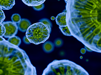
Co-Located Conference Agendas3D-Bioprinting "Track B" | 3D-Culture & Organoids | Organ-on-a-Chip and Body-on-a-Chip: In Vitro Systems Mimicking In Vivo Functions "Track A" | Stem Cells in Drug Discovery & Toxicity Screening 2018 | 

Thursday, 4 October 201807:30 | Conference Registration, Materials Pick-Up, Morning Coffee, and Breakfast Pastries | |
Session Title: Conference Opening Plenary Session |
| | |
Plenary Session Chair: Linda Griffith, Ph.D., Professor, Massachusetts Institute of Technology (MIT) |
| | 08:30 |  | Keynote Presentation Microphysiological Models for Metastatic Cancer
Roger Kamm, Cecil and Ida Green Distinguished Professor of Biological and Mechanical Engineering, Massachusetts Institute of Technology (MIT), United States of America
Circulating tumor cells form metastases by reaching a distant microcirculation, undergoing transendothelial migration, entering the remote tissue and proliferating. Microfluidic assays have been developed to visualize and quantify this process within vascular networks that recapitulate aspects of the in vivo microcirculation. Tumor cells, with or without accompanying immune cells, are streamed into a vascular network grown in a 3D matrix, some fraction of which arrest and extravasate into the surrounding matrix. These studies provide detailed information on the ability of different tumor cell types to extravasate, the adhesion molecules they use, and the effects of various other cell types in the intravascular and extravascular spaces. While these models are largely organ-independent, work has also begun to investigate the specificity of certain cancers to metastasize to organs such as the brain. For this purpose, a model of the blood-brain barrier has been produced, characterized in terms of its morphology and vascular permeability, and then used it to explore extravasation and tumor formation with the brain as the target organ.
|
| 09:00 |  | Keynote Presentation Intestine on a Chip for Basic Biology and Patient-Specific Medicine
Nancy Allbritton, Kenan Professor of Chemistry and Biomedical Engineering and Chair of the Joint Department of Biomedical Engineering, University of North Carolina and North Carolina State University, United States of America
Technical advances are making it possible to create tissue
microenvironments on platforms that are compatible with high-content
screening strategies. We have developed microfabricated devices to
enable culture of organized cellular structures which possess much of
the complexity and function of intact intestinal tissue. Stem-cell
culture enables single stem cells or intestinal crypts isolated from
primary small or large intestine from humans or mice to grow and persist
indefinitely as organotypic structures containing all of the expected
lineages of the intestinal epithelium. Our microengineered arrays and
fluidic devices build on this knowledge base to reconstruct
millimeter-scale primary intestinal epithelium that closely mimics the
polarized 3D in vivo microarchitecture of the intestine Chemical
gradients of growth and differentiation factors as well as cytokines are
readily applied across the tissues. These bioanalytical platforms are
envisioned as next generation systems for assay of microbiome-, drug-
and toxin-interactions with the intestinal epithelia. Finally intestinal
biopsy samples can be used to populate the constructs with cells
producing patient-specific tissues for personalized medicine. |
| 09:30 |  | Keynote Presentation Cell Fiber Technology For 3D Tissue-on-a-Chip Application
Shoji Takeuchi, Professor, Center For International Research on Integrative Biomedical Systems (CIBiS), Institute of Industrial Science, The University of Tokyo, Japan
The talk describes a 3D cell culture method using core-shell hydrogel microfibers (cell fibers). The core is filled with cells and ECM proteins, and the shell is composed of calcium alginate. Since the core diameter is about 100 microns, oxygen and nutrients can be diffused into the central area of the 3D tissue; therefore this culture system allows us to culture the tissue for a long period without central necrosis. Using this culture system, fiber-based tissues such as blood vessels, nerves, and muscles can be formed with in the core. Here, I will discuss the application of the cell fiber technology for various tissue-on-a-chip studies including cardiac tissue, neuro-muscular junctions, and vascularized skin etc. |
| 10:00 |  | Keynote Presentation 3D Bioprinting of Soft Tissue: Translation to Clinic
Paul Gatenholm, Professor, Director of 3D Bioprinting Center, Chalmers University of Technology, Sweden; CEO, CELLHEAL AS, Norway, Sweden
3D Bioprinting has a potential to revolutionize regenerative medicine due to unique ability to place multiple cell types in predetermined position and build bottom up the microenvironment to control cellular fate processes. Adult stem cells can be harvested in operating room and combined with biopolymeric hydrogels and 3D bioprinted into desire shape. We have focused our research onto translation of 3D bioprinting technology to clinic. Together with plastic surgeons we are studying preclinical transplantation of 3D bioprinted constructs with stem cells isolated from patient in operating room. The goal is to promote wound healing and repair soft tissue. Translation includes regulatory approved cell isolation protocols and use of regulatory compliant bioinks. We have invented cell-instructive bioinks to be able to control cellular fate processes and to grow vascularized tissue which is of great interest for bringing 3D Bioprinting technology to clinic and space applications. |
| 10:30 | Coffee Break and Networking in the Exhibit Hall | 11:00 |  | Keynote Presentation Industrial Adoption of Integrated Multi-Organ-Chip Solutions
Reyk Horland, CEO, TissUse GmbH, Germany
Microphysiological systems have proven to be a powerful tool for
recreating human tissue- and organ-like functions at research level.
This provides the basis for the establishment of qualified preclinical
assays with improved predictive power. Industrial adoption of
microphysiological systems and respective assays is progressing slowly
due to their complexity. In the first part of the presentation examples
of industrial transfer of single-organ chip and two-organ chip solutions
are highlighted. The underlying universal microfluidic Multi-Organ-Chip
(MOC) platform of a size of a microscopic slide integrating an on-chip
micro-pump and capable to interconnect different organ equivalents will
be presented. The second part of the presentation focusses on the
challenges to translate a MOC-based combination of four human organ
equivalents into a commercially useful tool for ADME profiling and
toxicity testing of drug candidates. This four-organ tissue chip
combines intestine, liver and kidney equivalents for adsorption,
metabolism and excretion respectively. Furthermore, it provides an
additional tissue culture compartment for a fourth organ equivalent,
e.g. skin or neuronal tissue for extended toxicity testing. Issues to
ensure long-term performance and industrial acceptance of such complex
microphysiological systems, such as design criteria, tissue supply and
on chip tissue homeostasis will be discussed. |
| 11:30 |  | Keynote Presentation Human Pluripotent Stem Cell-derived Cardiomyocytes/Neurons on 3D Micro-Tissue Devices for Matured and Functional Cell Products in Drug Discovery and Screening
Norio Nakatsuji, Chief Advisor, Stem Cell & Device Laboratory, Inc. (SCAD); Professor Emeritus, Kyoto University, Japan
Human pluripotent stem (PS) cells, including ES and iPS cells, are promising sources of various model cells for drug discovery and toxicology screening. However, there are important (but still unsatisfactory) needs of more matured and functional cells with reliable and low-cost production from human iPS or ES cells. We developed a method of robust and low-cost cytokine-free differentiation of cardiomyocytes from human ES/iPS cells by combination of small chemical compounds. We also developed low-cost and labor-saving methods for construction of 3D multi-cellular devices with aligned nanofibers for more matured and functional cell device products. Our human iPSC-derived cardiomyocytes or neurons seeded on nanofiber scaffolds form 3D multi-layered structures (Micro-Tissues), which show matured and stable cell/tissue functions. Our Micro-Tissue devices are easy to handle and useful for drug screening and toxicology assays. |
| 12:00 |  | Keynote Presentation Multi-MPS Interactions for Chronic Inflammatory Disease: A Scaling Challenge
Linda Griffith, Professor, Massachusetts Institute of Technology (MIT), United States of America
The pioneering work of Shuler and colleagues over 20 year ago demonstrated the potential for using interconnected MPS for pharmacology and toxicology applications by showing metabolic conversion of a compound in one MPS and downstream effects on a second MPS. As the role of immunological contributions to drug safety have become more appreciated, the need for more complex immunologically-competent MPS has grown. This has driven development of more complex MPS that are also potentially valuable for modeling inflammatory diseases. This talk will address technical challenges in modeling complex diseases with “organs on chips” approaches include the need for relatively large tissue masses and organ-organ cross talk to capture systemic effects, as well as new ways of thinking about scaling to capture multiple different functionalities from drug clearance to cytokine signaling crosstalk. An example of how gut-liver interactions can be parsed at these levels will be featured, along with new approaches for culturing complex 3D tissues with synthetic extracellular matrix and higher-order multi-organ interactions involving immunology. |
| 12:30 | Networking Lunch in the Exhibit Hall -- Meet the Exhibitors and View Posters | 14:00 | Afternoon of 4 October 2018: Track Consolidated with Organ-on-a-Chip Track ["Track A"] |
Friday, 5 October 201808:00 | Morning Coffee and Breakfast Pastries in the Exhibit Hall | |
Session Title: Deployment of Stem Cells in the Discovery and Development Process, circa 2018 |
| | 08:30 | Patient-Specific iPSC-derived Cardiomyocytes for Drug Development and Toxicity Testing
Ulrich Broeckel, Professor of Pediatrics, Medicine and Physiology - Chief, Section of Genomic Pediatrics, Medical College of Wisconsin, United States of America
Using iPSC-derived cell types holds the promise to revolutionize drug development and toxicity testing. In particular, these cells provide a platform to incorporate both phenotypic variability and genetic diversity. In the presentation, we will discuss the impact of phenotypic and genetic diversity on drug screening and toxicity testing. | 09:00 | | 09:30 | Evaluation of Human Stem Cell-derived Neurons for Neuropsychiatric Drug Discovery and Safety Testing
Robert Halliwell, Professor of Neuroscience and Clinical Pharmacology, University of The Pacific, United States of America
Studies in neuropharmacology and neurotoxicology have relied extensively on animal models that are inefficient, costly and of poor predictive validity; there are also ethical concerns about using large numbers of animals in biomedical research. Developments in stem cell science have provided new opportunities to generate human neurons from stem cells but characterization of these cells is still at an early stage.
Our lab is investigating several sources of adult (somatic) stem, induced pluripotent stem (iPS) and embryonic stem (ES) cells to differentiate into functional human neurons. We are using patch-clamp electrophysiology, immunocytochemistry and flow cytometry to determine the neuropharmacological and neurophysiological properties of neurons derived from iPS and somatic stem cells. In addition, we are addressing the potential value of these human stem cells in vitro to predict developmental neurotoxicity that can occur in vivo through exposure to environmental agents and therapeutic medicines, including anticonvulsant drugs.
Our results will illustrate the value of stem cell-derived neurons in the discovery and development of new neuropsychiatric medicines and in the evaluation of their safety. | 10:30 | Morning Coffee Networking in the Exhibit Hall | 11:00 |  | Keynote Presentation Polymers that Control Cells
Mark Bradley, Professor of Therapeutic Innovation, Precision Healthcare University Research Institute, Queen Mary University of London, United Kingdom
The focus of the Bradley group is the application of the tools of chemistry to allow the manipulation, control and understanding of specific biological processes and functions and to address specific biological questions, problems and needs. One piece of work which forms the basis of my talk is polymer microarray technology which has been developed in the Bradley group for over 15 years. For example my group was the first to use polymer microarray technology to discover new polymers that prevent bacterial adhesion (see J. Mater. Chem., 2011, 21, 96). In my talk I will introduce polymer microarray technology; including our inkjet mediated fabrication methodologies (which allows over 7,000 different substrates to be made on a single glass slide) and describe how this approach has been used in a large number of stem cell based applications. |
| 11:30 |  | Keynote Presentation Human-on-a-chip Systems For Use in Efficacy and Toxicological Investigations in Pre-clinical Drug Discovery Utilizing Stem Cell-derived Disease Models
James Hickman, Professor, Nanoscience Technology, Chemistry, Biomolecular Science and Electrical Engineering, University of Central Florida; Chief Scientist, Hesperos, United States of America
The utilization of multi-organ human-on-a-chip or body-on-a-chip systems for toxicology and efficacy, which ultimately should lead to personalized medicine applications, is a topic that has received much attention recently for drug discovery and subsequent regulatory approval. The considerable attrition-rate of drug candidates at all stages of development to a significant extent arises from the poor predictive nature of preclinical models for efficacy and toxicity, especially the inability to translate efficacy between preclinical and clinical situations. Systems capable of directly measuring organ function, biomarker release, and most importantly the synergistic interactions between organs, especially the generation of liver metabolites would be ideal. Our human-on-a-chip systems utilize function based cell models that accurately capture and predict multi organ complexity in response to administered compound within correctly scaled and physiologically relevant platforms. Our group, in collaboration with Dr. Michael Shuler from Cornell University, has been constructing these systems with up to 6 organs and have demonstrated long-term (>28 days) evaluation of drugs and compounds, that have shown similar response to results seen from clinical data or reports in the literature. Application of these systems for stem cell derived disease models of ALS and Alzheimer’s will be presented. Cardiac and skeletal muscle mechanistic toxicity and PDPK modeling will also be addressed. Additionally, this talk will give results of six workshops held at NIH to explore what is needed for validation and qualification of these new systems. |
| 12:30 | Networking Lunch in the Exhibit Hall -- Meet the Exhibitors and View Posters | 13:30 | Zika Virus Vulnerability and the Strain Differences in Trophoblasts From Pluripotent Stem Cells
Toshihiko Ezashi, Research Associate Professor, The University of Missouri, United States of America
The early stages of human placental development are not well understood, especially when embryo implants and primitive trophoblasts invade uterus to form primary villi. As many pregnancy complications, including early embryonic loss, preeclampsia, virus infections, and xenobiotic exposures appear to originate at this stage, models are desperately needed to study the molecular and cellular mechanisms controlling early placenta formation. In vitro trophoblast models from human pluripotent stem cells (hPSC) are now well established and provide valuable insights into the human trophoblast during the first trimester of pregnancy, which has been largely inaccessible to researchers. The risk of fetal microcephaly by Zika virus (ZIKV) infection is also associated with pregnancy in the first trimester. As consistent with the clinical observations, trophoblasts derived from hPSC, which represent the early stage of placental structure, are readily infected by ZIKV, whereas trophoblasts from term placental villi are highly resistant. This represents the fetus’ susceptibility to ZIKV changes over gestation and higher vulnerability which takes place in the early stage. The study with two genetically distinct ZIKV strains of African and Asian lineage showed a counterintuitive result that African strains were more cytopathic than Asian strains, which are implicated in fetal microcephaly. Although Asian strains were infectious and replicate similarly to African strains, the former was significantly less lytic. The less destructive Asian ZIKV may lead to fetal infection by allowing the pregnancy to continue and develop complications, while pregnancy infected by the African strain would terminate without the mother’s recognition. The trophoblasts derived from hPSC can be a valuable model for studying implantation and those events associated with trophoblast specification and their functions. It also provides a means for screening for pathogens, drugs, and xenobiotics as suspected threats to the conceptus. | 14:00 | Differentiating Arterial and Venous Endothelial Cells from Pluripotent Stem Cells
Guohao Dai, Associate Professor, Department of Bioengineering, Northeastern University, United States of America
In this talk, I will present the transcriptional regulation of arterial venous identity and our novel biomaterial approaches to control the vascular differentiation and functional specifications of pluripotent stem cells and their application in tissue regeneration and drug discovery. |
|


 Add to Calendar ▼2018-10-04 00:00:002018-10-05 00:00:00Europe/LondonStem Cells in Drug Discovery and Toxicity Screening 2018Stem Cells in Drug Discovery and Toxicity Screening 2018 in Coronado Island, CaliforniaCoronado Island, CaliforniaSELECTBIOenquiries@selectbiosciences.com
.png)