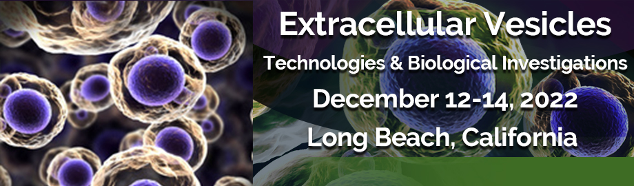
Co-Located Conference AgendasExtracellular Vesicles 2022: Technologies Driving Biological Investigations | Lab-on-a-Chip and Microfluidics World Congress 2022 | Organoids & Microphysiological Systems 2022 | Point-of-Care & Rapid Diagnostics 2022 | 

Monday, 12 December 2022 |
Please Refer to the Plenary Session Agenda in the Lab-on-a-Chip Website for Details of Programming on Monday, December 12, 2022 |
| |
Tuesday, 13 December 202208:00 | Conference Registration, Morning Coffee, Tea, and Pastries in the Exhibit Hall | 09:00 |  | Conference Chair Welcome and Introduction and Overview of EV Field by Conference Co-Chairperson
Mei He, Associate Professor, University of Florida, United States of America
|
| 09:15 |  | Conference Chair Welcome and Introduction by Conference Co-Chairperson and an Overview of the Field of EVs
Michael Graner, Professor, Dept of Neurosurgery, University of Colorado Anschutz School of Medicine, United States of America
|
| |
Session Title: A Deep Dive into Biological Investigations on Extracellular Vesicles (EVs) |
| | 09:30 |  | Keynote Presentation Developing Technologies for Single Vesicle Isolation and for Regenerative Medicine
Danilo Tagle, Director, Office of Special Initiatives, National Center for Advancing Translational Sciences at the NIH (NCATS), United States of America
Extracellular vesicles (EVs) are lipid membranous vesicles released from almost all cell types, and they provide a tremendous opportunity as sources of novel biomarkers from liquid biopsies, as well as agents for tissue repair and wound healing in regenerative medicine. EVs carry complex molecular cargoes, such as proteins, RNAs [e.g., mRNA and noncoding RNAs (microRNA, transfer RNA, circular RNA and long noncoding RNA)], and DNA fragments; these cargoes are delivered to recipient cells and serve as a cell-to-cell communication system. The molecular contents of EVs largely reflect the cell of origin and thus show cell-type specificity. Exosomes are endogenous nanoparticles that constitute a fraction of extracellular vesicles that are secreted by all cell types into the extracellular environment, and play an important role in intercellular signaling. Exosomes are being utilized in a variety of biomedical applications, including targeted drug delivery, gene therapy, diagnosis, and tissue regeneration. Despite significant efforts made in this relatively new field of research, progress has been held back by challenges such as inefficient separation methods, difficulties in characterization, and lack of specific biomarkers. This presentation will elaborate on the NIH support for exosome isolation, as well as its potential use in regenerative medicine. |
| 10:00 |  | Keynote Presentation Plasma Extracellular Vesicles For Cardiovascular Disease
Dominique PV de Kleijn, Professor Experimental Vascular Surgery, Professor Netherlands Heart Institute, University Medical Center Utrecht, The Netherlands, Netherlands
Cardiovascular Disease (CVD) is with the cardiovascular events of Ischemic Heart Disease and Stroke, the number 1 and 2 cause of death in the world and expect to increase especially in Asia. We use plasma extracellular vesicle (EV) protein content of vesicles from plasma sub-fractions on plasma of stroke and peripheral artery disease(PAD) patients, patients after carotid atherectomy (CEA) and patients suspected for chronic coronary syndrome (CCS). Using 25 ul of plasma, we developed an automated 96-well based protocol using sequential precipitation. Using samples of the AtheroExpress, the largest ongoing CEA biobank we try to early detect the risk of a second Major Adverse Cardiovascular Event (MACE: myocardial infarction, stroke or cardiovascular death) in PAD and CEA patients. Identification of such high risk patients is very important for possible (expensive) add-on pharmaceutical therapy or the decision to operate or not. Sequential precipitation for EV isolation is also used for the diagnosis of CCS. |
| 10:30 | Placental Cell-Specific Extracellular Vesicle (EV) Changes Throughout Gestation Quantitated by Nanoscale Flow Cytometry
Terry Morgan, Professor of Pathology and Laboratory Medicine, Oregon Health and Science University, United States of America
Placental alkaline phosphatase (PLAP) positive EVs increase with gestational age and may be relatively increased early in pregnancy in women who develop severe pregnancy complications. Importantly, PLAP alone is not specific for placental EVs and multiplex antibody labeling is required to measure cell-specific events in plasma. We employ nanoscale high resolution flow cytometry to reliably image, count, and isolate cell- and size-specific EVs in banked plasma. This is the first study to demonstrate that most of the PLAP+ EV events in pregnant plasma arise from EVTs, not floating villi. | 11:00 | Mid-Morning Coffee, Tea and Networking in the Exhibit Hall | 11:30 | | 12:00 |  | Keynote Presentation Regulation of Extracellular Vesicle Biogenesis In Tissue Repair
Brian Eliceiri, Professor, UC San Diego, United States of America
Extracellular vesicles (EVs) are released by defined intracellular trafficking machinery that can mediate paracrine signaling that affects immune responses and tissue repair. Here, specific examples of how specific pathways and genetic models affect EV payloads, and how these payloads can be used to design custom engineered therapeutics. |
| 12:30 | Networking Lunch in the Exhibit Hall -- Visit Exhibitors and View Posters | |
Session Title: Technologies Driving the Extracellular Vesicles (EVs) Field |
| | 13:30 |  Trends in EV Characterization Technologies Trends in EV Characterization Technologies
Jean-Luc Fraikin, CEO, Spectradyne
Methods for characterizing EVs are evolving, and new technologies are being developed that deliver size, concentration, and fluorescent phenotyping, each to a varying degree of success. Learn about how some of these new and cutting-edge technologies work, their strengths and weaknesses, and where we at Spectradyne see the technology landscape moving next.
| 14:00 |  Biomarker Co-localization Measurements (C-NTA) Using the New Particle Metrix ZetaView x30 Family Biomarker Co-localization Measurements (C-NTA) Using the New Particle Metrix ZetaView x30 Family
Sven Rudolf Kreutel, Chief Executive Officer, Particle Metrix GmbH and CEO, Particle Metrix Inc., USA
During the last decades, Nanoparticle Tracking Analysis (NTA) has emerged as a vital and fast characterization technology for biological nanoparticles like Extracellular Vesicles, Exosomes and Viruses. While classic NTA scatter operation feeds back particle size and total concentration, the fluorescence detection capability enables the user to gain specific biochemical information. Determination of biomarker co-localization however, is a challenging task for any analytical instrument. A new laser generation ensuring perfect overlap of the illumination volumes of individual channels paired with very fast switching times between fluorescence channels lay the foundation of co-localization nanoparticle tracking analysis (C-NTA) introduced recently by Particle Metrix. For the first time, we report results of co-localization measurements of reference material as well as real biological samples based on NTA technology on the new ZetaView® PMX-230 TWIN.
| 14:30 |  Nanoflow Cytometry: Sensitive Sizing and Phenotyping of Single EVs Nanoflow Cytometry: Sensitive Sizing and Phenotyping of Single EVs
Clayton Deighan, North American Sales and Applications Manager, NanoFCM
This presentation will review the principles and practice of nano flow cytometry. Covering the operational and experimental parameters important to resolving and counting single EVs. The latest updates on the technical capabilities of nano flow will be provided and multiple published examples of the data will be presented.
| 15:00 | Mid-Afternoon Coffee and Tea Break and Networking | 15:30 |  Paving the Way to Automation and Standardization for Scalable Isolation of EVs Paving the Way to Automation and Standardization for Scalable Isolation of EVs
Jared Lynch, Director of Business Development, IZON Science
As the potential for EV-based diagnostics and therapeutics continues to grow, so too does the need for scalable isolation and precise characterization. In this presentation, I will present how technologies and services offered by Izon Science fill this gap. I will introduce Izon’s newly released Gen 2 qEV SEC isolation product line as well as our recent work on scaling up EV isolation and purification using larger sized qEV columns with automated flow systems. Finally, you will be taken through an overview of Izon platforms and offerings, to gain a clear understanding of how Izon technologies can fit into a larger workflow.
| 16:00 |  Nanoscale Sorting with the CytoFLEX SRT Cell Sorter Nanoscale Sorting with the CytoFLEX SRT Cell Sorter
James McCracken, Product Manager, Beckman Coulter Life Sciences
Nanoscale flow cytometry is one of several techniques employed in the characterization of submicron particles. Biological particles often explored in this size range include bacteria, viruses, extracellular vesicles, and exosomes. This analysis tool has become more prevalent due to its high-throughput and multiparametric capabilities. In this presentation and accompanying application note we describe the ability of the CytoFLEX SRT cell sorter to sort nanoscale samples including bead controls, bacteria, and virus-like particles.
| 16:30 |  | Keynote Presentation EV-ID: Highly Multiplexed Affinity Proteomics of Single Extracellular Vesicles Using Interference and Label-based Imaging
David Juncker, Professor and Chair, McGill University, Canada
|
| 17:00 | Early Cancer Detection Powered by Extracellular Vesicles Using the Verita™ AC Electrokinetic Chip
David Searson, Senior Engineer, Biological Dynamics, United States of America
Utilizing the power of AC Electrokinetic (ACE) technology, Biological Dynamics’ Verita™ lab-on-a-chip platform provides a unique isolation of extracellular vesicles (EVs) directly from blood. By analyzing the isolated EV protein cargo and applying machine learning, multi-cancer early detection (MCED) is enabled. The detection performance at high specificity for multiple histologies enables future applications in cancer diagnostics. | 17:30 | The Benefits of Being Thin: How Ultrathin Silicon Membranes are Enabling New Technologies for Discovery in Biomedical Research
James McGrath, Professor of Biomedical Engineering, University of Rochester, United States of America
A decade-and-a-half after we first used silicon microfabrication to
create free-standing ultrathin nanoporous membranes, the materials are
being utilized by a growing number of laboratories as uniquely capable
tools for biomedical research. Today we manufacture a variety of
silicon-based nanoporous and microporous membranes with the common
characteristics that they are ultrathin (15 nm - 300 nm) with
well-defined pore sizes. The extremes thinness of 'nanomembranes' makes
them orders-of-magnitude more permeable to both diffusing molecules and
pressurized flow than conventional membranes. The ultrathin nature of
the membranes also gives them a glass-like imaging quality in optical
microscopy. These properties provide abundant opportunities for novel
membrane-based devices and assays. This talk will provide an overview of
two leading applications of our nanomembranes. First is their use to
create in vitro models of human tissue (i.e. 'tissue chips' or
'microphysiological systems' where they provide optically transparent
and highly permeable scaffolds to compartmentalize tissues including the
brain neurovascular unit, the blood-retinal-barrier, bone, and tendon.
The second application I will cover is the use of as tool for diagnostic
applications. Here we have leveraged our understanding of filtration to
develop a digital assay of extracellular vesicle biomarkers and
pressure-based sensor that detects virus in a point-of-care microdevice. | 18:00 | The Making of an Extracellular Vesicle Core
Paolo Neviani, Director - ExtraCellular Vesicle Core, Children’s Hospital Los Angeles, United States of America
The Children’s Hospital Los Angeles Extracellular Vesicle Core was founded in 2018, as the first core of this kind in the country, with the mission of providing the research community with expertise, optimized tools and up to date technologies dedicated to the field of EVs. The core provides isolation, characterization, and analysis of extracellular vesicles in an effort to facilitate EV research by adhering to the ever-evolving guidelines for standardization developed by the International Society of Extracellular Vesicles. | |
Evening Reception and Session on "Single EV Analysis via Flow Cytometry" Sponsored by Luminex Corporation -- All Conference Participants are Welcome to Attend |
| | 18:30 |  Applications for Small Particle Analysis Using High Sensitivity Flow and Imaging Flow Cytometry Applications for Small Particle Analysis Using High Sensitivity Flow and Imaging Flow Cytometry
Stephanie Brunelle, Senior Product Manager, Flow Cytometry, Luminex Corp.
Research in the fields of virology, microbiology, nanoparticles, and
extracellular vesicles (EVs) has shown tremendous growth over the past
few years. Viruses, which range in size from 17 nm to ~1.5 microns,
small bacteria, nanoparticles, and small EVs such as exosomes (less than
150 nm in diameter), are all on the subcellular scale. Quantifying and
characterizing small particles in a reproducible and reliable manner is
challenging due to their small size. In this presentation, we will
demonstrate the capabilities for small particle applications from Amnis®
CellStream® and Amnis® ImageStream®X
Mk II. The Amnis CellStream system has the advantage of high throughput
flow cytometry with higher sensitivity to small particles due to the
CCD-based, time-delay-integration image capturing system. Here, we will
present immunophenotyping data from EVs. The Amnis ImageStreamX Mk II
that combines the quantitative power of flow cytometry with microscopy
in one system has a High Gain mode to increase the sensitivity for small
particles by adjusting the CCD-camera to a higher gain setting,
increasing the signal obtained from the small particles while minimizing
the noise. In this portion of the presentation, examples of High Gain
mode using murine leukemia virus-sfGFP will be shown.
| 19:00 | EV Analysis and Flow Cytometry - A Perspective from Cellarcus
John Nolan, CEO, Cellarcus Biosciences, Inc., United States of America
| 19:30 | Detection and Enrichment of miRNA in Extracellular Vesicles
John Tigges, Flow Cytometry Science Center Director, Beth Israel Deaconess Medical Center, United States of America
In the past decade, basic research and clinical diagnostics involving extracellular vesicles has increased and shown progress. However, techniques, protocols, and instrumentation are lacking in the field. Furthermore, the ability to standardize techniques and reproduce research findings is problematic. Cytometry has become one of the most used instruments and efforts for standardization are centered around these technologies. Nano-flow cytometry has the potential to become a powerful tool in EV research. To this end, we have applied nano-flow cytometry to detect specific RNA populations within EVs called miRNA. miRNAs have become an important cargo material to study in the EV field. They have been associated with both disease diagnosis and prognosis. By using cytometry instruments to detect miRNAs, the field can move towards more reproducible and effective clinical results. | 20:00 | Evening Discussion Session on "Single EV Analysis via Flow Cytometry" Sponsored by Luminex Corporation -- Open to All Conference Participants | 20:30 | Close of Day 2 Programming |
Wednesday, 14 December 202208:00 | Morning Coffee, Tea and Networking in the Exhibit Hall | |
Session Title: Emerging Areas in the Extracellular Vesicles (EVs) Space Including EV-based Therapeutics Development, Single EV Analysis and Microfluidics for EV Studies |
| | 09:00 | Advancing Cancer Liquid Biopsy with Microfluidics and Nanoengineering
Yong Zeng, Associate Professor, University of Florida, United States of America
A primary focus of my research is on the development of innovative microfluidics and nanobiosensing technologies and molecular biomarkers for cancer diagnosis and treatment. Herein, we will discuss some of the nanoengineered microfluidic systems for efficient immunoisolation of circulating extracellular vesicles (EVs) and ultrasensitive profiling of EV biomarkers, including proteins and miRNAs. Adaptation of our new technologies to clinical studies of circulating EVs for non-invasive liquid biopsy of cancer will be discussed. | 09:30 |  Exosome Engineering for Active Targeting and Intracellular Delivery of Therapeutic Proteins: Realization from Concept Exosome Engineering for Active Targeting and Intracellular Delivery of Therapeutic Proteins: Realization from Concept
Chulhee Choi, CEO, ILIAS Biologics Inc.
As extracellular vesicles that play an active role in intercellular communication by transferring cellular materials to recipient cells, exosome offer great potential as a natural therapeutic drug delivery vehicle. Currently, both academia and industry try to develop exosome platform-based therapeutics for disease management, some of which are already in clinical trials. An opto-genetically engineered exosome system (EXPLOR®) that we previously developed was implemented for loading therapeutic cargo into exosomes which can deliver therapeutic cargos into target cells in free form. We are studying the clinical potential of ‘Exo-target®’, therapeutic exosomes with EXPLOR technology, in multiple disease areas including inflammatory diseases. NF-kB has been well accepted as master regulator for inflammation. The introduction of I-kB into target cells can inhibit inflammatory responses by restraining nuclear translocation of NF-kB. Therefore, we have developed Exo-target loaded with dominant active form of I-kB (Exo-srI-kB). We found that Exo-srI-kB treatment attenuates both local and systemic inflammation and animal mortality associated inflammatory disease including sepsis, ischemic reperfusion induced acute kidney injury and preterm birth in our preclinical studies. Especially, maternally injected Exo-srI-kB could cross-over placenta barrier to deliver therapeutic cargos to fetal side, which resulted in prolonged pregnancy by more than 24 hours and additional advantages. Altogether, these results suggest therapeutic value of Exo-target for various disease by delivering API intracellularly to the target cells.
| 10:00 | Single Particle Analyses Toolbox: The Key to Unlocking the Heterogeneity of Extracellular Vesicles
Randy Carney, Assistant Professor, University of California-Davis, United States of America
The heterogeneity in composition and structure of individual EVs strongly contributes to their varied functions with respect to therapeutics. Yet it remains a challenge to examine structure-function at the resolution of single vesicles. In this talk, I will introduce a suite of high throughput commercial and custom tools that probe the structure and composition of single EVs, with a focus on spectroscopic techniques. I will examine best practices and key advantages of each technique, and provide an outlook of what is needed still to advance their study. | 10:30 | Coffee Break and Networking in the Exhibit Hall | 11:00 |  Improving EV Measurement Sensitivity and Standardization in Flow Cytometry Research with the BD Small Particle Detector Improving EV Measurement Sensitivity and Standardization in Flow Cytometry Research with the BD Small Particle Detector
Scott Bornheimer, Associate Director, Medical and Scientific Affairs, BD Biosciences
| 12:00 |  | Keynote Presentation Affinity Selection of Extracellular Vesicles using Plastic-based Microfluidic Devices for the Molecular Sub-typing of Breast Cancer Patients
Steve Soper, Foundation Distinguished Professor, Director, Center of BioModular Multi-Scale System for Precision Medicine, The University of Kansas, United States of America
We have been developing tools for the diagnosis of a variety of diseases. The commonality in these tools is that they consist of microfluidic devices made from plastics via injection molding. Thus, our tools can be mass produced at low-cost to facilitate bench-to-bed side transition and point-of-care testing (PoCT). We have also been generating novel assays focused on using liquid biopsy samples that are enabled using microfluidics. In this presentation I will talk about the evolution of our fabrication efforts of plastic-based microfluidic and nanofluidic devices as well their surface modification to make the devices biocompatible for in vitro diagnostics. One tool that we have generated is a plastic device (38 × 42 mm) that consists of 1.5M pillars, which are surface decorated with affinity agents targeting certain disease-associated extracellular vesicles (EVs). The affinity agents are covalently attached to the surface of the microfluidic device using a hetero-bifunctional linker, which consists of a coumarin moiety to allow for the photolytic release of the captured EVs using a blue-light LED to minimize photodamage to the EVs’ molecular cargo. In this presentation, I will discuss the utility of these microfluidic devices using EVs as a source of mRNAs for molecular sub-typing of breast cancer patients. EVs were affinity selected from breast-cancer patients’ plasma by searching for both epithelial and mesenchymal expressing EVs to allow for highly efficient sub-typing using the PAM50 gene panel. |
| 12:30 | Tiny Packages, Enormous Potential: Exploiting Extracellular Vesicles for Early Detection, Disease Monitoring, and Molecular Subtyping of Cancer
Andrew Godwin, Professor and Division Director, Genomic Diagnostics, Founding Director, Kansas Institute for Precision Medicine, Deputy Director, KU Cancer Center, University of Kansas Medical Center, United States of America
Pathologic analysis of tumor tissue biopsies is the gold standard for the initial diagnosis of cancer. However, recently liquid biopsies, which analyze tumor-derived material circulating in the bloodstream and other bodily fluids, are rapidly gaining traction in the clinic. These tests offer considerable potential in oncology, which include early detection, monitoring treatment response and disease recurrence. Liquid biopsy biomarkers include circulating tumor cells, cell-free DNA, nanoparticles, and extracellular vesicles (EVs). Regarding the latter, EVs are showing great promise as circulating biomarkers. The International Society for Extracellular Vesicles define EVs as particles naturally released from the cell that are delimited by “a lipid bilayer and cannot replicate” Center among EVs are nano-sized vesicles (40 to 150 nm) of endocytic origin also known as small EVs/exosomes, which are produced and released by most cell types under normal physiologic and in diseased states. sEVs carry cargo representative of their originating cell including nucleic acids, cytokines, membrane-bound receptors, and a wide assortment of other, biologically active lipids and proteins. Since sEVs/exosomes travel systemically throughout the body, efforts are underway to exploit them as potential biomarkers to detect and monitor disease states. Ways to exploit sEVs for cancer diagnostics and monitoring response to therapy will be discussed. | 13:00 | Networking Lunch in the Exhibit Hall -- Visit Exhibitors and View Posters | 14:00 | Probing Life in Bubbles – Nanoplasmonic Quantification of Pathogen-derived Extracellular Vesicles in Blood
Tony Hu, Professor and Weatherhead Presidential Chair, Tulane University School of Medicine, United States of America
Extracellular vesicles (EVs) are small membrane-bound vesicles secreted by all cells, circulate at high levels, and convey nucleic acids, peptide/proteins, lipids. EVs secreted by microbial pathogens or infected or malignant cells represent an excellent source of biomarkers, but technical challenges have prevented development of EV-based assays. With the advanced technologies, we have identified and validated new biomarkers for rapid pathogen differentiation (e.g. SARS-CoV-2 and mycobacterial), early disease diagnosis (including cancer), and/or real-time evaluation of disease response. Our multidisciplinary expertise enables us to employ the characteristic properties of engineered nanodevices to improve the capture and detection of circulating biomarkers. | 14:30 | Microfluidic Technologies for the Isolation and Characterization of Extracellular Vesicles For Lung Cancer Theranostics
Sunitha Nagrath, Professor of Chemical Engineering and Biomedical Engineering, University of Michigan-Ann Arbor, United States of America
The extracellular vesicle (EV) has emerged as one of the diagnostic means of cancer through liquid biopsy. So far, ultracentrifugation has been accepted as gold standard for EV isolation in spite of its lengthy processing time and low recovery rate. Owing to the recent technological advances in microfluidics, several microfluidic devices for EV isolation have been developed and showed promising with better recovery rate and processing time. Here we present microfluidic based approaches to isolate and profile tumor derived EVs. To demonstrate the harvesting of EVs from specific cells for therapeutic use we have developed a microfluidic based strategy to harvest EVs from Natural Killer (NK) cells in blood circulation. In combination we have developed theranostics application of EVs in lung cancer. | 15:00 | Click Chemistry-Mediated Surface Protein Assay for Quantifying Subpopulations of Hepatocellular Carcinoma-Associated Extracellular Vesicles
Yazhen Zhu, Assistant Professor, David Geffen School of Medicine at UCLA, United States of America
Extracellular vesicles (EVs) are a heterogeneous group of phospholipid bilayer-enclosed particles that are released by all types of cells, and even more so by tumor cells. It is well known that tumor-associated EVs are present in circulation at a relatively early stage of disease. Since the surface proteins of tumor-associated EVs mirror those of the parental tumor cells, performing surface protein analysis on tumor-associated EVs offers a systemic and repeatable assessment of tumor cells that can offer substantial diagnostic value for early-stage diseases. Hepatocellular carcinoma (HCC) predominantly occurs in patients with underlying cirrhosis, and is the fourth most common cause of cancer-related deaths worldwide. Current clinical surveillance guidelines, which include biannual liver ultrasonography with or without serum alpha-fetoprotein (AFP), exhibit low sensitivity to detect early-stage HCC, limiting delivery of curative-intent treatments. Therefore, exploiting the diagnostic potential of HCC EVs’ surface protein signatures as a novel biomarker for detecting early-stage HCC holds great promise to augment current HCC diagnostic modalities. We demonstrated the feasibility of HCC EV Surface Protein Assay (SPA) for detecting subpopulations of HCC EVs by coupling two platform technologies: Click Chemistry-mediated EV Click Beads for isolation and enrichment of HCC EVs, and real-time immuno-PCR for HCC EV quantification. The resultant HCC EV Surface Protein Scores exhibit great diagnostic potential for early detection of HCC. | 15:30 | Afternoon Coffee Break | |
Session Title: Viruses and Extracellular Vesicles (EVs) |
| | 16:00 | Extracellular Vesicles and Viruses: Close Relatives or Enemies?
Leonid Margolis, Head, Section of Intercellular Interactions, Eunice Kennedy-Shriver National Institute of Child Health and Human Development, NIH, United States of America
| 16:30 | Perspectives and Open Discussion: The Nexus of Viruses and Extracellular Vesicles (EVs)
Michael Graner, Professor, Dept of Neurosurgery, University of Colorado Anschutz School of Medicine, United States of America
Leonid Margolis, Head, Section of Intercellular Interactions, Eunice Kennedy-Shriver National Institute of Child Health and Human Development, NIH, United States of America
| 17:00 | Close of Conference |
|


 Add to Calendar ▼2022-12-12 00:00:002022-12-14 00:00:00Europe/LondonExtracellular Vesicles 2022: Technologies Driving Biological InvestigationsExtracellular Vesicles 2022: Technologies Driving Biological Investigations in Long Beach, CaliforniaLong Beach, CaliforniaSELECTBIOenquiries@selectbiosciences.com