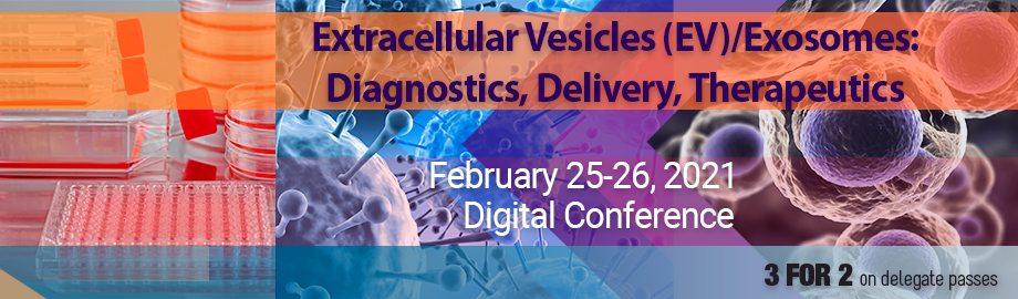
Co-Located Conference AgendasCirculating Biomarkers 2021 | Extracellular Vesicles (EV)-Exosomes: Diagnostics, Delivery and Therapeutics | 

Thursday, 25 February 2021 |
Conference is Powered by the Zoom Platform. All Times Listed are US Eastern Time. |
| | 08:30 |  | Conference Chair Welcome, Introduction, Topics Addressed and Conference Opening by Conference Chairperson
Michael Graner, Professor, Dept of Neurosurgery, University of Colorado Anschutz School of Medicine, United States of America
|
| 08:45 |  | Conference Chair Welcome, Introduction, Topics Addressed and Conference Opening by Conference Chairperson
Lucia Languino, Professor of Cancer Biology, Thomas Jefferson University, United States of America
|
| |
Session Title: Current Themes in Extracellular Vesicle (EV) Research |
| | |
Session Chairperson: Lucia Languino |
| | 09:00 |  | Keynote Presentation Plasma Extracellular Vesicle Sub-fractions as Biomarker Source for Cardiovascular Disease
Dominique PV de Kleijn, Professor Experimental Vascular Surgery, Professor Netherlands Heart Institute, University Medical Center Utrecht, The Netherlands, Netherlands
Cardiovascular Disease (CVD) is with the cardiovascular events of Ischemic Heart Disease and Stroke, the number 1 and 2 cause of death in the world and expect to increase especially in Asia.
Ischemic heart disease (IHD) comprises 3 entities: Chronic Coronary Syndrome (CCS or Stable Angina), unstable angina (UA) and myocardial infarction (MI). Because IHD is associated with an increased risk of adverse clinical events such as heart failure and death, early recognition of IHD is of utmost importance.
However, to diagnosis CCS is challenging, as many patients present with atypical symptoms. Troponins are the main diagnostic tool for detection of MI. Blood biomarkers for CCS (typically causing stable angina) and UA, however, are not available. These diagnoses frequently require hospital visits/admissions for time-consuming and costly (non)invasive tests. We use plasma extracellular vesicle protein content of vesicles from plasma sub-fractions as an accurate source for early diagnosis of CCS. This plasma sub-fraction technology is also used for prognosis of a second MI or stroke to identify the high risk patient that can be treated with more intensive and often more costly medication. |
| 09:30 |  | Keynote Presentation Extracellular Vesicles in Synapse Formation
Alissa Weaver, Cornelius Vanderbilt Professor of Cell and Developmental Biology, Vanderbilt University Medical Center, United States of America
Neuronal synapse formation is critical for brain development and depends on secreted factors from astrocytes. We investigated the role of small extracellular vesicles (EVs) in this process and found that small EVs secreted from primary astrocytes, but not from neurons or C6 glioma cells, greatly enhance spine and synapse formation by primary cortical neurons. I will discuss the molecular mechanisms by which this occurs.
|
| 10:00 | Mid-Morning Break | |
Session Title: Current Themes in Extracellular Vesicle (EV) Cancer Research |
| | |
Session Chairperson: Alissa Weaver |
| | 10:30 |  | Keynote Presentation Extracellular Vesicles as Prognostic Markers in HNSCC in Response to Immunotherapy: From Bedside to Bench
My Mahoney, Professor and Vice Chair, Thomas Jefferson University, United States of America
We recently demonstrated that the pro-proliferative and -survival stem cell marker desmoglein 2 (Dsg2) enhances squamous cell carcinoma (SCC) tumor development through small extracellular vesicles (sEVs) in an IL-8/miR-146a dependent mechanism. In head and neck SCC (HNSCC), a linear correlation between Dsg2 and IL-8 was detected by RNAseq of tumor tissues. Here, in a 4-week window of opportunity trial, human papillomaviruses (HPV)+ and HPV- HNSCC patients received 2 doses of the immune checkpoint inhibitor anti-PD1 mAb, Nivolumab. Patients were stratified according to pathologic treatment response as demonstrated by reduction in tumor size. EVs isolated from post treatment tumor culture supernatant by FACS analysis, confirmed the downregulation of immune checkpoint receptors in responders. Transcriptomic analysis of the tumor tissues showed downregulation of Dsg2 in response to treatment. ELISA analysis of plasma showed significantly lower levels of IL-8 in responders as compared to non-responders. Interestingly, higher level of IL-8 was mainly in the HPV+ non-responders. Among HPV+ patients, approximately 30% have high IL-8 and all were non-responders. Understanding non-response to immunotherapy in HPV positive cohort has been elusive and the discovery of an association with IL-8 in the periphery could speak to an active HPV viral influence that may overshadow the effects of PD-1 inhibition or other immune mechanisms. Further investigation into a resistance to immunotherapy and the potential for confounding systemic and local factors is necessary to further understand this observation. |
| 11:00 | The Role of Core Facilities and Emerging Technologies in Maximizing Rigor and Reproducibility of EV Quantification and Characterization and Following MISEV Guidelines
Rachel DeRita, Director, Extracellular Vesicle Core Facility, University of Pennsylvania School of Veterinary medicine, United States of America
It remains very clear in the field of extracellular vesicle (EV) research that the rapid rate of increase in publications and expansion of interdisciplinary clinical EV interest has created the need for increased standardization and access to the appropriate technologies to uphold these standards. As the first core facility in the United States with the sole intention of creating a space where users can both isolate and characterize EVs, we provide a central location for the facilitation of EV research via access to multiple technologies (both established and emerging) such as resistive pulse sensing, nanoparticle tracking analysis, ultracentrifugation, high-performance liquid chromatography, flow cytometric analysis of EVs and additional immune or fluorescence-based EV characterization techniques. We surveyed a group of leading scientific investigators and researchers in varying stages of their scientific careers in the Mid-Atlantic region of the US. The survey data demonstrate applications of greatest current and future interest to be employed in a shared lab resource. The current demand is highest for isolation services, ultracentrifugation and NTA, with a gradually increasing demand for immunophenotying analyses such as the ExoView chip array, fluorescent NTA and flow cytometry. We additionally present strategies and data-based examples of how shared resource facilities can facilitate multifactorial and rigorous EV characterization in accordance with MISEV guidelines, and encourage collaboration among EV researchers. In order to answer the larger remaining questions in the EV field such as the isolation of specific EV subsets, EV tracking between cells and the use of EVs for biomarker discovery and drug delivery, it is essential that shared resource facilities interact not only with investigators, but with each other to integrate the necessary resources to progress.
| 11:30 |  | Keynote Presentation Extracellular Vesicle-Derived Analytes for Cancer Progression
Dolores Di Vizio, Professor, Cedars Sinai Medical Center, United States of America
Cancer-derived extracellular vesicles (EVs) are highly heterogenous and include atypically large particles (1-10 microns), known as large oncosomes that are abundant in PC patients with metastatic PC and are enriched with tumor-derived cargo. Next Generation Sequencing studies (whole genome and whole transcriptome) as well as Mass Spectrometry studies using different approaches, including SILAC, have enabled us to refine the methodologies that allow to isolate and characterize these vesicles in human plasma samples from patients with prostate cancer with high rigor and reproducibility. We are building upon our studies and moving forward with a deep characterization of the protein content of large oncosomes and the development of a preclinical platform that can help translating our laboratory investigations to the clinic. One of our long term goals is to undertake an integrative approach that takes advantage of all the macromolecules carried in these tumor-derived particles and ultimately identify epigenomic, metabolic, transcriptional and other functional pathways that can be extracted from plasma vesicles and provide tumor information in real time. |
| 12:00 | Lunch Break | |
Session Title: Single EV Analysis, Flow Cytometry and Technologies for EV Analysis 2021 |
| | |
Session Chairperson: Michael Graner |
| | 13:00 | Revealing the Diversity of EVs with Single Vesicle Analysis
John Nolan, CEO, Cellarcus Biosciences, Inc., United States of America
Extracellular vesicles (EVs) appear to be involved in many physiological processes and have attracted great attention as potential diagnostic and therapeutic targets. The realization of this potential has been hampered by the lack of analytical methods with the necessary specificity, sensitivity, and quantitative rigor needed for robust hypothesis testing, biomarker development, and EV engineering. New analytical tools with improved specificity and sensitivity are revealing the diverse constituents of biofluid preparations that had been previously referred to as “exosomes” and “microvesicles,” and are forcing a re-evaluation of both methods and nomenclature. Single vesicle analysis represents the ultimate level of EV analysis, and we have been using single vesicle flow cytometry (vFC) to count, size, and measure the molecular cargo on individual EVs at a large scale. I will review the state of EV analytics, highlight critical elements of single EV analysis, and share insights into EV diversity that have emerged from the application of quantitative analytical methods to the study of EVs. | 13:30 |  Accurate Concentration of Biological Nanoparticles – Why and How? Accurate Concentration of Biological Nanoparticles – Why and How?
Jean-Luc Fraikin, CEO, Spectradyne
Extracellular vesicles (EVs), virus and non-viral drug delivery vehicles are rapidly moving from the research lab into real-world applications. Why should we care about measuring the concentration of these biological nanoparticles accurately? In the case of a COVID vaccine for example, we should care because particle concentration is the dose! In a broad range of applications, particle concentration is a critical parameter that must be carefully controlled in order to develop safe and effective products. This talk will include a comparison of methods and examples to illustrate how accurate concentration measurements are important for good quality science.
| 14:00 |  Know What’s In Your Sample: Phenotyping of Extracellular Vesicles (EVs) with Fluorescent Nanoparticle Tracking Analysis (f-NTA) Know What’s In Your Sample: Phenotyping of Extracellular Vesicles (EVs) with Fluorescent Nanoparticle Tracking Analysis (f-NTA)
Sven Rudolf Kreutel, Chief Executive Officer, Particle Metrix GmbH and CEO, Particle Metrix Inc., USA
In the last decade, Nanoparticle Tracking Analysis (NTA) has emerged as a vital and fast characterization technology for Extracellular Vesicles, Exosomes and Microvesicles. in the size range from 30 nm to 1 µm. While getting a particle size distribution, the user typically cannot discriminate whether the particle is a vesicle, protein aggregate, cellular trash or an inorganic precipitate from scatter data alone. The fluorescence detection capability of NTA enables the user to gain specific biochemical information about the particle surface and increases the resolution and the discrimination power. However, traditional NTA instruments are equipped with one laser wavelength in operation. This limits the detection of biomarkers to only a couple of fluorescence dyes. For change of excitation wavelength, it was necessary to change the laser source, a time and sample consuming step. To overcome this issue Particle Metrix developed a unique QUATT laser technology and now allows the rapid analysis of biomarker concentrations or ratios with four excitation lasers on the same sample. Here we report the multi-fluorescence detection of four independent biomarkers (CD63, CD81, CD9 and CD41) in one NTA sample with the new Particle Metrix ZetaView® QUATT for the first time.
| 14:30 |  Analyzing Extracellular Vesicles With The Amnis® ImageStream®X Mk II High Gain Mode Analyzing Extracellular Vesicles With The Amnis® ImageStream®X Mk II High Gain Mode
Haley Pugsley, Manager and Senior Scientist, Luminex Corporation
The importance of EVs has become apparent, as they are key mediators of intercellular communication. However, quantifying and characterizing EVs in a reproducible and reliable manner is challenging due to their small size—exosomes range from 30 to 100 nm in diameter. Flow Cytometry is an attractive method for measuring EVs; however, this has been challenging since fl ow cytometers were designed to measure and detect cells. High Gain mode on the Amnis® ImageStream®X Mk II Imaging Flow Cytometer was designed to address the challenges of measuring particles that are much smaller than cells. This presentation demonstrates the improved small particle detection, including EVs, using this new High Gain mode.
| 15:00 | Mid-Afternoon Break | |
Session Title: Where is the EV Field Heading, circa 2021? |
| | |
Session Chairperson: Dolores Di Vizio |
| | 15:30 |  | Keynote Presentation Designing Engineered EVs for Virus X
Chris Baldwin, Chief Commercial Officer, Exopharm Ltd., Australia
Exopharm will report on its early work of conferring tropism to exosomes using SARS-CoV-2 spike protein to deliver siRNA to impede coronavirus replication and its implication for future pandemic responses. |
| 16:00 | Nuclear Transport of Extracellular Vesicles
Aurelio Lorico, Professor of Pathology, Touro University Nevada School of Medicine, United States of America
We have recently identified a novel nuclear path, where Rab7+ late endosomes enter into nuclear envelope invaginations and mediate the nuclear translocation of EV-derived biomaterials, such as proteins and RNA. The close association of three proteins; endoplasmic reticulum (ER)-localized vesicle-associated membrane protein (VAMP)-associated protein VAP-A, oxysterol-binding protein (OSBP)-related protein ORP3 and small GTPase Rab7 is essential for the entrance and/or maintenance of Rab7+ late endosomes into NEI and the nuclear transfer of EV-derived biocomponents (e.g., proteins and nucleic acids) that in turns may regulate the gene expression in host (or EV-targeted) cells (Santos et al., J. Biol. Chem. 2018). | 16:30 |  | Keynote Presentation Biology and Function of Exosomes in Cancer
Raghu Kalluri, Professor and Chairman, Department of Cancer Biology, University of Texas MD Anderson Cancer Center, United States of America
Humans circulate trillions of exosomes at all times. Exosomes are extracellular vesicles with a size range of 40-150 nm and a lipid bilayer membrane; and are released by all cell types. Normal exosomes contain DNA, RNA and proteins. Some proteins such as CD9, CD63 and flotillin are often detected on the surface of exosomes and serve as markers. Exosomes likely remove excess and/or unnecessary constituents from the cells, functioning like garbage bags. Although their precise physiological role remains largely unknown, is it suggested that exosomes may mediate specific cell-cell communication and activate signaling pathways in cells they fuse or interact with. Exosomes are detected in the tumor microenvironment and emerging evidence suggests that they play a role in facilitating tumorigenesis by regulating angiogenesis, immunity and metastasis. Circulating exosomes could be used as liquid biopsies and non-invasive biomarkers to potentially inform on early detection and diagnosis of cancer patients. Exosomes can be used for the treatment of cancer. This lecture will highlight some of the recent advances in the area of exosomes biology and their utility in the diagnosis and treatment of cancer. Additionally, the use of exosomes in personalized therapy for cancer patients will be discussed. |
| 17:00 | Close of Day 1 of the Conference |
Friday, 26 February 2021 |
Conference is Powered by the Zoom Platform. All Times Listed are US Eastern Time. |
| | |
Session Title: Viruses and Extracellular Vesicles (EVs): Similarities, Differences and Current Research Efforts |
| | |
Session Chairperson: Michael Graner |
| | 08:00 |  | Keynote Presentation Exosomes and Viruses: A Tale of Two Overlapping Worlds
Fatah Kashanchi, Professor and Director of Research, Lab of Molecular Virology, George Mason University, United States of America
Extracellular vesicles (EVs) play a significant role in intercellular communication by serving as a carrier for the transfer of membrane and cytosolic proteins, lipids, and RNA between cells. In recent years, using state of art technologies such as RNA seq, RPMA, and single cell omics, we have found that virally infected cells including HIV-1, HTLV-1, Rift Valley Fever, Zika, Ebola, and Coronavirus infected cells secret exosomes that contain biomarker of these infections in urine, saliva, CSF, and blood. We have been able to separate and characterize EVs from several different viruses including HIV-1. These EVs are not infectious and have a different density than infectious virions using gradients. They contain various viral RNAs including TAR (non-coding RNA), Nef, gp120/160 and Tat. The origin of these EVs are infected cells, especially when treated with cART or Interferons. They are present in patient samples tested (plasma and CSF, 33%-95%) to date (4 cohorts of 5-20 patients each). The EVs contribute to pro-inflammatory signals in the naïve recipient cells using TLR3 signaling. Recently, we have asked about the timing difference between EV and virus release from infected cells using serum starvation experiments from cells followed by release. Results from supernatants of uninfected cells showed a peak of tetraspanin proteins (CD63, CD81, and CD9) at 6 hours and a gradual decrease of all EV associated proteins by 24 hours. However, the EV from HIV-1 infected cells showed all three tetraspanins present at 3 hours and expression gradually increased up to 24 hours. HIV-1 viral proteins (p24, gp120, Nef) expression was present at 6 hours and continued to increase and peaked at 24 hours. HIV-1 supernatant 6- hour sample was found not to be infectious. However, infectious HIV-1 was successfully rescued from 24-hour sample.
Our data indicates that EV release may occur prior to viral release in infected cells, thereby implicating a potentially significant effect of EVs on uninfected recipient cells prior to subsequent viral infection. |
| 08:30 | Extracellular Vesicles in Cytokine Regulation
Leonid Margolis, Head, Section of Intercellular Interactions, Eunice Kennedy-Shriver National Institute of Child Health and Human Development, NIH, United States of America
Extracellular vesicles play an important role in human immuno-modulation mediated by cytokines both in normal condition and in pathologies. We found that both myocardial infarction and in HIV-1 infection extracellular vesicles are associated with immune activation, an important driver of the disease. | 09:00 | Coordination of Functionally Related mRNAs and Proteins in Granules, GW/P bodies, and Vesicles
Jack Keene, James B. Duke Professor, Duke University Medical Center, United States of America
Gene expression is determined by transcriptional and post-transcriptional mechanisms that coordinate regulatory layers and are localized in many cellular sub-environments. Post-transcriptional regulatory layers consist of trans RNA-binding proteins and multiple copies of cis messenger RNAs (mRNAs) and noncoding RNAs (miRNAs and other ncRNAs) that together form modular RNA regulons (RBPs) that in turn, coordinate the latter state of gene expression on a global level. The multi-targeting of mRNAs in RNA regulons include cis binding sites that are: a) RBP sequence specific, b) miR binding sites, c) modified mRNAs (e.g. methylated), that together produce functionally related proteins that can be found in cellular vesicles, bodies or granules by translation. Thus, RNA regulons are found in nearly all species and coordinate RNA turnover, localization, and/or translation globally in response to biological activation or repression. RNA regulons have myriad ramifications for biological coordination. Over time, dozens of RNA regulons have been reported in many biological systems and in dozens of species. For examples from the field: 1) PUF RNA regulons coordinate fungal metabolism and pathogenesis dating back over 500 million years; 2) trypanosome RNA regulons coordinate parasite differentiation in the blood at a time when transcription is silenced. 3) dozens of mammalian RNA regulons have biological and physiological ramifications in which mRNAs encoding functionally related proteins bound to several of the ~1,500 RBPs now known RBPs. Thus, RNA regulons can efficiently utilize and locate protein building blocks and regulatory factors that function as complex traits. Moreover, these overlapping mRNA subsets are highly dynamic and responsive to intrinsic and extrinsic signals. To understand these dynamic processes our lab developed Digestion Optimized-RIP-seq that is quantitative and applicable to understanding how combinatorial mechanisms regulate dynamic coordination of RNA targets during growth and differentiation, as well as revealing novel RNA regulons in cancer, neurodegeneration and liver development/regeneration. Our goal is to teach these technologies to students and fellows in order to decipher the remodeling of RNA regulons so to reveal underlying mechanisms of disease.
| 09:30 | Single Particle Detection of Extracellular Vesicles and Native SARS-CoV-2 Virions by Microfluidic Resistive Pulse Sensing
Zoltan Varga, Biological Nanochemistry Research Group, Research Centre for Natural Sciences (RCNS), Hungary
Microfluidic resistive pulse sensing (MRPS) is the nanoscale implementation of the Coulter principle in a microfluidic cartridge. As such, MRPS is a non-optical, single-particle detection method to measure the size distribution and concentration of nanoparticles. MRPS can be used to characterize extracellular vesicles (EVs). Enveloped viruses such as SARS-CoV-2 are spherical biological nanoparticles in the hundred nanometer size range, therefore MRPS is the method of choice for their characterization. In this study, MRPS was used to measure the size distribution of various EVs and SARS-CoV-2. The analysis of the supernatant of infected Vero cells indicated an envelope diameter of approximately 80 nm for the SARS-CoV-2 virions, which is in good agreement with previous cryo-EM studies. The detection of more than ten thousand virions made it possible to determine the size distribution of SARS-CoV-2 and paves the way for the MRPS-based detection of COVID-19 infection. | 10:00 | Mid-Morning Break | 10:30 | Extracellular Vesicle Glycosylation
Johnathon Anderson, Assistant Professor, University of California-Davis, United States of America
Up to 50% of the proteins encoded in the human genome are predicted to contain glycosylation sites. Aberrant glycosylation is a hallmark of several diseases, including many types of cancer from different origins. Although the functional significance of such patterns of glycan expression and presentation are not yet well characterized, tumor growth, metastases and immunoregulation have all been associated with such abnormal displays of glycosylation within the tumor microenvironment. Our group is interested in understanding glycosylation dynamics in the context of cancer-associated fibroblasts (CAFs), and mesenchymal stromal cells (MSCs), which serve as a source of CAFs in some types of cancer. One of the most remarkable changes in cancer glycosylation is the aberrant expression of sialic acid–bearing glycans called sialoglycans. Cells within the tumor bed are often covered with a dense layer of sialoglycans which have emerged in recent years as potent immune modulators that promote tumor immune evasion. Sialoglycans serve as the cognate ligands for a family of immune checkpoint receptors called sialic acid–binding immunoglobulin-like lectins (Siglecs), which are expressed by various populations of leukocytes. Preclinical studies have shown that tumor associated sialoglycans negatively influence immune cell function by interacting with the immune-inhibitory Siglec family members. Our group is interested in how sialoglycan expression is regulated in MSCs and CAFs, and what functional role they may play in their derived extracellular vesicles. | 11:00 | EVs and Viruses Panel Discussion by Lucia Languino and Michael Graner, Conference Chairpersons
- Status of the Field
- What have we learnt and what are the research trends
- The Panelists will weigh-in on these topics -- The Panelists Are:
Esther Nolte-‘t Hoen
Fatah Kashanchi
Jack Keene
Jan Lötvall
Jennifer Jones
Leonid Margolis
| 11:45 | Switch to Circulating Biomarkers Session | |
Please View Details of Circulating Biomarkers Session under the Circulating Biomarkers Website Agenda |
| |
|


 Add to Calendar ▼2021-02-25 00:00:002021-02-26 00:00:00Europe/LondonExtracellular Vesicles (EV)-Exosomes: Diagnostics, Delivery and TherapeuticsExtracellular Vesicles (EV)-Exosomes: Diagnostics, Delivery and Therapeutics in Virtual ConferenceVirtual ConferenceSELECTBIOenquiries@selectbiosciences.com