Co-Located Conference AgendasLab-on-a-Chip and Microfluidics Europe 2018 | Organ-on-a-Chip, Tissue-on-a-Chip Europe 2018 | Point-of-Care Diagnostics & Biosensors Europe 2018 | 

Tuesday, 5 June 201807:30 | Conference Registration, Materials Pick-Up, Morning Coffee and Tea | |
Session Title: Conference Opening Plenary Session |
| | |
Plenary Session Chairman: Richard Spero, Ph.D., Co-Founder and CEO, Redbud Labs: Advanced Microfluidic Technologies |
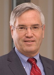
| | 08:15 |  | Conference Chair Conference Chairman Welcome and Opening Remarks
Richard Chasen Spero, CEO, Redbud Labs, United States of America
|
| 08:30 |  | Keynote Presentation Nanoplasmonic Biosensors: From Innovative Materials to Multimode Sensing with Integrated Microdevices
Amy Shen, Professor and Provost, Okinawa Institute of Science and Technology Graduate University, Japan
Gold nanostructures are a highly attractive class of materials with
unique electrochemical and optical sensing properties. Recent
developments have greatly improved the sensitivity of optical sensors
based on metal nanostructured arrays. We introduce the localized surface
plasmon resonance (LSPR) sensors and describe how its exquisite
sensitivity to size, shape and environment can be harnessed to detect
molecular binding events. We then describe recent progress in three
areas representing the most significant challenges: integration of LSPR
with complementary electrochemical techniques, long term live-cell
biosensing and practical development of sensors and instrumentation for
routine use and high-throughput detection. As an example we will
demonstrate a novel refractive index and charge sensitive device
integrated with nanoplasmonic islands to develop
nano-metal-insulator-semiconductor (nMIS) junctions. The developed
sensor facilitates simultaneous detection of charge and mass changes on
the nanoislands due to biomolecule binding. A brief insight on
microcontact printing to functionalize proteins on nanoplasmonic sensors
will also be discussed. The developed nanosensors can readily be
adopted for multiplexed and high throughput label-free immunoassay
systems, further driving innovations in biomedical and healthcare
research. |
| 09:00 |  | Keynote Presentation Towards Wearable Real-Time Clinical Monitoring Using Microfluidic Devices
Martyn Boutelle, Professor of Biomedical Sensors Engineering, Imperial College London, United Kingdom
Modern acute critical care medicine is increasingly seeking to protect
vulnerable tissue from damage by monitoring the patterns of physical,
electrical and chemical changes taking place in tissue – so called
multimodal monitoring. Such patterns of molecular changes offer the
exciting possibility of allowing clinicians to detect changes in patient
condition and to guide therapy on an individualized basis in real time.
Microfluidic lab-on-chip devices coupled to tissue sampling using
microdialysis provide an important new way for measuring real-time
chemical changes as the low volume flow rates of microdialysis probes
are ideally matched to the length scales of microfluidic devices. In
this presentation, I will describe the combination of miniature
electrochemical sensors and biosensors with 3D printed microfluidic
devices for transplant organ and patient monitoring. Concentrations of
key biomarker molecules can then be determined continuously using either
optically or electrochemically, using amperometric, potentiometic and
array sensors. Wireless devices allow analysis to take place close to
the patient. Droplet-based microfluidics, by digitizing the dialysis
stream into discrete low volume samples, both minimizes dispersion
allowing very rapid concentration changes to be measured, and allows
rapid transport of samples between patient and analysis chip. This talk
will overview successful design, optimization, automatic-calibration and
use of both continuous flow and droplet-based microfluidic analysis
systems for real-time clinical monitoring, using clinical examples from
our recent work. |
| 09:30 |  | Keynote Presentation Silicon Nanotechnology Meets Biology (Smaller and Wetter is Better)
Gregory Timp, Keough-Hesburgh Professor of Electrical Engineering & Systems Biology, The University of Notre Dame, United States of America
According to Moore’s law, the scaling of silicon integrated circuits is
supposed to reach the 5 nm-node sometime after 2020, although the
schedule is still problematic due to the astronomical cost and
atomically precise line-rules. On the other hand, biology has been
performing cost-effectively using proteins the size of 5 nm (and
smaller) that fold with atomic precision for 4.28 billion years now—it
is a robust and proven technology, albeit wet. In this talk, it is
argued that there is still “plenty of room at the bottom” for improving
performance if silicon nanotechnology is adapted to biology. With
silicon nanotechnology it is now within our grasp to create an interface
to biology on a nanometer-scale. Three examples of such interfaces are
proffered. The first is a liquid flow cell that works like an envelope
made from 30 nm-thick silicon nitride membranes, which can hold and
sustain living cells in medium and yet fits inside a Scanning
Transmission Electron Microscope (STEM). In a STEM, the liquid cell can
be used to visualize and track live cell physiology like a phage
infecting a bacterium with nucleic acids at 5 nm resolution. The second
is a nanometer-diameter pore sputtered through a silicon nitride
membrane 10-nm-thick that can be used to transfect cells precisely with
nucleic acids to affect gene expression in them and, under different
bias conditions, detect protein secretions from single cells with single
molecule sensitivity. The secretions inform on the cell phenotype and
offer a molecular diagnosis of disease. Finally, the third interface is a
sub-nanometer-diameter pore, which is about the size of an amino acid
residue, in either silicon dioxide or silicon nitride membranes ranging
from 6 to 10 nm-thick. Sub-nanopores like this have been used to read
the primary structure of a protein, i.e. the amino acid sequence, with
low fidelity, but with single molecule sensitivity, vastly outstripping
the sensitivity of conventional methods for sequencing such as mass
spectrometry. Taken altogether, the prospects are dazzling for a new
type of integrated circuit that incorporates biology with
state-of-the-art silicon electronics. |
| 10:00 | Morning Coffee Break and Networking in the Exhibit Hall | 10:45 |  | Keynote Presentation Extracellular Vesicles (EVs) and Cell Free DNA (cfDNA) as Blood-based Biomarkers: Plastic-based Microfluidics for their Enrichment and Analysis
Steve Soper, Foundation Distinguished Professor, Director, Center of BioModular Multi-Scale System for Precision Medicine, The University of Kansas, United States of America
While there are a plethora of different blood-based markers, EVs are
generating significant interests due to their relatively high abundance
(~1013 particles per mL of blood) and the information they
carry. EVs contain a diverse array of nucleic acids, such as mRNA,
lncRNA, and miRNA that can be used for disease management. In addition
to EVs, cfDNA also are biomarkers that can be used to help manage
different disease states using the mutations they possess that can have
high diagnostic value. In spite of the relatively high abundance of
cfDNA in diseased patients (~160 ng/mL), the extraction and enrichment
of cfDNA has been inefficient, even by commercial kits, due to the low
abundance of the tumor bearing DNA fragments (<0.01%) and the short
nature of these fragments, especially cancer-related cfDNA (as small as
50 bp). In this presentation, we will discuss the design, fabrication
and analytical figures-of-merit of a microfluidic device that can serve
the dual purpose for the affinity-based selection of EVs and the solid
phase extraction of cfDNA directly from plasma using the same device.
The microfluidic is made from a plastic that can be injection molded to
produce high quality devices at low cost. For EVs, the device is made
cyclic olefin copolymer (COC) is UV/O3 activated to allow for the
efficient immobilization of affinity agents to the surface of the
device. In the case of cfDNA, the device is made from COC as well, but
is only UV/O3 activated (i.e., no affinity agents used). Information
will be provided as to the ability to molecularly profile the cargo
contained within the affinity-selected EVs, in particular mRNA
expression profiling. We will also discuss the use of this microfluidic
to isolate with high recovery cfDNA from plasma samples with size
selection capabilities. The isolated cfDNA could be queried for
mutations using an allele-specific ligation detection reaction at a
mutant to wild-type ratio <0.1%. |
| 11:15 |  | Keynote Presentation Microphysiological Models Relying on Emergence of Multi-Cellular Engineered Living Systems
Roger Kamm, Cecil and Ida Green Distinguished Professor of Biological and Mechanical Engineering, Massachusetts Institute of Technology (MIT), United States of America
Recent work from many labs has demonstrated the unique capability of
cells placed in 3D culture to self-organize into functional units and
organ-like systems. In some cases, pluripotent cells can be induced to
differentiate down independent pathways, leading to an organoid. In
others, interacting units can be generated, often from iPS cells, and
‘engineered’ to interact in a way that recapitulates certain aspects of
in vivo function or disease. Such models have tremendous potential both
to gain new insight into disease processes and for moderate throughput
drug screening. In this talk I will describe several models developed
in our lab including a neuromuscular junction, blood-brain barrier, and
vascularized skeletal muscle, addressing some of the design principles
they have in common, the future potential, and barriers to progress. |
| 11:45 |  | Keynote Presentation Microscale Flow Patterning
Moran Bercovici, Associate Professor, Faculty of Mechanical Engineering; Head, Technion Microfluidic Technologies Laboratory, Technion, Israel Institute of Technology, Israel
The ability to manipulate fluids at the microscale is a key element of
any lab-on-a-chip platform, enabling core functionalities such as liquid
mixing, splitting and transport of molecules and particles.
Lab-on-a-chip devices are commonly divided in two main families:
continuous phase devices, and discrete phase (droplets) devices. While a
large number of mechanisms are available for precise control of
droplets on a large scale, microscale control of continuous phases
remains a substantial challenge. In a traditional continuous-flow
microfluidic device, fluids are pumped actively (e.g. by pressure
gradients, electro-osmotic flow) or passively (e.g. capillary driven)
through a fixed microfluidic network, making the device geometry and
functionality intimately dependent on one another (e.g. DLD, inertial
mixer, H-separator, etc.). The advent of on-chip microfluidic valves
brought more flexibility in routing fluids through microfluidic
networks, adding a dynamic dimension to the static geometrical network.
However, the number of degrees of freedom of valve-based systems is
restricted by their dependence on bulky pneumatic lines (regulators,
pressure systems, controllers), which are difficult to scale down in
size and cost. In this talk I will present our ongoing work leveraging
non-uniform EOF and thermocapillary flows to control flow patterns in
microfluidic chambers. By setting the spatial distribution of surface
potential or a spatial temperature distribution, we demonstrate the
ability to dictate desired flow patterns without the use of physical
walls. We believe that such flow control concepts will help break the
existing link between geometry and functionality, bringing new
capabilities to on-chip analytical methods. |
| 12:15 | Networking Lunch in the Exhibit Hall -- Meet the Exhibitors and View Posters | |
Session Title: Emerging Themes in Point-of-Care, Rapid and Cost-Effective Diagnostics |
| | 13:30 |  | Keynote Presentation Integrating Aptamer Technology with Paper-Based Point-of-Care Devices for Biomedical Monitoring
John Brennan, Professor and Director, Biointerfaces Institute, McMaster University, Canada
The talk will focus on the development of printed point-of-care paper sensors with integrated aptamer-based recognition, amplification and signaling technologies for detection of a range of clinical analytes. Examples will be provided to demonstrate multi-step reactions on paper for ultra-sensitive detection of infectious organisms and cancer biomarkers. |
| 14:00 |  | Keynote Presentation Point-of-Care Diagnostics 2.0
Emmanuel Delamarche, Manager Precision Diagnostics, IBM Research - Zürich, Switzerland
Diagnostics are ubiquitous in healthcare because they support prevention, diagnosis and treatment of diseases. Specifically, point-of-care diagnostics are particularly attractive for identifying diseases near patients, quickly, and in many settings and scenarios. One of our contribution to the field of microfluidics is the development of capillary-driven microfluidic chips for highly miniaturized immunoassays. In this presentation, I will review how to program capillary flow and encode specific functions to form microfluidic elements that can easily be assembled into self-powered devices for immunoassays, reaching unprecedented levels of precision for manipulating samples and reagents. This technology can also be augmented using peripherals and smartphones for flow control and monitoring with sub-nanoliter precision. Is the next generation of point-of-care devices finally coming? |
| 14:30 |  | Keynote Presentation Nanobiosensors Design and Applications in Diagnostics
Arben Merkoçi, ICREA Professor and Director of the Nanobioelectronics & Biosensors Group, Institut Català de Nanociencia i Nanotecnologia (ICN2), Barcelona Institute of Science and Technology (BIST), Spain
There is a high demand to develop innovative and cost effective devices with interest for health care beside environment diagnostics, safety and security applications. The development of such devices is strongly related to new materials and technologies being nanomaterials and nanotechnology of special role. We study how new nanomaterials such as nanoparticles, graphene nano/micromotors can be integrated in simple sensors thanks to their advantageous properties. Beside plastic platforms physical, chemical and mechanical properties of cellulose in both micro and nanofiber-based networks combined with their abundance in nature or easy to prepare and control procedures are making these materials of great interest while looking for cost-efficient and green alternatives for device production technologies. Both paper and nanopaper-based biosensors are emerging as a new class of devices with the objective to fulfil the “World Health Organization” requisites to be ASSURED: affordable, sensitive, specific, user-friendly, rapid and robust, equipment free and deliverable to end-users. How to design simple paper-based biosensor architectures? How to tune their analytical performance upon demand? How one can couple nanomaterials such as metallic nanoparticles, quantum dots and even graphene with paper and what is the benefit? How we can make these devices more robust, sensitive and with multiplexing capabilities? Can we bring these low cost and efficient devices to places with low resources, extreme conditions or even at our homes? Which are the perspectives to link these simple platforms and detection technologies with mobile communication? I will try to give responses to these questions through various interesting applications related to protein, DNA and even contaminants detection all of extreme importance for diagnostics, environment control, safety and security. |
| 15:00 | Afternoon Coffee Break and Networking in the Exhibit Hall | 16:00 |  | Keynote Presentation Novel Isolate of Extracellular Nanovesicles
Jong Wook Hong, Professor of Bionano Technology and Bionano Engineering, Hanyang University, Korea South
Extracellular vesicles (EVs) are the cell-secreted nano- and micro-sized particles consisted of lipid bilayer containing nucleic acids and proteins for diagnosis and therapeutic applications. The inherent complexity of EVs is a source of heterogeneity in various potential applications of the biological nanovesicles including analysis. To diminish heterogeneity, EV should be isolated and separated according to their sizes and cargos. However, current technologies do not meet the requirements. Here we introduce new way that provides noninvasive and precise separation of EVs based on their sizes without any recognizable damages. We believe the system and methodology would open new windows in precision medicine and personalized medicine. |
| 16:30 | Using Lasers to Create Affordable Point-of-Care Diagnostic Solutions on Paper Platforms
Collin Sones, Principal Research Fellow/ Associate Professor, University of Southampton, United Kingdom
The talk will summarize the group’s work relating to the field of affordable point-of-care diagnostic devices on porous materials such as paper. The presentation will detail the use of unique laser-based direct-write technique developed to pattern microfluidic devices in paper and paper-like materials, and will further explain how this platform-technology lends to the incorporation of the various newer attributes to the ever popular paper-based analytical devices and the widely used lateral flow devices or dipsticks. | 17:00 |  Point of Need Testing & Liquid Biopsy Technology & Market Overview: Challenges, Market Trends and New Solutions Point of Need Testing & Liquid Biopsy Technology & Market Overview: Challenges, Market Trends and New Solutions
Asma Siari, Technology and Market Analyst, Yole Développement
As healthcare becomes more consumer-focused, the need for convenient diagnosis, monitoring, and screening tests is expanding worldwide. Disruptive innovations can substantially change industries and make their products and services cheaper, more accessible and provide benefits for patients. With integration and decentralization of healthcare delivery systems and shift of diagnostics from reference laboratories to locations closer from the patient, Point of Need testing based on microfluidic technologies is gaining ground, making a major impact on the medical practices in recent years. Innovative companies in this market have respond to the unmet need for non-invasive and decentralized testing by undertaking technological approaches to develop diagnostics tests for an array of applications. Liquid biopsy is also an emerging technology enabling a novel approach to cancer testing. Considered as an attractive alternative to invasive and expensive solid biopsies, liquid biopsy tests are helping cancer therapy selection and monitoring. Liquid biopsy testing involves the molecular characterization of analytes such as ctDNA, CTCs, or exosomes, enabling better understanding of cancer mechanisms. However, an extensive validation of these tests in clinical trials is definitely required before they are expected to gain widespread adoption in clinical practice. Yole will provide an overview of these devices, their applications and the challenges that industries will face to commercialize their technologies.
| 17:30 |  | Keynote Presentation Single Molecule Screening
Joshua Edel, Professor, Imperial College London, United Kingdom
Analytical Sensors play a crucial role in today’s highly demanding
exploration and development of new detection strategies. Whether it be
medicine, biochemistry, bioengineering, or analytical chemistry the
goals are essentially the same: 1) improve sensitivity, 2) maximize
throughput, 3) and reduce the instrumental footprint. In order to
address these key challenges, the analytical community has borrowed
technologies and design philosophies which has been used by the
semiconductor industry over the past 20 years. By doing so, key
technological advances have been made which include the miniaturization
of sensors and signal processing components which allows for the
efficient detection of nanoscale object. One can imagine that by
decreasing the dimensions of a sensor to a scale similar to that of a
nanoscale object, the ultimate in sensitivity can potentially be
achieved - the detection of single molecules. |
| 18:00 | Networking Reception in the Exhibit Hall with Beer and Wine. Engage with the Exhibitors, View Posters in a Relaxed Setting at the De Doelen Rotterdam | 19:00 | Close of Day 1 of the Conference |
Wednesday, 6 June 201807:00 | Morning Coffee, Tea and Networking | |
Session: POC Research, Technologies and Trends, circa 2018 |
| | 08:30 |  BioDot, Inc. Provides Non-contact Low Volume Dispensing Solutions Valuable in the Biosensor Industry BioDot, Inc. Provides Non-contact Low Volume Dispensing Solutions Valuable in the Biosensor Industry
Chris Fronczek, Director of Applications, BIODOT, Inc.
Overview of BioDot, Inc., it’s technology, solutions, and some specific biosensor applications.
| 09:00 | Thin Film Electrophoresis – Sample Preparation in the Mail
Rosanne Guijt, Professor, Deakin University, Australia
Simplification of laboratory techniques has underpinned the development of diagnostic devices deployable outside a laboratory setting. Besides instrumental challenges, the dependence of most assays on liquid reagents has been challenging. Recently, we demonstrated a new opportunity in the field of point of care diagnostics, demonstrating the electrophoretic separation of cationic analytes in a polymer inclusion membrane in absence of liquid reagents. This concept was translated to a light-weight platform and demonstrated for the extraction of a pharmaceutical from a dried blood spot in the mail. | 09:30 |  Making Molecular Diagnostics More Sensitive, Cheaper with Microfluidic Mixing Making Molecular Diagnostics More Sensitive, Cheaper with Microfluidic Mixing
Richard Chasen Spero, CEO, Redbud Labs
Molecular diagnostics are rapidly moving from the central lab to point-of-care. Several new rapid, compact platforms are coming on the market, but poor limit of detection (LOD) limits their clinical utility. We report on recent case studies where our microfluidic mixing chip, MXR, was added to existing systems to dramatically accelerate molecular assays, improving LOD by up to 100x and enabling detection at single-digit copy number. Surprisingly, MXR did not increase cartridge cost. To the contrary, a design revision reduced cartridge cost by 2x.
| 10:00 | Morning Coffee Break and Networking in the Exhibit Hall | 10:30 |  | Keynote Presentation High Content Single Cell Analysis Using the Programmable Bio-Nano-Chip System: New Tools for Cancer Diagnosis
John McDevitt, Chair, Department Biomaterials, New York University College of Dentistry Bioengineering Institute, United States of America
Over the past few decades, use of biomarkers has become increasingly intrinsic to practice of medicine and clinical decision-making. Diagnosis and management of oral cancer is a promising area whereby biomarker driven testing has potential to provide significant impact on patient care. Oral cancer is sixth most common cancer worldwide and has been marked by high morbidity and poor survival rates with little over the past few decades. Beyond prevention, early detection is the most crucial determinant for successful treatment and survival of oral cancer. This talk will feature details related to a new ‘cytology-on-a-chip’ platform capable of high-content single-cell measurements. This methodology permits concurrent analysis of molecular biomarker expression and cellular/nuclear morphology using over 200 fluorescence intensity and shape parameters for each region of interest extracted from multi-spectral fluorescence images. Molecular biomarkers: EGFR, avß6, CD147, ß-catenin, MCM2, and Ki67 were selected based on their capacity, through prior immunohistochemistry studies, to distinguish stages of disease progression towards oral cancer. Measurement time to complete this chip-based image analysis is approximately 20 minutes vs. about 1-3 days for gold standard pathology exam. This new clinical decision tool has been developed and validated in context of major clinical study involving 714 prospectively recruited patients. These efforts have led to collection of data across 6 diagnostic categories and assembly of one of largest well-qualified cytology database (confirmed by tissue biopsy) ever collected for prospectively recruited potentially malignant oral lesions. The application of statistical machine learning algorithms exploiting this large database has led to development of robust classification models with validated and stable parameters. High sensitivity and high specificity adjunctive diagnostic aids have been developed through these efforts. |
| 11:00 |  | Keynote Presentation A New Platform for Point-of-Care Testing of Cardiac Biomarkers Meeting Current Guidelines
Eloisa Lopez-Calle, Head Assay Formats, Roche Diagnostics GmbH, Germany
Cardiovascular diseases, such as acute myocardial infarction (AMI) and heart failure (HF), are the leading cause of death globally. A rapid and accurate diagnosis of these diseases is crucial for the immediate initiation of the treatment for the patients; and biomarkers, such as troponin and NT-proBNP, received the highest clinical guideline recommendation for that use. The measurement of these biomarkers with point-of-care (POC) devices has the unique benefits to (i) reduce the turn-around-time compared to laboratory testing, by avoiding the time-consuming process of sample transportation and pre-analytics (plasma/serum generation), (ii) to use it in the pre-hospital, ambulance settings, and (iii) to rule-out AMI/HF faster, thus allowing an improved flow of patients through the emergency department. Today, however, only few of the commercially available POC-systems show a guideline-compliant analytical performance for troponin testing, which is a recommended imprecision (coefficient of variation, CV) of <10% at the clinical cutoff, given as the 99th percentile upper reference limit of a healthy population. We have developed an easy-to-use and portable prototype platform for the POC-testing of cardiac troponin T (cTnT) and NT-proBNP using a 30 µL whole blood sample with a time-to-result of 12 minutes or less. The immunoassay is run in a ready-to-use disposable cartridge with fully integrated reagents. After sample application of whole blood, plasma is generated by centrifugation, followed by incubation with immuno-reagents and washing. Finally, the fluorescence is measured, followed by calculating the biomarker amount. This presentation shows features of the new, CLIA-waivable platform matching with POC settings as well as the proof-of-concept for the guideline-compliant determination of cTnT and NT-proBNP with lab-like performance (cTnT: 3.8% as CV at the cutoff (14 ng/L), 6.8 ng/L as functional sensitivity (CV<10%), 8300 ng/L as upper-end measuring range; NT-proBNP: 4.1% as CV at 125 pg/mL-cutoff, 23 pg/mL as functional sensitivity, upper-end measuring range: 17000 pg/mL; excellent correlation of both assays with the commercial Elecsys® tests, hs-TnT 5th Gen. and proBNP II). |
| 11:30 | Rapid Diagnosis of Breast Cancer - iInnovative Approaches with a Focus on Low and Middle-Income Countries
Jane Brock, Chief of Breast Pathology, Brigham and Women’s Hospital, Harvard Medical School, United States of America
Breast Cancer care includes prevention, early detection, diagnostics and therapeutics. Therapeutic decisions are made based on traditional prognostic factors including tumor size, lymph node status, and factors obtained from pathological assessment including tumor grade, immunohistochemical profile of Estrogen and Progesterone Receptor (ER and PR) and Her2/neu gene amplification status. Point of care technology is not currently used in this routine pathological assessment, but there are new opportunities to expedite and facilitate diagnosis, primarily driven by the need to provide breast cancer diagnoses in low-resource settings to the tens of thousands of women who develop breast cancer worldwide. This presentation will discuss alternative methods of tissue biopsy handling and imaging and prognostic marker evaluation that can obviate the need for expensive traditional processing equipment and microscopes, and can allow for more rapid cancer diagnosis and biomarker evaluation compared with current traditional methods. | 12:00 |  A Unique Collaboration For Rapid Prototyping Injection Molded Microfluidic Devices A Unique Collaboration For Rapid Prototyping Injection Molded Microfluidic Devices
Markus Ebster, Vice President Sales & Marketing, z-microsystems®
z-microsystems is located in Austria and Canada specializing in micro-injection moldmaking and injection molding of microfluidic devices with over 15 years experience in the industry. Their unique core competence is how to help design a microfluidic device that is injection moldable, prototype it, manufacture the micro-injection molds in-house, transition into a pre-production/pilot phase before taking the process into full mass production under stringent clean room conditions. Following several years of study and establishing a strong relationship with the University of Toronto, they identified the need to replace PDMS chip manufacture with a faster, better quality, more repeatable and cost effective method, to rapid prototype new microfluidic device designs. Collaborating with the university, z-microsystems Canadian office was eligible for federal funding and started a unique development project in November 2015. The development combines the universities know-how in their Centre for Microfluidics Systems with z-microsystems injection molding experience, to deliver a viable and value-added alternative to rapid prototype new microfluidic devices.
| 12:30 | Networking Lunch in the Exhibit Hall -- Meet the Exhibitors and View Posters | 13:30 | Lab on a Chip System for Blood Coagulation Measurement using Disposable Optical Sensor
Gökhan Saglam, Project Manager, KOÇ University, Turkey
A novel Point of Care blood coagulation measurement platform, presenting its results and what can be achieved more with this technology. | 14:00 | Toward Highly Sensitive Point-of-Care Testing Using Linear Cryogel Arrays
Thomas Brandstetter, Group Leader, University Freiburg, Germany
Linear cryogel arrays, which consist of an assembly of functional cryogel monoliths anchored in a transparent capillary, are presented as a novel microfluidic platform to perform highly sensitive immunochromatographic assays. | 14:30 | Molecular Motors and Cytoskeletal Filaments in Biosensing and Parallel Biological Computation
Alf Månsson, Professor, Linnaeus University and Lund University Sweden, Sweden
Actin and myosin are the contractile proteins underlying muscle contraction and key aspects of motile phenomena in non-muscular cells. Here, I report how isolated contractile proteins are used on flat surfaces and in nanofabricated networks for applications in biosensing and biocomputation. More specifically, the motor system underlies transportation of analytes for biocomputation, nanoseparation and concentration at a nanoscale detector as well as biomarker-detection by aggregation of actin filaments. | 15:00 | Micro-Analytical Devices for Therapeutic-Drug Quantification in Whole Blood at the Point of Care
Jean-Manuel Segura, Professor, University of Applied Sciences and Arts Western Switzerland Valais, Switzerland
Therapeutic Drug Monitoring (TDM) allows for personalized dosage during therapeutic treatments and is often mandatory for modern potent drugs against cancer, infections or in organ transplantation cases. A prototypical example is the antibiotics tobramycin, which is often prescribed to neonates in case of bacterial infection and requires TDM to ensure efficacy while avoiding oto- and nephrotoxicity. Currently, the process of TDM is demanding for the patient as several milliliters of blood are required, is slow and costly due to the transfer of sample to a central laboratory, and suffers of limited efficacy owing to the difficulty to interpret the results for a non-specialist. In a first project, we aimed at circumventing these problems by developing a point-of-care device enabling the quantification of therapeutic drugs in blood using fluorescence-polarization immunoassays (FPIA). We showed that FPIA can be downsized with reduced requirements in blood sample (only 1 µL) and number of steps, without compromising assay reliability, and can be successfully integrated within paper-like micro-chambers. Whole-blood measurements were made possible by further using the paper-like micro-chambers as a filtering device. The final TDM point-of-care test requires minute amounts of blood and minimal handling steps. In a second project, we addressed cases where single measurements are not sufficient like during cancer chemotherapies. Here patients are subjects to administration of high doses of drugs during long periods of time which can last up to several days. Ideally, drug doses should be continuously adjusted to keep blood concentrations within the therapeutic range. This requires regular blood tests, typically every 15 to 30 minutes. I will present the latest results of a project aiming to develop an autonomous monitoring system able to continuously measure drug concentrations in blood. | 15:30 | ON-A-CHIP BIOSENSING WITH OPTICAL NANO-RESONATORS
Ozlem Yavas, PhD Student, ICFO-Institute of Photonic Sciences, Spain
We present our latest advances in the optical, label-free detection of different biomarkers based on gold and silicon nanoantennas integrated into a state-of-the-art microfluidic setting, enabling highly specific and multiplexed sensing. | 16:00 | Close of Day 2 of the Conference. |
|


 Add to Calendar ▼2018-06-05 00:00:002018-06-06 00:00:00Europe/LondonPoint-of-Care Diagnostics and Biosensors Europe 2018Point-of-Care Diagnostics and Biosensors Europe 2018 in Rotterdam, The NetherlandsRotterdam, The NetherlandsSELECTBIOenquiries@selectbiosciences.com