Tuesday, 14 June 201608:00 | Coffee and Registration | |
Day One Session | | Session Chair: Lori Passmore, Group Leader, MRC Laboratory of Molecular Biology, University of Cambridge, United Kingdom |
| | 09:00 | Super-Resolution Fluorescence Microscopy: Where To Go Now?
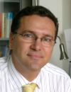
Bernd Rieger, Quantitative Imaging Group Leader, Delft University of Technology, Netherlands
| 09:30 |  | Keynote Presentation From Molecules To Whole Organs
Francesco Pavone, Principal Investigator, LENS, University of Florence, Italy
Some examples of correlative microscopies, combining linear and non linear techniques will be described. Particular attention will be devoted Alzheimer disease or to neural plasticity after damage as neurobiological application. |
| 10:15 | Super-Resolution Imaging by dSTORM
Markus Sauer, Professor, Julius-Maximilians-Universität Würzburg, Germany
In this presentation I will demonstrate the advantageous use of dSTORM for quantitative imaging of synaptic proteins, the study of plasma membrane organization, and the molecular architecture of multiprotein complexes. Finally, I will outline how dSTORM can be used advantageously to improve next generation medical therapies. | 10:45 | Coffee and Networking | 11:15 | Correlated Fluorescence And X-Ray Tomography: Finding Molecules In Cellular CT Scans
Carolyn Larabell, Professor, University of California San Francisco, United States of America
Fluorescence microscopy reveals the distribution of specific molecules, soft x-ray tomography generates 3D views of cell structures, and overlaying the two data sets enhances the information obtained. I will describe these technologies and present examples of their applications. | 11:45 | Integrating Advanced Fluorescence Microscopy Techniques Reveals Nanoscale Architecture And Mesoscale Dynamics Of Cytoskeletal Structures Promoting Cell Migration And Invasion
Alessandra Cambi, Assistant Professor, University of Nijmegen, Netherlands
This lecture will describe our efforts to exploit and integrate a variety of advanced microscopy techniques to unravel the nanoscale structural and dynamic complexity of individual podosomes as well as formation, architecture and function of mesoscale podosome clusters. | 12:15 | Multi-Photon-Like Fluorescence Microscopy Using Two-Step Imaging Probes
George Patterson, Investigator, National Institutes of Health, United States of America
We present a multi-photon like approach to non-linear imaging based on fluorescent molecules which require sequential photon absorption steps to produce fluorescence. | 12:45 | Lunch and Networking | 13:30 | Poster Viewing Session | 14:00 | 3D Single Particle Tracking: Following Mitochondria in Zebrafish Embryos
Don Lamb, Professor, Ludwig-Maximilians-University, Germany
Single particle tracking (SPT) allows one to follow objects as they perform their normal tasks. I will give a general introduction to SPT and exemplify what I introduce with research from my group on tracking of Mitochondria in Zebrafish Embryos. | 14:30 | Visualizing Mechano-Biology: Quantitative Bioimaging Tools To Study The Impact Of Mechanical Stress On Cell Adhesion And Signalling
Bernhard Wehrle-Haller, Group Leader, University of Geneva, Switzerland
In my talk I will present the use and design of fluorescent probes for quantitative live cell imaging, in order to study the function of specific proteins of the cytoskeleton or cell-matrix adhesions, relevant to adhesion, migration and survival of normal and cancer cells. | 15:00 | Optical Imaging of Molecular Mechanisms of Disease
Clemens Kaminski, Professor, University of Cambridge, United Kingdom
I will present dynamic and superresolved imaging techniques to study protein misfolding and aggregation in the context of neurodegeneration. | 15:30 | Coffee and Networking | 16:00 | 3-D Optical Tomography For Ex Vivo And In Vivo Imaging
James McGinty, Professor, Imperial College London, United Kingdom
During my presentation I will give example applications, including immunohistology of excised whole mouse pancreas, in vivo fluorescence lifetime OPT of zebrafish embryos, mapping of tumour progression and vascularisation in live adult zebrafish and resolving a FRET interaction in a mouse model. | 16:30 | Imaging Gene Regulation in Living Cells at the Single Molecule Level
James Zhe Liu, Group Leader, Janelia Research Campus, Howard Hughes Medical Institute, United States of America
I will discuss our latest efforts on utilizing advanced live imaging modalities to study molecular dynamics underlying stem cell pluripotency and neurological disorders. | 17:00 | End Of Day One |
Wednesday, 15 June 2016 |
Day Two Session |
| | 09:30 |  | Keynote Presentation Super-Resolution Microscopy With DNA Molecules
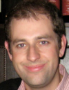
Ralf Jungmann, Group Leader, Max Planck Institute of Biochemistry, Germany
Using DNA-based barcoding and super-resolution imaging approaches, we eventually want to unravel the location and interplay of a multitude of genes, RNAs, and proteins in a truly quantitative fashion with highest spatial resolution in single cells and on cell surfaces. |
| 10:15 | A Revolutionary Miniaturised Instrument For Single-Molecule Localization Microscopy And FRET
Achillefs Kapanidis, Professor, University of Oxford, United Kingdom
The development, capabilities and applications of a robust, versatile, and automated desktop instrument for single-molecule fluorescence studies will be discussed. | 10:45 | Coffee and Networking | 11:00 | Democratising Live-Cell High-Speed Super-Resolution Microscopy
Ricardo Henriques, Group Leader, University College London, United Kingdom
Democratising live-cell high-speed low-illumination super-resolution microscopy. | 11:30 | Single Nano-Object Tracking And Localization Microscopy For Applications In Live Brain Tissues
Laurent Cognet, CNRS Research Director, LP2N - Institut d'Optique, University of Bordeaux, France
Super-resolution microscopy and single particle tracking are presented in live neurons and acute brain slices through the development of different approaches based on uPAINT microscopy, single quantum dot tracking or single carbon nanotube tracking. | 12:00 | Information In Localisation Microscopy
Patrick Fox-Roberts, PostDoc, Kings College London, United Kingdom
The information flow through the microscope limits the speed of localisation microscopy. Higher speeds lead to errors which can be spotted using Bayesian analysis. | 12:30 | Lunch and Networking | 13:00 | Poster Viewing Session | 13:25 | Poster Prize Presentation Sponsored by ePosters | 13:30 | High-Content Imaging Approaches For Drug Discovery For Neglected Tropical Diseases
Manu De Rycker, Team Leader, University of Dundee, United Kingdom
The development of new drugs for intracellular parasitic diseases is hampered by difficulties in developing relevant high-throughput cell-based assays. Here we present how we have used image-based high-content screening approaches to address some of these issues. | 14:00 | High Resolution In Vivo Histology: Clinical in vivo Subcellular Imaging using Femtoseceond Laser Multiphoton/CARS Tomography
Karsten König, Professor, Saarland University, Germany
We report on a certified, medical, transportable multipurpose nonlinear microscopic imagingsystem based on a femtosecond excitation source and a photonic crystal fiber with multiple miniaturized time-correlated single-photon counting detectors. | 14:30 | Coffee and Networking | 14:45 | Lateral Organization Of Plasma Membrane Constituents At The Nanoscale
Gerhard Schutz, Professor, Vienna University of Technology, Austria
It is of interest how proteins are spatially distributed over the membrane, and whether they conjoin and move as part of multi-molecular complexes. In my lecture, I will discuss methods for approaching the two questions, and provide biological examples. | 15:15 | Correlative Light And Electron Microscopy In Structural Cell Biology
Wanda Kukulski, Group Leader, University of Cambridge, United Kingdom
Correlating fluorescence microscopy with electron tomography allows combining protein localization and dynamics with 3D cellular ultrastructure. I will present how we use this approach to understand vesicle budding at the plasma membrane. | 15:45 | Close of Conference |
|

 Add to Calendar ▼2016-06-14 00:00:002016-06-15 00:00:00Europe/LondonBioimaging: From Cells To Molecules 2016 Bioimaging: From Cells To Molecules 2016 in Cambridge, UKCambridge, UKSELECTBIOenquiries@selectbiosciences.com