Co-Located Conference AgendasBiosensors & Biosecurity Summit 2017 | POC Diagnostics, Global Health-Viral Diseases 2017 |  Other Track Agendas Other Track AgendasLab-on-a-Chip and Microfluidics: Companies, Technologies and Commercialization | Lab-on-a-Chip and Microfluidics: Emerging Themes, Technologies and Applications |

Monday, 2 October 201712:45 | Conference Registration, Materials Pick-Up, and Networking | |
Session Title: Opening Plenary Session -- Convergence of Microfluidics, POC Diagnostics and Biosensors |
| | 13:30 |  | Keynote Presentation Micro & Nanotechnologies For Physical Phenotyping of Cells
Dino Di Carlo, Armond and Elena Hairapetian Chair in Engineering and Medicine, Professor and Vice Chair of Bioengineering, University of California-Los Angeles, United States of America
My lab uses microtechnology to interface at the scale of biology to aid in scientific investigation, develop new approaches to diagnose and monitor disease, and engineer therapies. A part of our laboratory develops tools to assay and exploit physical properties of cells in diagnostics and drug screening. Physical properties of cells can provide integrative, rapid, and low-cost information about disease. My discussion will focus on instruments we have developed that quantify single-cell physical phenotypes such as deformability, size, and contractility. These new tools show promise to enable diagnostic and screening approaches that assay immune cell function at the point-of-care, tumor cell malignancy with higher confidence than molecular markers alone, and many other cell phenotypes associated with disease. |
| 14:00 |  | Keynote Presentation Re-envisioning Point-of-Care Pathogen Diagnostics for the Developed and Developing Worlds
Paul Yager, Professor, Department of Bioengineering, University of Washington and CSO, UbiDX, Inc., United States of America
Whether the purpose is to guide treatment of an individual’s infection, or to control the outbreak of a pandemic, there is an urgent need for low-cost rapid diagnostic devices capable of identifying the cause of infectious disease that free testing from the centralized laboratory. “Ubiquitous diagnostics”, aided by the revolution that brought us today’s distributed computing and communications, can bring the best diagnostic capabilities to physicians’ office laboratories and pharmacies in the developed world, or to places in the developing world where nothing is available now. The practice of medicine itself can be improved if diagnostic tests could be carried out more rapidly and pervasively. The Yager lab has been engaged in a 20-year pursuit of microfluidics-based tools and complete systems for pathogen identification in human samples, most recently in an inexpensive instrument-free single-use disposable format that could be used by consumers in their homes and by healthcare workers in low-resource settings in the developing world. The central effort today is to utilize capillary action in porous materials to eliminate the need for pumps and other equipment to control fluid flow. By coupling the chemical testing with cell phones, critical health data can be analyzed rapidly and anywhere, and the best healthcare decisions can be made for patients, for regional healthcare systems, and for global health. There have been 3 analytical approaches pursued in our group: Identification and quantification of 1) antibodies specific to pathogens, 2) proteins of the pathogens, and 3) nucleic acid sequences derived from the pathogens. We report on recent results on particularly simple paper-based systems supported by NIH (detection of influenza proteins; improvement of specificity of Zika virus serology), DARPA (detection of DNA and RNA from bacterial and viral pathogens) and DTRA (detection of proteins from the Ebola virus). All projects involve close collaboration with partners inside and outside the university environment, and are aimed at producing clinically useful tools for point-of-care medicine. |
| 14:30 |  | Keynote Presentation Paper-based Nanobiosensors: Diagnostics Going Simple
Arben Merkoçi, ICREA Professor and Director of the Nanobioelectronics & Biosensors Group, Institut Català de Nanociencia i Nanotecnologia (ICN2), Barcelona Institute of Science and Technology (BIST), Spain
Biosensors field is progressing rapidly and the demand for cost
efficient platforms is the key factor for their success. Physical,
chemical and mechanical properties of cellulose in both micro and
nanofiber-based networks combined with their abundance in nature or easy
to prepare and control procedures are making these materials of great
interest while looking for cost-efficient and green alternatives for
device production technologies. Both paper and nanopaper-based
biosensors are emerging as a new class of devices with the objective to
fulfill the “World Health Organization” requisites to be ASSURED:
affordable, sensitive, specific, user-friendly, rapid and robust,
equipment free and deliverable to end-users. How to design simple
paper-based biosensor architectures? How to tune their analytical
performance upon demand? How one can ‘marriage’ nanomaterials such as
metallic nanoparticles, quantum dots and even graphene with paper and
what is the benefit? How we can make these devices more robust,
sensitive and with multiplexing capabilities? Can we bring these low
cost and efficient devices to places with low resources, extreme
conditions or even at our homes? Which are the perspectives to link
these simple platforms and detection technologies with mobile phone
communication? I will try to give responses to these questions through
various interesting applications related to protein, DNA and even
contaminants detection all of extreme importance for diagnostics,
environment control, safety and security. |
| 15:00 |  | Keynote Presentation Microfluidic Devices – Key Technologies to Enable Real-Time Patient Monitoring and Treatment
Martyn Boutelle, Professor of Biomedical Sensors Engineering, Imperial College London, United Kingdom
Clinical practice is beginning to wake upto the potential of real-time molecular information from venerable tissue as a means to understand the progression in the tissue of injury or disease. Such patterns of molecular changes, particularly when combined with paternal of physical of electrical signatures, offer the exciting possibility of allowing clinicians to guide therapy on an individualized basis in real time. In this presentation I will describe the development of 3D printed microfluidic devices connected to wireless electronics for transplant organ and patient monitoring. Tissue sampling is via integrated microdialysis probes. Concentrations of important biomarkers are measured using microscale amperometric biosensors (energy metabolites, and excitatory neurotransmitters) and ion-selective electrodes (ISE) for tissue ionic balance. Detailed patterns of ionic responses can be revealed using a high density ISE array within the flow stream. High time resolutions can be achieved using a novel droplet based microfluidic system. The presentation with describe the design and optimization challenges and include clinical examples from our recent work. |
| 15:30 | Coffee Break and Networking | 16:15 |  | Keynote Presentation Microfluidics for Bottom-Up Tissue Engineering
Shoji Takeuchi, Professor, Center For International Research on Integrative Biomedical Systems (CIBiS), Institute of Industrial Science, The University of Tokyo, Japan
In this presentation, I will talk about several Microfluidic-based approaches for the rapid construction of 3D-cellular construct. Large-scale 3D tissue architectures that mimic microscopic tissue structures in vivo are very important for not only in tissue engineering but also drug development without animal experiments. We demonstrated a construction method of 3D tissue structures by using cell beads and cell fibers. To prepare the cellular beads, we used an axisymmetric flow focusing device (AFFD) that allows us to encapsulate cells within monodisperse collagen beads. By molding these cell beads into a 3D chamber and incubating them, we successfully obtained complicated and milli-sized 3D cellular constructs. As the cell fibers, a cell-encapsulating core-shell hydrogel fiber was produced in a double coaxial laminar flow microfluidic device. When with myocytes, endothelial, and nerve cells, they showed the contractile motion of the myocyte cell fiber, the tube formation of the endothelial cell fibers and the synaptic connections of the nerve cell fiber, respectively. By reeling, weaving and folding the fibers using microfluidic handling, higher-order assembly of fiber-shaped 3D cellular constructs can be performed. Moreover, the fiber encapsulating beta-cells is used for the implantation of diabetic mice, and succeeded in normalizing the blood glucose level. |
| 16:45 |  | Keynote Presentation Mixed-Scale Fluidic System: Searching for Drug-induced DNA Damage in Circulating Tumor Cells
Steve Soper, Foundation Distinguished Professor, Director, Center of BioModular Multi-Scale System for Precision Medicine, The University of Kansas, United States of America
Improved therapies that yield more cures and better overall survival for cancer patients are needed. For example, women with breast cancer have a 5-year survival rate of 22% (Stage IV) and 72% (Stage III). Doxorubicin, cisplatin, paclitaxel, and tamoxifen are examples of drugs used for treating breast cancer with selection of therapy typically based on the classification and staging of the patient’s cancer. While treatment regimens assigned to some patients may be optimal using the current classification model, others within certain breast cancer sub-types fail therapy. New assays must be developed to determine how a patient’s physiology and genetic makeup affects drug efficacy. In this presentation, a series of chips are used for the isolation and processing of circulating tumor cells (CTCs). The chips quantify response to therapy using three pieces of information secured from the CTCs; (1) CTC number; (2) CTC viability; and (3) the frequency of DNA damage (abasic (AP) sites) in genomic DNA (gDNA) harvested from the CTCs. Microscale chips are used for CTC selection, CTC enumeration and viability determinations. The chip to read AP sites is a nanosensor chip made via nano-imprinting in plastics and contains a nanochannel with dimensions less than the persistence length of double-stranded DNA (~50 nm). Labeling AP sites with fluorescent dyes and stretching the gDNA in the nanochannel to near its full contour length allows for the direct readout of the AP sites, even from a single CTC. This information is used to determine how a patient is responding to therapy. |
| 17:15 | 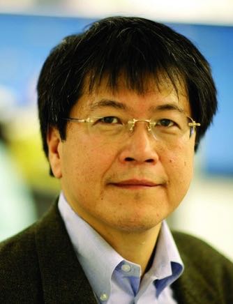 | Keynote Presentation PCR-Free MicroRNA Quantification Based on Ion-Selective Nanoporous Membranes and Nanopores
Hsueh-Chia Chang, Bayer Professor of Chemical and Biomolecular Engineering, University of Notre Dame, Interim Chief Technology Officer, Aopia Biosciences, United States of America
We report an integrated biochip platform that can identify and quantify low copy numbers of microRNA biomarkers in a heterogeneous physiological sample like blood, saliva or urine. This quantification assay is done without PCR amplification, reporter labeling, extensive off-chip pretreatment and expensive optical sensors. Consequently, it does not introduce PCR/ligation bias and limits analyte loss during pretreatment. The main components of the integrated biochips are nanoporous membranes and solid-state nanopores with pore radii smaller than the nm-scale Debye length. We use the ion concentration and charge polarization features of the ion-selective membranes to control the on-chip ionic strength, actuate pH by splitting water, lyse exosomes and isolate/concentrate the target molecules. The final nanopore sensor or sensor array utilizes surface modification and pore geometry to preferentially delay the translocation time of the target microRNAs to achieve single-molecule identification and quantification. The integrated chip achieves a translocation frequency (throughput) that is at least one hundred times higher than any literature or commercial nanopore technology. |
| 17:45 | 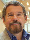 | Keynote Presentation Nanomotor-Microchip Diagnostics: Moving the Receptor in Microchannel Networks
Joseph Wang, Chair of Nanoengineering, SAIC Endowed Professor, Director at Center of Wearable Sensors, University of California-San Diego, United States of America
This presentation will describe new motion-based microchip assays based on autonomously moving receptor-functionalized nanomotors. The new motor-based sensing approach relies on new capabilities of modern nano/microscale motors. Particular attention will be given to catalytic nanowire and microtube motors propelled by the electrocatalytic decomposition of a chemical fuel, The increased cargo-towing force of new man-made nanomotors, along with their precise motion control within microchannel networks, versatility and facile functionalization, can be combined for developing advanced microchip systems based on active transport. The motion-based biosensing strategy relies on the continuous movement receptor-modified microengines through complex samples in connection to diverse ‘on-the-fly’ biomolecular interactions of nucleic acids, proteins, bacteria or cancer cells. A variety of receptors, attached to self-propelled nanoscale motors, can thus move around the sample and, along with the generated microbubbles, lead to greatly enhanced fluid transport and accelerated recognition process. Selective capture and transport of target DNA and cancer cells from raw complex body fluids will be demonstrated. Key factors governing such motion-based sensing will be covered. The resulting assays add new and rich dimensions of analytical information and offer remarkable sensitivity, coupled with simplicity, speed and low costs. We will discuss the challenges of implementing molecular recognition into the nanomotor movement and for generating well-defined distance signals. New microengines with a ‘built-in’ recognition capability, based on boronic-acid or molecularly-imprinted outer layers, will also be discussed. The latter obviated the need for the receptor immobilization. The greatly improved capabilities of chemically-powered artificial nanomotors could pave the way to exciting and important bioanalytical applications and to sophisticated nanoscale and microchip devices performing complex tasks. |
| 18:15 | Networking Cocktail Reception with Beer, Wine and a Light Dinner in the Exhibit Hall. Engage with Colleagues and Visit the Exhibitors | 19:45 | Close of Day 1 of the Conference |
Tuesday, 3 October 201708:00 | Conference Registration, Materials Pick-Up, Morning Coffee and Breakfast Pastries | |
Session Title: Emerging Approaches and Applications in the Microfluidics and Lab-on-a-Chip World |
| | 09:30 |  | Keynote Presentation Microfluidic Technologies for Liquid Biopsy & Precision Medicine
Chwee Teck Lim, NUS Society Chair Professor, Department of Biomedical Engineering, Institute for Health Innovation & Technology (iHealthtech), Mechanobiology Institute, National University of Singapore, Singapore
Capturing of circulating Tumor Cells (CTCs) from the peripheral blood of patients (a.k.a. liquid biopsy) can potentially lead to applications in cancer diagnosis and personalized medicine. Using a label-free approach via our developed microfluidic biochips, we not only show successful detection and retrieval of CTCs, but also how selective capturing of each single CTCs can lead to downstream single cell analysis and enable personalized treatment through the detection of specific actionable or druggable mutation. |
| 10:00 |  | Keynote Presentation Instrumented Tissue-in-a-Chip: A Bridge from In Vitro to In Vivo
Charles Henry, Professor and Chair, Colorado State University, United States of America
Microfluidic methods provide a promising path to mimicking human organ function with applications ranging from fundamental biology to drug metabolism and toxicity. The vast majority of these systems use dissociated, immortalized, or stem cells to create two and three-dimensional models in vitro. While these systems can provide valuable information, they are fundamentally incapable of recreating the three-dimensional complexity of real tissue. As a result, an important gap exists between in vitro models and in vivo systems. To address this gap, we have created instrumented Tissue-In-a-Chip (iTIC) system that combines microfluidic devices with ex vivo tissue slices or explants to recreate model systems that capture the cellular diversity of real tissue and bridge the gap between in vitro models and in vivo systems. In this presentation two systems will be discussed. The first uses a high-density electrode array equipped with microfluidic flow to image chemical release profiles from living adrenal slices. The second uses a 3D printed microfluidic device with removable inserts to hold and perfuse fluids over intestinal tissue, enabling generation of differential chemical conditions on either side of this important barrier tissue. |
| 10:30 | Coffee Break and Networking in the Exhibit Hall | 11:15 |  Microfluidic Mixing: Generic Solutions to a Ubiquitous Problem Microfluidic Mixing: Generic Solutions to a Ubiquitous Problem
Richard Chasen Spero, CEO, Redbud Labs
Some microfluidic challenges are common across applications. Perhaps none is more pervasive than microfluidic mixing, which is a necessary but confounding problem in many devices. Done effectively, microfluidic can dramatically accelerate molecular assays, improve analytic precision, and boost sensitivity. Here we survey applications where mixing is essential to assay success, including 10x faster microarray assays, 200x faster reagent dispersal, and 5x reduction in hybridization time. We will discuss the prognosis for achieving a generic solution for microfluidic mixing, with a focus on methods that can be combined with practical cartridge manufacturing and assembly methods.
| 11:45 | Constructing Microfluidic Analytical Systems from 3D-Printed Blocks with Integrated Functional Components
Noah Malmstadt, Professor, Mork Family Dept. of Chemical Engineering & Materials Science, University of Southern California, United States of America
The integration of functional components into microfluidic systems is necessary for building compact analytical devices. Optical detection and measurement, electromechanical fluid switching, active mixing, and magneto- and electrophoretic separations all require the integration of components on top of the underlying fluidic architecture. There has been significant work towards integrating such functional components by including them directly in the microfabrication process. This approach, however, leads to expensive fabrication workflows that are often limited to a narrow set of applications. To overcome these limits, we are designing a microfluidic architecture that is easily adaptable to a wide variety of applications. This architecture is based on 3D-printed elements that can be assembled into analytical devices. Functional components are integrated into these elements; the structures are printed to directly accommodate off-the-shelf components including photodiodes, heaters, sensors, and fiber optic fittings. We have demonstrated the utility of this approach by assembling a system capable of performing automated ELISA assays from 3D-printed blocks with integrated functional components. | 12:15 | Networking Lunch in the Exhibit Hall -- Meet the Exhibitors and View Posters | |
Session Title: The Convergence of Microfluidics, 3D-Printing and Tissues-on-Chips |
| | 13:30 | Hydrogel Microarrays and Microfluidics – A Different Spin to Traditional SLA- and Extrusion-based 3D Printing Methods
Luiz Bertassoni, Associate Professor, Biomaterials and Biomechanics, School of Dentistry, Cancer Early Detection Advanced Research, Knight Cancer Institute, Oregon Health & Sciences University, United States of America
Fabrication of three-dimensional tissues with controlled microarchitectures has been shown to enhance tissue functionality. 3D printing can be used to precisely position cells and cell-laden materials to generate controlled tissue architectures. Therefore, it represents an exciting alternative for organ fabrication. Our group has been interested in developing innovative printing-based technologies to improve our ability to regenerate tissues with improved function, as well as to engineer hydrogel based microfluidic devices. In this seminar, we will present SLA/DLP-based 3D printing methods to fabricate high-throughput screening platforms to probe mechanotransduction and geometry-controlled stem cell differentiation. Further, we will discuss recent methods our lab has developed to engineer vascularized tissue constructs, and 3D printed magnetically-gated hydrogel microfluidic chips. The use of these technologies in various regenerative applications will be discussed. | 14:00 |  Latest Advancements in Microfluidic Flow Control and Microfabrication Technologies for Diagnostic, Organ-on-Chip and MEMS Applications Latest Advancements in Microfluidic Flow Control and Microfabrication Technologies for Diagnostic, Organ-on-Chip and MEMS Applications
Marko Blom, Chief Technical Officer, Micronit Microtechnologies
We will present the latest advances in flow control, together with commercially and scientifically relevant examples. Flow control will be discussed in a broad sense showing membrane-based valves and capillary flow control. We will show integrated membrane valve examples realized via adhesive-free techniques, combining a range of validated thermoplastic and elastomeric materials, with actuation via pneumatic but also via other means. Furthermore, we will present integration of the electrostatic triggering mechanism of a capillary burst valve in a low-cost, thermoplastic chip, enabling sequential capillary flow in fully autonomous Point-of-Care devices. On the chip design and fabrication side we will show workflow automation for the chip and process design process, partially enabled by recently initiated ISO standardization efforts. Combined with this we will present standardized top-down and edge-connect micro-to-micro and micro-to-macro fluidic and electrical interfacing. Finally, we will show new fabrication technologies using laser-based processing, next to integration techniques particularly relevant to the Organ-on-Chip field, integrating membranes and sensing elements, among others enabling TEER (transepithelial electrical resistance) and oxygen sensing.
| 14:30 |  | Keynote Presentation Digital Droplet Generation: From Digital PCR to Digital Pipetting
Tingrui Pan, Professor, Department of Biomedical Engineering, University of California-Davis, United States of America
Digital droplet generation enabled by microfluidic impact printing has been recently introduced, benefiting from the nature of simple device architecture, digital metering, low cost, non-contamination, scalable multiplexability and high throughput. In this talk, we will review this novel microfluidic-based droplet generation platform, utilizing modular microfluidic cartridges and expandable combinatorial printing capacity controlled by plug-and-play multiplexed actuators. Such a customizable microfluidic printing system allows for ultrafine control of the droplet volume from picoliters (~10pL) to nanoliters (~100nL), a 10,000 fold variation. The high flexibility of droplet manipulations can be simply achieved by controlling the magnitude of actuation (e.g., driving voltage) and the waveform shape of actuation pulses, in addition to nozzle size restrictions. Importantly, we will discuss the recent exciting developments of the microfluidic droplet generation into an array of emerging biomedical applications, including digital PCR and digital pipetting. |
| 15:00 | Coffee Break and Networking in the Exhibit Hall | 15:45 | Free-Surface Microfluidics and SERS for High Performance Sample Capture and Analysis
Carl Meinhart, Professor, University of California-Santa Barbara, United States of America
Nearly all microfluidic devices to date consist of some type of fully-enclosed microfluidic channel. The concept of ‘free-surface’ microfluidics has been pioneered at UCSB during the past several years, where at least one surface of the microchannel is exposed to the surrounding air. Surface tension is a dominating force at the micron scale, which can be used to control effectively fluid motion. There are a number of distinct advantages to the free surface microfluidic architecture. For example, the free surface provides a highly effective mechanism for capturing certain low-density vapor molecules. This mechanism is a key component (in combination with surface-enhanced Raman spectroscopy, i.e. SERS) of a novel explosives vapor detection platform, which is capable of sub part-per-billion sensitivity with high specificity. | 16:15 |  | Keynote Presentation Microfluidic Chip System Combined with Tetrahedron DNA Nanostructures For Circulating Tumor Cell Capture
Xianqiang Mi, Professor, Shanghai Advanced Research Institute Chinese Academy of Sciences, China
As the typical liquid biopsy technology for cancer prevention and control, circulating tumor cells (CTCs) analysis shows great prospects. However, the extremely low content of CTCs in blood brings great difficulty for its capture and detection. Microfluidic chip technology and DNA nano technology have made great progress in CTCs capture, but there are still some shortcomings, such as low capture efficiency, narrow spectrum for target and easily leading to cell inactivation. Here, based on the principle of microfluidic immuno-affinity, we propose a new CTCs capture method with the combination of microfluidic chip and DNA nanostructure technology. The performance of the developed microfluidic chip was verified by the tumor cell suspension and real blood samples. This new CTC capture method has the advantage of high capture efficiency, broad spectrum for target and good ability to keep cell viability. |
| 16:45 | Bioprinting Pancreas-on-a-Chip
Ibrahim Ozbolat, Hartz Family Associate Professor of Engineering Science and Mechanics, The Huck Institutes of the Life Sciences, Penn State University, United States of America
Despite the recent achievements in cell-based therapies for curing type-1 diabetes (T1D), vascularization of beta (ß)-cell clusters is still a major roadblock as it is essential for long-term viability and function of ß-cells in vivo. In this research, we report micro-vascularization within engineered pancreatic islets (EPIs) made of rodent cells. EPIs cultured in fibrin constructs maintained their viability and functionality over time while non-vascularized EPIs could not retain their viability nor functionality. We then bioprinted the EPIs along with bioprinted macro-vascular network in a pancreas-on-a-chip model. Here we demonstrate a proof-of-concept study for a vascularized pancreas-on-a-chip model for the first time, where patient specific stem cell-derived human beta cells can be vascularized in the near future for an effective treatment of T1D. | 17:15 |  | Keynote Presentation Bioprinting: The State of the Field
Gabor Forgacs, Professor, University of Missouri-Columbia; Scientific Founder, Organovo; CSO, Modern Meadow, United States of America
The modern era of bioprinting commenced in 2000 with the work of Thomas Boland and his re-engineered Hewlett Packard desktop inkjet printer. Since then a number of other technologies have been developed utilizing extrusion, acoustic waves, laser assisted delivery and lately “liquid bioprinting”. Meanwhile the field has also matured from its purely academic roots into successful commercial ventures. Overall bioprinting has seen spectacular progress in the past 17 years and a number of market analyses have predicted a bright future for the field. I will provide here a hype-free overview of the technology, where it stands today, what it has specifically accomplished and what can be expected in the years to come. |
| 17:45 | Networking Cocktail Reception with Beer, Wine and Appetizers in the Exhibit Hall. Engage with Colleagues and Visit the Exhibitors | 19:30 | Close of Day 2 of the Conference. |
Wednesday, 4 October 201708:00 | Morning Coffee, Breakfast Pastries and Networking in the Exhibit Hall | |
Session Title: Novel End-User Applications of Microfluidics/LOAC |
| | 08:30 |  | Keynote Presentation Controlled Incremental Filtration: A Novel Microfluidic Approach for High-Throughput Separation of Circulating Cells from Whole Blood
Sergey Shevkoplyas, Associate Professor, Biomedical Engineering, University of Houston, United States of America
Separation of circulating cells from whole blood and other blood-derived samples is an important part of a broad range of medical applications. A versatile microfluidic approach that is suitable for a large range of particle sizes and high levels of enrichment, with a volumetric throughput sufficient for large-scale applications, has yet to emerge. Most microfluidic technologies developed for separating circulating cells from blood have been of very little use for large volume processing because of relatively low volumetric throughputs, small minimal feature sizes, and the need for exceedingly dilute samples. Controlled incremental filtration (CIF), a new microfluidic approach we recently developed, enables a microfluidic device with a relatively small footprint to operate at practical throughputs (>15mL/min), have distinctly manufacturable minimal feature sizes (>20µm), process blood samples without dilution, and concentrate cells of interest >40-fold. This talk will describe the use of CIF-based devices for leukoreduction of platelet rich plasma, and extraction of lymphocytes from whole blood. |
| 09:00 |  | Keynote Presentation A Novel Platelet Assay Device for Bleeding Screening and Drug Response
Sehyun Shin, Professor & Director, Nano-Biofluignostic Engineering Research Center, Korea University and Anam/Guro Hospital of Korea University, Korea South
|
| 09:30 | Microfluidic On-Demand Harvesting of Therapeutic Exosomes
Mei He, Assistant Professor, University of Kansas and Chief Science Officer, Clara Biotech, United States of America
The finding of exosomes opens new opportunities for developing nature, non-toxic therapeutic delivery systems. Exosomes play important biological roles via transferring selectively enriched proteins, RNAs, and mitochondrial DNA. The nano-sized exosomes are highly biocompatible with intrinsic payload and exhibit much stronger antigen loading flexibility, compared to other polymer nano-platforms. In spite of the significant roles in therapeutics and diagnostics, the study and development of the utility of exosomes is hampered by substantial technical difficulties in obtaining sufficient and pure immunogenic exosomes. Current production protocols (e.g, ultracentrifugation and filtration) are un-scalable, often labor-intensive and time-intensive, and in low-yield and purity (< 25%). In this work, we report a versatile, scalable microfluidic approach for processing exosomes with precise control and specificity, and on-demand harvesting. Current microfluidic exosome isolation approaches either handle limited quality of exosomes in microliter scale, or the processed exosomes are bound to solid surface/particles and unable to stay intact. We report continuous-flow, light-triggered on-demand harvesting of exosomes over milliliter scale of volumes. We foresee that microfluidic technology will provide game changer roles for exploring the utility of exosomes in therapeutics. | 10:00 |  | Keynote Presentation Flow Control and Configurable Microstructures Using Non-Uniform Electroosmosis
Moran Bercovici, Associate Professor, Faculty of Mechanical Engineering; Head, Technion Microfluidic Technologies Laboratory, Technion, Israel Institute of Technology, Israel
Electro-osmotic flow (EOF) is the motion of a liquid due to interaction of an externally applied electric field with the net charge in the diffuse part of an electrical double layer.
I will present our ongoing work leveraging non-uniform EOF to control flow patterns in a Hele-Shaw microfluidic chamber (two parallel plates separated by a small gap). By setting the spatial distribution of surface potential, we demonstrate the ability to dictate desired flow patterns without the use of physical walls. Furthermore, by replacing the rigid ‘ceiling’ (top plate) of the chamber with a thin elastic sheet, we demonstrate that internal pressure gradients formed in the liquid can be used to drive local deformations in the sheet. This opens the door to the implementation of configurable microstructures and microfluidic devices. In addition to experimental demonstrations, I will present key elements of the analytical models we developed, and discuss both future applications and limitations of the technology. |
| 10:30 | Coffee Break and Networking in the Exhibit Hall | 11:15 | HP Advanced Microfluidics and Applications
Alexander Govyadinov, Senior Technologist, HP Incorporated, United States of America
The inkjet technology platform utilizing microfabrication processes and
well established materials can be repurposed for development of variety
of advanced microfluidic devices. We demonstrated advanced microfluidic
components such as integrated micropumps and fragile components
transport systems, micromixers, microfluidic valves and switch boards
and other components of advanced microfluidics. This presentation
describes operating principles of microfluidic elements, examples of
their integration in functional devices and discusses inkjet technology
potential for broad range of microfluidic life science applications. | 11:45 | .jpg) | Keynote Presentation CD Fluidics for Extreme Point of Care (EPOC)
Marc Madou, Chancellor's Professor, University of California-Irvine, United States of America
In this contribution, we review the usefulness of centrifugal microfluidic technologies applied to point-of-care diagnosis in under-resourced or extreme environments. The various challenges faced in these extreme point-of-care (EPOC) settings are showcased, using areas in India and Africa as examples. Measures for point-of-care devices to effectively address these challenges are highlighted, and centrifugal or CD-based microfluidic technologies are presented as a promising solution to accomplish these criteria. We describe the advantages of centrifugal fluidic handling, as well as the ability of a standard CD player to perform several standard laboratory tests, fulfilling the role of an integrated lab-on-a-CD. Innovative centrifugal approaches for extreme point-of-care are highlighted, including sensing and detection strategies, smart power sources and biomimetic inspired solutions for environmental control. The evolution of centrifugal microfluidics, along with examples of commercial and advanced prototype centrifugal microfluidic systems, is presented, illustrating the success of deployment at the point-of-care. A close fit of emerging centrifugal systems to address a critical panel of tests for under-resourced clinic settings, formulated by medical experts, is demonstrated. This emphasizes the potential of centrifugal microfluidic technologies to be applied effectively to extreme point-of-care scenarios and plays a role in improving primary care in resource-limited settings across the world. |
| 12:15 | Networking Lunch in the Exhibit Hall -- Meet the Exhibitors and View Posters | |
Session Title: Late-Breaking Technologies and Approaches in the Microfluidics and Lab-on-a-Chip Fields |
| | 13:30 | Nanotechnology for a Genomic Revolution
Shane Bowen, Director, Scientific Research; Dept. of Research & Technology Development, Illumina, United States of America
Over the duration of the human genome project (HGP) from 1990 to 2003, more than $2.7 billion dollars was spent across 20 global institutions resulting in the cataloging of a human genome at 8 -9 X coverage. This was a monumental achievement completed through the use of hierarchical shotgun method, which set the foundation upon which modern methods of genetic analysis stand. Since the completion of the HGP, numerous efforts have been put forth to increase the accuracy, simplify the process reduce the amount of time and cost associated with measuring the sequence of any organism. This is now the foundation of modern medicine and is on the verge of a changing the way not only healthcare is performed but also revolutionizing agriculture, forensics and pharmaceutical development around the world. Over the years of 2009 through 2014, myself and a team of research scientists at Illumina developed a method to sequence a human genome in less than 48 hours at a coverage of 30X and at the cost of $1000. During this talk, I will give an overview of the technologies developed to make this happen as well as set the stage for the next iteration of innovations paving the path to the $100 genome, being enabled by the NovaSeq platform commercialized by Illumina this year. | 14:00 | Integrating Microfluidics and Tissue Engineering for Organ-on-a-Chip Applications
Kennedy Okeyo, Senior Lecturer, Institute for Frontier Life and Medical Sciences, Kyoto University, Japan
Organ-on-a-chip continues to attract a great deal of attention owing to their potential application in drug development, disease modeling and basic biological studies. For these applications, it is important to develop realistic on-chip organ models which recapitulates tissue-specific, differentiated functions of many cell types for accurate prediction of in vivo tissue functions and drug activities. This talk will introduce our strategy of combining microfluidics and tissue engineering approaches toward fabrication of tissue/organ models in which several cell types are in direct interaction, hence enabling the realization of tissue-level cell-cell interaction, which is important for recapitulating in vivo tissue functions. We will also highlight recent progress in step-by-step fabrication of higher-order tissue models achieved by the mesh culture technique, and on-going integration with microfluidics to enable disease modeling and, potentially, drug activity monitoring. | 14:30 | Nanofluidics Enables Applications in Ionic Flow Control and Water Desalination
Shaurya Prakash, Associate Professor, Department of Mechanical and Aerospace Engineering, The Ohio State University, United States of America
Nanofluidics targets the science and technology for flow in and around structures with critical dimensions in the 1-100 nm range. Over the past decade, several advances in science of nanofluidic transport has enabled several novel applications relevant to the microfluidics and nanofluidics device community. In this talk, I will showcase the scientific advances pioneered by my group at the Microsystems and Nanosystems Laboratory at The Ohio State University. Moreover, I will discuss how these scientific advances in understanding aqueous electrolyte transport in glass nanochannels have enabled the possibility for several technological advances. As successful technological examples, I will show two technologies being developed by my team for (i) manipulating aqueous electrolytes in nanochannels for ionic flow control analogous to the solid state electronic transistor characteristics, and (ii) for generating engineered hydrophobic regions within nanochannels for high flux separations of water and ions, leading to novel platforms for high salinity water desalination. | 15:00 | Close of Conference Track |
|


 Add to Calendar ▼2017-10-02 00:00:002017-10-04 00:00:00Europe/LondonLab-on-a-Chip and Microfluidics: Emerging Themes, Technologies and ApplicationsSELECTBIOenquiries@selectbiosciences.com