Co-Located Conference AgendasFlow Chemistry Asia 2019 | Microfluidics & Organ-on-a-Chip Asia 2019 | 

Thursday, 14 November 201908:00 | Conference Registration, Materials Pick-Up, Morning Coffee and Tea | |
Session Title: Conference Opening Session -- Emerging Trends and Themes in Microfluidics |
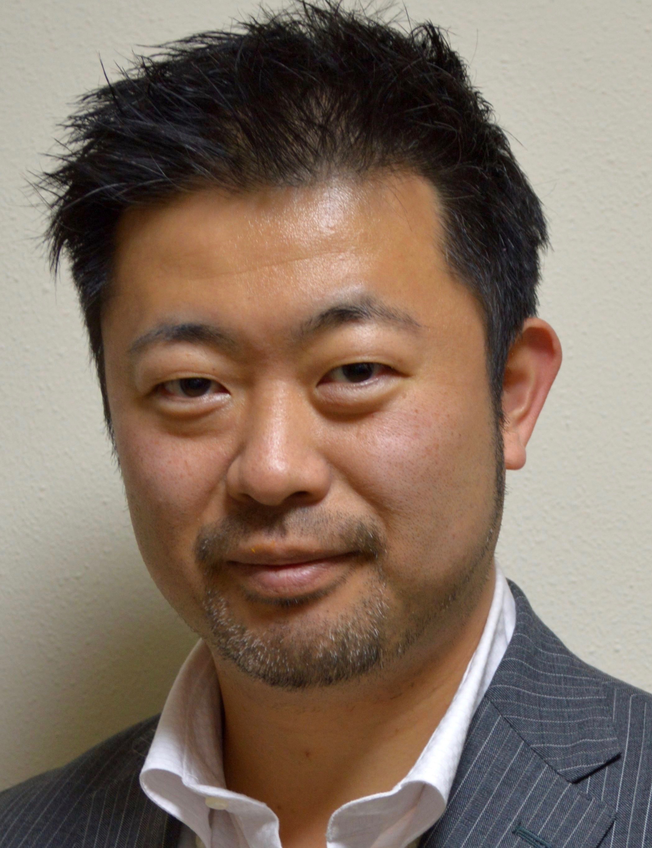
| | 09:00 |  | Conference Chair Image-Activated Cell Sorting and Beyond
Keisuke Goda, Professor of Chemistry, University of Tokyo and Adjunct Professor, Wuhan University, Japan
I introduce a newly developed technology known as “Image-Activated Cell Sorting” [Cell 175, 266 (2018)] that realizes real-time image-based intelligent cell sorting at an unprecedented rate. It integrates high-throughput cell microscopy, focusing, sorting, and deep learning on a hybrid software-hardware data-management infrastructure, enabling real-time automated operation for data acquisition, data processing, intelligent decision-making, and actuation. I also show the broad utility of the technology to real-time image-activated sorting of microalgal and blood cells for studying photosynthesis and atherothrombosis, respectively. The technology is highly versatile and expected to enable machine-based scientific discovery in biological, pharmaceutical, and medical sciences. |
| 09:30 |  | Keynote Presentation Promoting Microfluidics From the Lab to Real Biomedical Applications
An-Bang Wang, Distinguished Professor, Institute of Applied Mechanics, National Taiwan University, Taiwan
Microfluidics could bring significant advantages in the applications of chemical reaction, process control and bio-medical detection. In the past seventeen years, the research topics have been shifted from different microfluidic components in our lab, including micropump, microvalve, and micromixer etc., to various integration systems, e.g., micro-multiphase flow/multiemulsion generators, microreactor, high-throughput fluid/flow properties detection, and the maskless pattern coating technology by two-phase microfluidic method, etc. Recently, special efforts were made for the extension of the microfluidic applications in the life science studies and clinical diagnoses with advantages of easy use, compact size and excellent cost performance, e.g., a lab-on-a-chip with sequential control for biomedical point-of-care testing and a new extremely material- and time-saving Western blotting method that is widely used in analytical chemistry. |
| 10:00 | Microfluidic Systems For 3D Tissue Engineering of Cancer and Vascular Tissues
Ryo Sudo, Associate Professor, Keio University, Japan
Advances in microfluidic device technologies enabled us to control microenvironments in cell culture. In particular, microfluidic devices are useful platforms for 3D tissue engineering of cancer and vascular tissues. In this presentation, I will introduce our recent works on cancer invasion and vascular formation using microfluidic systems. | 10:30 | Morning Coffee and Tea Break Plus Networking | 11:30 |  | Keynote Presentation From Model Membranes to Single Cells: Microfluidic Approaches to Sensing and Diagnostics
Stephen Evans, Professor, University of Leeds, United Kingdom
This presentation will cover two aspects of our recent work. The first will look at a novel biosensor based on phospholipid-coated nematic liquid crystal (LC) droplets and demonstrate the detection of a model antimicrobial peptide (AMP). Monodisperse lipid-coated LC droplets were generated and trapped in a bespoke microfluidic structure and were treated with AMP. The disruption of the lipid monolayer by the AMP was detected at concentrations well within its biologically active range, and was indicated by a dramatic change in the appearance of the droplets associated with the liquid crystal transition from a typical radial configuration to a bipolar configuration. This suggests the system has feasibility as a drug-discovery screening tool and for toxin detection.
The second area of research to be described is on the investigation of cell deformability and its importance for diagnostic applications. In particular we use cell deformability in both shear-dominant and inertia-dominant microfluidic flow regimes to probe different aspects of the cell structure. In the inertial regime, we follow cellular response from (visco-)elastic through plastic deformation to cell structural failure and show a significant drop in cell viability for shear stresses >11.8 kN/m2. Comparatively, a shear-dominant regime requires lower applied stresses to achieve higher cell strains. From this regime, deformation traces as a function of time contain a rich source of information including maximal strain, elastic modulus, and cell relaxation times and thus provide a number of markers for distinguishing cell types and potential disease progression. These results emphasize the benefit of multiple parameter determination for improving detection and will ultimately lead to improved accuracy for diagnosis. |
| 12:00 | Construction of Artificial Cell-Membrane System Using Microfluidics
Ryuji Kawano, Associate Professor, Tokyo University of Agriculture and Technology, Japan
Artificial cell membranes have emerged as a biomimetic tool in such areas as membrane protein study, synthetic biology, and drug discovery. Planar lipid bilayers are used for functional studies of ion channels and nanopore sensing. The stability of lipid bilayers and the reproducibility of bilayer formation are important and it still remains challenging. To address these issues, we have used microfabrication and microfluidic technologies that have a major advantage: easy to handle lipid molecules or solution at the micron scale using microfluidics. Applying these advantages, we propose a stable and reproducible preparation procedure for the planar lipid bilayers using “droplet contact method”, and they are applying to measure pore-forming peptides and nanopore sensing. | 12:30 | Networking Lunch, Exhibit and Poster Viewing | |
Session Title: Microfluidics, 3D-Printing and Tissue Engineering |
| | 13:30 | Fabrication of Bilayer Cellular Systems for Organ-on-a-chip Applications Enabled by the Micromesh Culture Technique
Kennedy Okeyo, Senior Lecturer, Institute for Frontier Life and Medical Sciences, Kyoto University, Japan
Organ-on-chips are promising for their potential application to drug screening, disease modeling and basic biological studies. However, challenges still persist with regard to fabricating organ-on-chip platforms which can faithfully recapitulate the in vivo architecture and functionality. A major limitation lies in the use of semipermeable or porous artificial materials such as PDMS (polydimethylsiloxane) to interface the different cell layers, such as epithelial-endothelial bilayer commonly used in organ-on-chip models. Such interfacing materials are by far too thick (> 5 micron in thickness) and rigid compared with the in vivo basement membrane (< 0.2 micron in thickness), and they limit cross-layer cell-cell interaction leading to poor mimicry of in vivo functionality. This talk will highlight our micromesh culture technique and demonstrate its application toward the realization of bilayer cellular systems interfaced with a fabricated hydrogel basement membrane mimic. The thickness of the basement membrane mimic can be tuned to less than 1 micron, allowing us to fabricate bilayer cellular systems with better mimicry of the in vivo architecture and functionality for possible application to drug assay, disease modeling and other organ-on-chip studies. | 14:00 |  Recent Advancements in Research Based on 3D Bioprinting Technology – From Single Cells to Miniature Organs Recent Advancements in Research Based on 3D Bioprinting Technology – From Single Cells to Miniature Organs
Haruka Yoshie, Application Specialist, CELLINK
One of the biggest challenges in biological and medical research is to translate scientific discoveries from the bench to clinical applications. Recently in an effort to bridge this gap, 3D bioprinting has been used to mimic native tissues. Utilizing relevant bioinks and 3D printers, one can form cell-containing small droplets with an inkjet technique, create physiologically relevant microenvironments for cells, and even print miniature organs with more sophisticated printing methods. In this talk, I will present recent developments in biomimetic architectures from single cell systems to multi-cellular tissue structures. In particular, I will focus on microfluidic and vasculature models, organoid formations, and small organ productions to recapitulate the in vivo environment. Finally, I will discuss some of the challenges to be overcome when utilizing bioprinting-based research for clinical applications of microfluidic and organ-on-a-chip technologies.
| 14:30 | Vascularized Tissue Engineering
Tatsuya Shimizu, Director and Professor, Institute of Advanced Biomedical Engineering and Science, Tokyo Women’s Medical University, Japan
Most tissues and organs need perfusable micro blood vessels. We have succeeded in introduction of micro blood vessels in three-dimensional functional tissues. Recent advances in vascularized tissue engineering will be presented and discussed. | 15:00 | Afternoon Coffee and Tea Break Plus Networking | 15:30 | High Throughput Error-Correction Code DNA Sequencing
Yanyi Huang, Professor, Peking University, China
Eliminating errors in next-generation DNA sequencing has proved challenging. I am going to talk about our newly developed error-correction code (ECC) sequencing, a method to greatly improve sequencing accuracy by combining fluorogenic sequencing-by-synthesis (SBS) with an information theory–based error-correction algorithm. ECC embeds redundancy in sequencing reads by creating three orthogonal degenerate sequences, generated by alternate dual-base reactions. This is similar to encoding and decoding strategies that have proved effective in detecting and correcting errors in information communication and storage. We showed that, when combined with a fluorogenic SBS chemistry with raw accuracy of 98.1%, ECC sequencing provides single-end, error-free sequences up to 200 bp. ECC approaches should enable accurate identification of extremely rare genomic variations in various applications in biology and medicine. | 16:00 | Cell Assay For Evaluation of Initial Action of Cell-Cell Adhesion Using Microfluidic Platform
Masahiro Motosuke, Professor, Department of Mechanical Engineering, Tokyo University of Science, Japan
Adhesion of blood cell and endothelial cell in blood stream is an initial event of cell-cell adhesion causing the arteriosclerosis and cancer metastasis. We developed a microfluidic system for the characterization of initial action of cell-cell adhesion. In this presentation, I will introduce our work relating microfluidic cell assay for evaluating the rolling behavior of blood cell.
| 16:30 | Precise Cell Manipulation Using Highly Focused Acoustic Fields
Ye Ai, Associate Professor, Singapore University of Technology and Design, Singapore
Precise manipulation of particles and biological cells is an essential process in various biomedical research fields, industrial and clinical applications. Among various force fields applied for microfluidic cell manipulation, acoustic waves have superior propagating properties in solids and fluids, which can readily enable non-contact cell manipulation in long operating distances. In addition, acoustic fields are advantageous to high power laser beams for non-contact optical tweezing in terms of biocompatibility, throughput and setup simplicity. Exploiting acoustic waves for fluid and cell manipulation in microfluidics has led to a newly emerging research area, acoustofluidics. In this presentation, I will talk about particle and cell manipulation in microfluidics using highly focused surface acoustic waves (SAW). In particular, I will discuss a unique design of a focused interdigital transducer (FIDT) structure, which is able to generate a highly localized SAW field on the order of 30-50 µm wide. This highly focused acoustic beam has an effective manipulation area size that is comparable to individual micron-sized particles. Here, I demonstrate the use of this highly localized SAW field for fluorescence activated single cell level sorting with high cell viability. | 17:30 |  Modular Organ-on-a-Chip Solutions Combine 3D Microtissues and Flow to Accelerate Drug Discovery Modular Organ-on-a-Chip Solutions Combine 3D Microtissues and Flow to Accelerate Drug Discovery
Jan Lichtenberg, CEO and Co-Founder, InSphero AG
Liver and pancreas constitute key organs in the metabolic syndrome and are highly interacting through different endocrine factors to maintain glucose homeostasis in the human body. An impaired function of one of the organs can cause metabolic diseases, such as diabetes or NASH. Studying these diseases requires a systemic model that can reproduce organ-organ-interactions. The practical implementation of human in-vitro multi-tissue systems in a scalable format includes several challenges. Key aspects encompass biological and technical reproducibility, availability of tissue models, possibility of on-demand production, their usability in suitable treatment windows, access to clinically relevant readouts, and system compatibility with standard lab processes. We implemented a human in-vitro multi-tissue system in a scalable format using 3D organotypic microtissues for establishing organ-organ interactions. The liver model consisted of a primary human hepatocyte/Kupffer cell co-culture with preserved metabolic and inflammatory function over at least two weeks. The primary human islet microtissues comprised all pancreatic endocrine cells at physiological ratio and remained glucose responsive over the same culturing period. The different microtissues were assembled in a microfluidic chip using a pipetting robot enabling an adequate number of replicates at minimal operational complexity for compound testing with access to a wide range of readouts.
| 18:00 |  | Keynote Presentation The NIH Microphysiological Systems Program: In Vitro Tools for Drug Development and Precision Medicine
Danilo Tagle, Director, Office of Special Initiatives, National Center for Advancing Translational Sciences at the NIH (NCATS), United States of America
Approximately 30% of drugs have failed in human clinical trials due to adverse reactions despite promising pre-clinical studies, and another 60% fail due to lack of efficacy. One of the major causes in the high attrition rate is the poor predictive value of current preclinical models used in drug development despite promising pre-clinical studies in 2-D cell culture and animal models. The NIH Microphysiological Systems (Tissue Chips) program led by NCATS is developing alternative approaches and tools for more reliable readouts of toxicity and efficacy during drug development. Tissue chips are bioengineered microphysiological systems utilizing human primary or stem cells seeded on biomaterials manufactured with chip technology and microfluidics that mimic tissue cytoarchitecture and functional units of human organs. These platforms can be a useful tool for predictive toxicology and efficacy assessments of candidate therapeutics. Effective partnerships with stakeholders, such as regulatory agencies, pharmaceutical companies, patient groups, and other government agencies is key to widespread adoption of this emerging technology. Tissue chips can also contribute to studies in precision medicine, environment exposures, reproduction and development, infectious diseases, microbiome and countermeasures agents. |
| 18:30 | Close of Day 1 of the Conference |
Friday, 15 November 201908:00 | Morning Coffee and Tea Plus Networking | |
Session Title: Organs-on-Chips 2019, Emerging Themes and Technologies |
| | 08:30 | Factors To Consider When Choosing Organoids As a Fit-For-Purpose Model
Terry Riss, Senior Product Manager, Cell Health, Promega Corporation, United States of America
There is a broad spectrum of 3D cell culture models available to perform biological research ranging from individual tumor spheroids to complex microfluidic representations of a human-on-a-chip. Choosing an appropriate model must include a consideration of exactly what you want to know at the end of the experiment, the capabilities of the model, the investment you are willing to make and the limitations of available assay technologies. We will provide an overview of factors to consider when choosing an appropriate model system that is driving to an increased use of organoids. | 09:00 | Reverse Bioengineering of Living Systems For Drug Discovery
Ken-ichiro Kamei, Associate Professor, Institute for Integrated Cell-Material Sciences (WPI-iCeMS), Kyoto University, Japan
One of the ultimate goals of bioengineering is to re-create natural living systems by means of synthetic biology and tissue engineering. The long-term mission of our laboratory is to recapitulate the in vivo physiology and pathology on a microfluidic device, such as “Body-on-a-Chip” (BoC) or “Microphysiological Systems” (MPSs). Indeed, OOC/MPSs exhibit great potential as alternatives for pre-clinical animal tests to assess drug efficacy and safety. Although several chips and systems have been reported in the last decade, there are still some important issues that need to be addressed; these include: 1) the use of functional tissue cells derived from human pluripotent stem cell (hPSC), 2) the alternatives of polydimethylsiloxane (PDMS) to prevent chemical absorption, and 3) the integration of in situ monitoring systems to monitor cellular responses. Here, our interdisciplinary approach of stem cell biology, material science, and micro/nano-engineering will be introduced to address the aforementioned issues involved in drug discovery and precision medicine. | 09:30 | Microphysiological Systems Based on Microfluidic Device For Pharmacokinetic Studies
Hiroshi Kimura, Professor, Micro/Nano Technology Center, Tokai University, Japan
In this presentation, we will present integrated microfluidic platforms, which allow precise control of the cell culture environment on Microphysiological systems. Maintenance and replication of physiological functions of cells cultured in vitro are extremely difficult in conventional cell-based assay methods during life science and medical applications. Microfluidics is an emerging technology with the potential to provide integrated environments for maintenance, control, and monitoring the environment around cells. We work mainly in fundamental technologies of microfluidic devices and systems, and their applications to biological sciences including Microphysiological systems. A physiological environment in vitro can be replicated by fabrication of microstructures and control of microfluidics in these devices. Moreover, functional components, such as sensors, valves and pump, can be integrated into the devices by microfabrication methods. We performed cell-based assays to evaluate the function of these devices. Because cells cultured in vitro could be maintained and measured, these devices may be applied to medical, pharmaceutical and biological sciences. | 10:00 | Morning Coffee and Tea Break Plus Networking | 10:30 | Fundamental Consideration Points For the Liver Organ-on-a-Chip
Seiichi Ishida, Section Chief, Division of Pharmacology, National Institute of Health Sciences, Japan
Hepatocytes, the main parenchymal cell in the liver, has many functions and is widely used in in vitro cell-based assays. However, maintaining their physiological function is not easy. Same is the case for hepatic stellate cells, which are involved in hepatic fibrosis. Reconstitution of complicated physiological structure of the liver is thought to be necessary to reproduce adequate hepatic functions in in vitro culture model. Liver organ-on-a-chip is expected to solve these difficulties. Fundamental consideration points for the construction of liver orga-on-a-chip are discussed, with the introductory description of basic liver physiology, in the presentation. | 11:00 | Engineering of a Vascularized 3D Cell Construct On-Chip Using Human iPSC-derived Cells
Yu-suke Torisawa, Associate Professor, Hakubi Center for Advanced Research, Kyoto University, Japan
Vascular networks are essential to maintain cellular viability and function; however, current 3D culture models lack vascular systems. Engineering perfusable vascular networks that can deliver reagents and blood cells to 3D cell constructs could be a powerful platform to recapitulate cellular microenvironments and tissue-level functions. We have developed a microfluidic method to form vascularized tissue-like cell constructs to model cellular interactions through blood vessels. When a tumor-like cell spheroid containing human umbilical vein endothelial cells (HUVECs) and fibroblasts was cultured in our microfluidic device, a perfusable vascular network was formed through the cancer spheroid. We confirmed that peripheral blood mononuclear cells can be perfused inside a cancer spheroid through a vascular network. Thus, we used this system to model the interaction between cancer cells and immune cells. To study the interaction between cytotoxic T cells and cancer cells thorough blood vessels, allo-reaction between endothelial cells and T cells by mismatching of their HLA will be problematic. Therefore, we engineered 3D vascular networks using human induced pluripotent stem cell-derived endothelial cells (hiPSC-ECs). CD8+ T cells primed by HUVECs exhibited higher cytotoxic activity toward HUVECs than autologous hiPSC-ECs and MHC class I KO-hiPSC-ECs, demonstrating the potential value of this vascularized cancer-on-a-chip for modeling the interaction between T cells and a tumor-like tissue through blood vessels. Generation of in vivo-like vascularized 3D cell constructs using hiPSC-ECs would provide a novel platform to develop organs-on-chips as well as human disease models. | 11:30 | On-Chip Vasculature for Engineering Three-Dimensional Cell Culture Environment of Spheroids and Organoids
Ryuji Yokokawa, Professor, Department of Micro Engineering, Kyoto University, Japan
In vivo, healthy vasculature has a hollow structure to supply oxygen and nutrients to tissues. However, tissues cultured in vitro frequently lack such functional vasculature, and thus result in a necrotic core of the cell aggregate. To extend the culture period of the three-dimensional tissue, perfusable vasculature is crucial. It will contribute not only to the long-term culture for regenerative applications of tissues but also to deepen our understanding of organ morphogenesis.
In this presentation, I will explain a method to use angiogenic sprouts to connect the inside of a tissue model, spheroid or organoid, with microfluidic channels via vessel. Three types of tissue models were used: co-cultured spheroids of hLFs and HUVECs, and tri-cultured spheroids of hLFs, HUVECs, and tumor cells, and kidney organoids. This enables us to perfuse the inside of spheroids to deliver oxygen and nutrients. Moreover, some preliminary evaluation of tumor growth and drug evaluation will be presented.
We have extended the on-chip vascular formation method to a three-dimensional spheroid containing tumor cells and kidney organoids. Although we successfully optimized conditions to induce sprouting toward the co-cultured spheroid and found the lumen formation, it was not readily applicable to other cell types. Therefore, we took advantage of the angiogenic factors from hLFs even in the tumor spheroid model for vascularization. Future work includes many applications such as a high-throughput assay for drug screening and a long-term culture of organoids for studying organogenesis. The model provides a new assay platform for the tissue-culture with vasculatures at in vivo-like high cell density. | 12:00 |  | Keynote Presentation Approaches to Wearable Microfluidic Sensor Devices for Well-Being and Personalized Healthcare
Martyn Boutelle, Professor of Biomedical Sensors Engineering, Imperial College London, United Kingdom
A new generation of portable, wearable biomedical devices is becoming possible through recent advances in microfluidic technologies, microelectronics, sensors and biosensors. Key to this integration is the phenomenal growth in mobile computing power provided by current tablets and mobile phones. Through miniaturization and carefully engineered smart designs we can embed computer control and analytical best practice into portable even wearable devices that are able to compensate for shortcomings such as falling performance. These hybrid microfluidic systems appear to their target users as simple stable systems that tell them what they want to know. My group specializes in designing and building such microfluidic systems to meet the needs of acute critical care medicine. Key molecular markers are measured using both optical and electrochemical sensors and biosensors. We then work with clinical care teams to show proof of concept of the real-time continuous chemical information that microfluidic systems can produce. Our ultimate goal is that such systems can be used to monitor patients and guide therapy in a patient-specific, personalized way. |
| 12:30 |  Overview of Promising Applications of Microfluidic Technologies: Organs-on-Chips, Sequencing, and Biotechnologies Overview of Promising Applications of Microfluidic Technologies: Organs-on-Chips, Sequencing, and Biotechnologies
Sébastien Clerc, Technology & Market Analyst, Microfluidics & Medical Technologies, Yole Développement
Microfluidics has plenty of applications, some of which are now established. However, new applications arise as the technology matures and open exciting possibilities in various fields. For example, organs-on-chips show promising results for toxicology and efficacy testing in the pharmaceutical and other industries. In the field of biotechnology, microfluidics are increasingly used to isolate, analyze and culture cells. DNA/RNA sequencing sounds like a more mature application, but recent technology improvements make it appear like this field is only in its infancy and that great things are coming next. In this talk, Sébastien Clerc will present an overview of microfluidic technologies for these three applications along with main market and technology trends.
| 13:00 | Networking Lunch, Exhibit and Poster Viewing | |
Session Title: Innovations in Microfluidics Technologies and Applications |
| | 13:30 | Microfluidic Mass Production and Bio-Applications of Liquid Metal Nanoparticles
Shiyang Tang, Vice-Chancellor's Postdoctoral Research Fellow, School of Mechanical, Materials, Mechatronic and Biomedical Engineering, University of Wollongong, Australia
Functional nanoparticles comprised of liquid metals, such as eutectic gallium indium (EGaIn) and Galinstan, present exciting opportunities in the fields of flexible electronics, sensors, catalysts, and drug delivery systems. Methods used currently for producing liquid metal (LM) nanoparticles (NPs) have significant disadvantages as they rely on both bulky and expensive high-power sonication probe systems, and also generally require the use of small molecules bearing thiol groups to stabilize the nanoparticles. In addition, LM NPs in aqueous solutions tend to oxidize and precipitate over time, which hinders their utility in systems that require long-term stability. In this presentation, innovative microfluidics-enabled acoustic platforms are described as inexpensive, easily accessible methods for the on-chip mass production of bio-functional EGaIn nanoparticles with tunable size distributions in an aqueous medium. Several novel nanoparticle-stabilization approaches are demonstrated using polymer macromolecules such as brushed polyethylene glycol and poly(1-octadecene-alt-maleic anhydride), negating the requirements for thiol additives while imparting a “stealth” surface layer. Furthermore, a surface modification of the nanoparticles is demonstrated using galvanic replacement and conjugation with antibodies, and the application of such bio-functionalized LM NPs in positron-emission tomography imaging is demonstrated. It is envisioned that the developed microfluidic techniques and the produced liquid metal-based nanoparticles can enable a wide range of biomedical applications. | 14:00 | Microfluidic Preparation of Flexible Micro-Grippers with Precise Delivery Function
Yu Hao Geng, First Author, The State Key Lab of Chemical Engineering, Department of Chemical Engineering, Tsinghua University, China
We firstly propose a one-step preparation method of micro-gripper by using gas-liquid Janus emulsions and thermo-sensitive hydrogels, and then characterize the behavior of oriented and precise delivery. | 14:30 |  | Keynote Presentation Culture-Independent Method for Identification of Microbial Enzyme-Encoding Genes by Single-Cell Sequencing Using a Water-in-Oil Microdroplet Platform
Takashi Funatsu, Professor, University of Tokyo, Japan
Environmental microbes are a major source of industrially valuable enzymes with potent and unique catalytic activities. Unfortunately, the majority of microbes remain unculturable or difficult to cultivate and thus are not accessible by culture-based methods. Recently, culture-independent metagenomic approaches have been successfully applied, opening access to untapped genetic resources. Here we present a methodological approach for the identification of genes that encode metabolically active enzymes in environmental microbes in a culture-independent manner. Our method is based on activity-based single-cell sequencing, which focuses on microbial cells exhibiting specific enzymatic activities. First, environmental microbes were encapsulated in water-in-oil microdroplets with a fluorogenic substrate for the target enzyme to screen for microdroplets that contain microbially active cells at the single cell level. Second, the microbial cells were recovered and subjected to whole genome amplification. Finally, the amplified genomes were sequenced to identify the genes encoding target enzymes. This method was successfully used to identify 14 novel beta-glucosidase genes from uncultured bacterial cells in marine samples. Our method contributes to the screening and identification of genes encoding industrially valuable enzymes. |
| 15:00 | Microtechnologies For Immune Health Profiling In Type 2 Diabetes Mellitus
Han Wei Hou, Assistant Professor, School of Mechanical and Aerospace Engineering, Nanyang Technological University, Singapore
Metabolic disorders such as diabetes mellitus and cardiovascular diseases (CVD) are becoming the main public health challenges in Singapore and globally with rising premature morbidity and associated mortality, as well as escalating healthcare costs. In this talk, I will present our recent research efforts in the development of microfluidics technologies for “label-free” isolation and phenotyping of circulating leukocytes in type 2 DM (T2DM). We demonstrated that diabetic leukocytes exhibited significant immune dysfunctions which were well-associated with traditional CVD risk factors, clearly illustrating their potential as novel surrogate biomarkers for point-of-care testing. Overall, this multi-disciplinary research greatly facilitates rapid and quantitative analysis of immune and vascular health using liquid biopsy, with progress towards clinical diagnostics and precision medicine in the treatment of vascular dysfunctions. | 15:30 | On-the-Fly Optofluidic Imaging Platforms For Deep and Large-Scale Single-Cell Analysis
Kevin Tsia, Associate Professor, Department of Electrical & Electronic Engineering, The University of Hong Kong, Hong Kong
This talk will introduce our recently developed ultrafast optofluidic imaging techniques (multi-ATOM and FACED imaging) that practically allow ultralarge-scale single-cell imaging (>millions of adherent or suspended cells) in real-time with the unprecedented combination of imaging resolution and speed. These techniques generate multiple single-cell image contrasts from which not only deep single-cell biophysical and biomechanical phenotypic profiles can be obtained (based on quantitative phase and other label-free contrasts), but also biochemical signatures of single-cells (based on fluorescence contrast). Combined with high-performance computing hardware and high-throughput computational methods (particularly machine learning), their utilities in large-scale cell-based assays using cell-lines, in-vivo mouse models, as well as primary human cells (e.g. for circulating tumor cell detection, routine blood analysis, drug screen) will be discussed.
| 16:00 | High-Throughput Large-Droplet Sorting
Akihiro Isozaki, Researcher of Kanagawa Institute of Industrial Science and Technology (KISTEC) and Research Assistant Professor, University of Tokyo, Japan
Droplet microfluidics is an attractive tool for large-scale single-cell analysis. However, there is a trade-off between sorting throughput and droplet volume that is related to the incubational capacity of droplets. Our method allows us to overcome this trade-off and achieve high-throughput (~5,000 droplets/sec) large-droplet (0.1 nL) sorting. In this talk, I present the details of our method and its applications. | 16:30 |  | Keynote Presentation Medical Diagnosis and Gene Therapy Using Microfluidic Devices
Manabu Tokeshi, Professor, Hokkaido University, Japan
We are working on the development of microfluidic devices and their measurement systems for rapid diagnosis based on immunoassays, and are also challenging the microfluidic based lipid nanoparticles production for drug delivery systems. In this presentation, I will introduce those latest achievements and discuss the future prospects. |
| 17:00 | High-Throughput Raman Flow Cytometry
Kotaro Hiramatsu, Assistant Professor, The University of Tokyo, Japan
I present the development of a high-throughput (>2,000 cells/s) and broadband (400-1600 cm-1) Raman spectroscopic flow cytometry and its application to large-scale single-cell analyses of the astaxanthin productivity and photosynthetic dynamics of Haematococcus lacustris. | 17:30 |  | Keynote Presentation Conference Closing Keynote Presentation: Quantum Diagnostics - From Single-Cells to Single-Molecules
Dino Di Carlo, Armond and Elena Hairapetian Chair in Engineering and Medicine, Professor and Vice Chair of Bioengineering, University of California-Los Angeles, United States of America
The ultimate limits of diagnostics in biology are the “quantum” units that convey information, e.g. single nucleic acids, proteins, and cells. Microfluidics has emerged as a powerful tool to compartmentalize single cells and molecules into sub-nanoliter droplets as individual bioreactors to enable sensitive detection and analysis down to this quantum limit. However, the current systems for quantum diagnostics have not been widely adopted, partly due to the requirement of specialized instruments and microfluidic chips to generate uniform droplets and perform adequate manipulations. I will discuss the platforms we are developing to fractionate volumes in simplified, instrument-free ways using 3D-shaped microparticles. Each “lab-on-a-particle” can be analyzed using widely available flow cytometers. These new lab-on-a-particle reagents eliminate the need for specialized new equipment for microfluidic compartmentalization and readout and promise to democratize single-molecule and single-cell technologies. |
| 18:00 | Close of Conference |
|


 Add to Calendar ▼2019-11-14 00:00:002019-11-15 00:00:00Europe/LondonMicrofluidics and Organ-on-a-Chip Asia 2019Microfluidics and Organ-on-a-Chip Asia 2019 in Tokyo, JapanTokyo, JapanSELECTBIOenquiries@selectbiosciences.com