Other Track Agendas3D-Printing in the Life Sciences, Biofabrication & Synthetic Biology | Tissue Engineering |

Thursday, 17 March 201608:00 | Conference Registration, Materials Pick-Up and Continental Breakfast in the Exhibit Hall | |
Session Title: Opening Plenary Session |
| | |
Plenary Session Chairman: Gabor Forgacs, Ph.D. |
| | 09:00 |  | Keynote Presentation Bioprinting: Challenges to Commercialization of Academic Research -- The Story of Organovo
Gabor Forgacs, Professor, University of Missouri-Columbia; Scientific Founder, Organovo; CSO, Modern Meadow, United States of America
This talk will present the story behind the first commercial bioprinting company, Organovo. Each attempt to commercialize academic research has its own challenges. In the case of bioprinting, a paradigm shifting innovation at the time, the pitfalls and hurdles were above the average. The talk will briefly overview the science underlying Organovo’s technology, the process to the establishment of the company, its evolving business model, the beginning of the commercial operation and the path to the Initial Public Offering (IPO). We will also briefly overview the present status of the commercial bioprinting space. It is hoped that the lessons from this story will provide useful input to others in the field. |
| 09:30 | 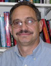 | Keynote Presentation Silk-based Inks and Medical Devices via 3D Printing
David L. Kaplan, Stern Family Endowed Professor of Engineering, Professor & Chair -- Dept of Biomedical Engineering, Tufts University, United States of America
Silk proteins provide unique high molecular weight, amphiphilic, protein polymers useful in 3D printing. Importantly all aqueous printing at ambient conditions, without the need for chemical or photoactivated crosslinkers, offers simple, mechanically robust and biocompatible outcomes using the silk system. |
| 10:00 | 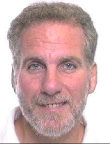 | Keynote Presentation Organ Engineering and Storage
Martin Yarmush, Founding Director of the Center for Engineering in Medicine, Massachusetts General Hospital and Harvard Medical School, United States of America
|
| 10:30 | Coffee Break and Networking | 11:15 |  | Keynote Presentation Microfluidics-based 3D Tissue Fabrication
Shoji Takeuchi, Professor, Center For International Research on Integrative Biomedical Systems (CIBiS), Institute of Industrial Science, The University of Tokyo, Japan
3D tissue construct is important not only in regenerative medicine but also drug testing without animal experiments. Here, I will discuss several MEMS/Microfluidics-based approaches for the rapid construction of 3D tissue. We demonstrated a bottom-up tissue construction method using different types of cellular modules that serve as building blocks for thick and dense 3D tissues (eg., cell beads and cell fibers). |
| 11:45 | 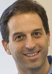 | Keynote Presentation Mammalian Synthetic Biology: From Parts to Modules to Therapeutic Systems
Ron Weiss, Director, MIT Synthetic Biology Center; Professor, Massachusetts Institute of Technology (MIT), United States of America
Synthetic biology is revolutionizing how we conceptualize and approach the engineering of biological systems. Recent advances in the field are allowing us to expand beyond the construction and analysis of small gene networks towards the implementation of complex multicellular systems with a variety of applications. In this talk I will describe our integrated computational / experimental approach to engineering complex behavior in a variety of cells, with a focus on mammalian cells. In our research, we appropriate design principles from electrical engineering and other established fields. |
| 12:15 | Networking Lunch, Discussions with Exhibitors and View Posters | |
Session Title: Emerging Themes in Tissue Engineering |
| | 14:00 | 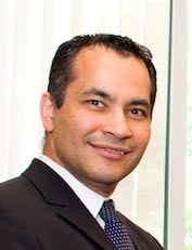 | Keynote Presentation Emerging Organ Models and Organ Printing for Regenerative Medicine
Ali Khademhosseini, Professor, Department of Medicine, Brigham and Women’s Hospital; Wyss Institute for Biologically Inspired Engineering, Harvard University, United States of America
Engineered materials that integrate advances in polymer chemistry, nanotechnology, and biological sciences have the potential to create powerful medical therapies. Our group aims to engineer tissue regenerative therapies using water-containing polymer networks called hydrogels that can regulate cell behavior. Specifically, we have developed photo-crosslinkable hybrid hydrogels that combine natural biomolecules with nanoparticles to regulate the chemical, biological, mechanical and electrical properties of gels. These functional scaffolds induce the differentiation of stem cells to desired cell types and direct the formation of vascularized heart or bone tissues. Since tissue function is highly dependent on architecture, we have also used microfabrication methods, such as microfluidics, photolithography, bioprinting, and molding, to regulate the architecture of these materials. We have employed these strategies to generate miniaturized tissues. To create tissue complexity, we have also developed directed assembly techniques to compile small tissue modules into larger constructs. It is anticipated that such approaches will lead to the development of next-generation regenerative therapeutics and biomedical devices. |
| 14:30 | Cell Sheet Engineering: Building Tissues Layer by Layer
Joyce Wong, Professor of Biomedical Engineering and Materials Science & Engineering, Boston University, United States of America
It is well known in materials science and engineering that the structural organization of a material system is instrumental in determining its functional properties. Similarly, biological tissues are characterized by unique architectures that perform specific functions. Here I will describe strategies to recapitulate the hierarchical organization using small diameter blood vessels as an example. These approaches can be applied to other laminate and layered systems such as skin. Our approach is to combine micropatterning techniques with thermoresponsive (poly-N-isopropylacrylamide) surfaces that enable the release of cell sheets that mimic the alignment of vascular smooth muscle cell (VSMC) layers of the artery. While tissue engineering can provide solutions for replacing diseased tissues, it would also be highly desirable to develop methods that would detect cardiovascular disease early –before the disease has progressed to the point requiring surgical intervention. We have therefore also been developing contrast agents that would enable molecular targeting of cardiovascular disease. In addition, we have been developing theranostic agents can serve to simultaneously image and release a drug payload to targeted areas. Tissue-engineered models such as angiogenesis-on-a-chip can be used to optimize these theranostic agents with the aim of early disease detection. | 15:00 | Microvasculature-on-a-chip for Tissue Engineering and Precision Medicine
Rong Fan, Harold Hodgkinson Professor of Biomedical Engineering, Yale University, United States of America
One of the major challenges in tissue engineering for transplantation and regenerative medicine is revascularization. Due to the complexity of angiogenesis and vasculogenesis mediated by a host of cellular and soluble mediators, it remains a daunting challenge to recapitulate the perivascular microenvironment in vitro for effective neotissue vascularization prior to transplantation. Stromal cells, such as fibroblasts and pericytes, provide not only mechanical support but also paracrine signals to regulate vascular morphogenesis and function. We are working to develop integrative microsystems that combine various stromal cell components and soluble signals derived therefrom to engineer high quality microvasculature network in vitro with the ultimate goal to achieve revascularization of large-sized neotissues and thus improve the performance post transplantation. First, we are harnessing the paracrine signals from phenotypically modified stromal cells (e.g., fibroblasts) to create endothelialized microvessel network in native extracellular matrix (e.g., collagen, fibrin, etc) hydrogels. Second, we are applying this platform to revascularization of mouse pancreatic islets in vitro, which can accelerate functional anastomosis with host tissue after implantation. The vascularized islets are characterized for glucose sensing and insulin production in response to glucose. Third, leveraging the success of in vitro tissue revascularization to build a brain tumor perivascular niche (PVN)-on-a-chip model to investigate the dynamics of glioma stem/progenitor cells in PVN and their resistance or response to treatment. All together, these are to address a critical need in tissue engineering and open new opportunities for in vitro modeling of tumor microenvironment for precision medicine. | 15:30 | Coffee Break and Networking in the Exhibit Hall | 16:00 |  | Keynote Presentation Manipulating Cells for Bioprinting and Use in Tissue Engineering
David Weitz, Professor, Harvard University, United States of America
This talk will describe the use of drop-based microfluidic techniques for encapsulating cells in small, highly uniform spheres of gel. This can be easily manipulated for further analysis of the cells, and for using the cells for further processing, such as 3D additive assembly of larger scale structures. |
| 16:30 | .png) | Keynote Presentation Clinical Applications of SVF and Tissue Engineering from a Plastic Surgeon’s Perspective: Past, Present and Future
Lewis J. Obi, Medical Director, Cell Surgical Network of Florida; Medical Director of Obi Plastic Surgery Clinic, United States of America
The history of plastic surgery dates back 5000 years to the Ancient Egyptian text known as the Edwin Smith Papyrus which describes treatments for a broken nose. The term plastic surgery derived from the Greek “plastike” implies the art of modeling of malleable flesh. Fast forward into the twentieth century when the modern day specialty of plastic surgery was established. The use of autologous grafts and flaps combined with microsurgery and synthetic materials led to the need for advanced imaging technology. The Canfield 3D imaging system made this technology affordable for plastic surgeons. Lipo suction led to a parallel science with the use of processed fat as a source of stem cells for regenerative surgery. The natural evolution of all of these methods in plastic surgery was to work with stem cell and bioprinting scientists. Dr Hee Young Lee of Korea recently developed a stem cell expansion and storage system producing enough cells for bioprinting. Paul Gaetenholm of Chalmers University has developed novel bio inks and bio printers which provides the final piece of the puzzle in this vast new science of bio printing. After 5000 years, plastic surgeons may not only repair broken noses but actually print new ones. |
| 17:00 |  | Keynote Presentation The Quest for the Tissue Engineering Grail: Vascularized Tissue Constructs
Jason Spector, Professor of Plastic Surgery and Otolaryngology, Weill Cornell Medical College, United States of America
The ultimate purpose of nearly all tissue engineering and Bioprinting is the clinical translation thereof. However, the actual clinical needs and specific parameters required are not always clearly defined. As such, this talk provides an overview (from a surgeon’s perspective) of the clinical scenarios in which tissue engineering and bioprinting might have their greatest impact with a particular focus on the current clinical strategies utilized in reconstruction including the use of grafts (non-vascularized) and flaps (vascularized). The discussion will also include an overview of the shortcomings of currently available tissue engineered products as well as highlight strategies for achieving the “Holy Grail” of tissue engineering—constructs with their own inherent hierarchical vascularity. |
| 17:30 | Directed Differentiation of Human Stem Cells to Kidney Organoids and Disease Modeling
Joseph Bonventre, Samuel A. Levine Professor of Medicine, Brigham and Women's Hospital, Harvard Medical School, United States of America
I will discuss recent advances in the directed differentiation of human ES cells and IPS cells into nephron progenitors and then kidney organoids. The efficiency with which nephron progenitor cells are generated is 80-90%. The kidney organoids are characterized by multi-segmented structures with characteristics of podocytes, proximal tubules, loop of Henle, and distal tubules. The tubular structures in the organoids express many transporters that are characteristic of mature nephrons. The organoids can be used for modeling a variety of both genetic and non-genetic disease processes. | 18:00 | Networking Cocktail Reception for All Delegates, Speakers, Sponsors and Exhibitors on the 15th Floor with a View of Boston and the Charles River | 19:30 | Close of Day 1 of the Conference. Microfluidics in Bioprinting Dinner Training Course Begins. |
Friday, 18 March 201608:00 | Morning Coffee, Breakfast Pastries, and Networking | |
Session Title: The Use of Additive Manufacturing and Other Platforms in the Generation of Organs for Tissue Engineering |
| | 08:30 | 3D Printing Complex Biological Scaffolds using Freeform Reversible Embedding of Suspended Hydrogels (FRESH)
Adam Feinberg, CTO and Co-founder, FluidForm Inc., Professor of Biomedical Engineering, Carnegie Mellon University, United States of America
3D printing of hydrogels is an additive fabrication approach that has the potential to create complex construct architectures not possible using other tissue engineering methods. While there have been a number of advances, the 3D printing of soft hydrogels such as alginate, collagen and fibrin remains challenging because these materials typically have an elastic modulus <100 kPa and deform during the layer-by-layer printing process in air. To address this we have developed a Bingham plastic support bath into which these hydrogels can be printed with high fidelity and subsequently released, using a process we term freeform reversible embedding of suspended hydrogels (FRESH). | 09:00 |  | Keynote Presentation Versatile Approaches to Extrusion-based Additive Manufacturing of Scaffolds and Tissue Engineering Constructs
Michael Gelinsky, Professor and Head, Center for Translational Bone, Joint and Soft Tissue Research, Faculty of Medicine, Technische Universität Dresden, Germany
By means of extrusion-based additive manufacturing (3D plotting) scaffolds and cell-loaded constructs can be fabricated easily. We have developed several pasty biomaterials based on biopolymer blends and polymer/mineral composites which can be utilized for 3D plotting and bioprinting with human cells and microalgae. By extruding two different materials through a coaxial double needle strands with a core/shell morphology and 3D scaffolds thereof can be manufactured which allow for dual growth factor loading and release. Combining two or more materials in a layered fashion leads to complex constructs suitable for the treatment of defects at tissue interfaces. |
| 09:30 | Layer-by-Layer Assembly of Cellularized Poly-(lactic acid) Constructs for Bone Tissue Engineering
Vera Guduric, Researcher, University of Bordeaux, France, France
Bone Tissue Engineering (BTE) requires tissue-specific cells and a porous biocompatible scaffold to support cell proliferation and differentiation. Rapid prototyping technique allows fabrication of custom-made 3D scaffolds with high resolution. Layer-by-Layer (LBL) microfabrication is based on assembly of small seeded blocks. The aim of this work was to evaluate proliferation and differentiation of human bone marrow stromal cells (HBMSCs) and endothelial progenitor cells (EPCs) in 2D and 3D using a LBL assembly of cellularized polylactic acid (PLA) membranes. PLA membranes were fabricated by direct 3D printing. HBMSCs and EPCs were seeded onto membranes as mono- and co-cultures in aim to perform experiments in 2D (1 seeded membrane) and 3D (LBL constructs). 2D experiments have shown high cell viability on PLA during 21 days as well as adequate morphology of seeded cells. Early osteoblastic and endothelial differentiations were observed after 14 days. Cell proliferation was increased in all experiments after 7 days. The migration of EPCs between layers of LBL constructs was observed as well as an osteoblastic differentiation of HBMSCs. These results indicate that LBL approach could be suitable for BTE, in order to promote homogenous cell distribution inside the scaffold and gene expression specific to the cells implanted. | 10:00 | Multi-scale Porosity Biomaterials for Bone-on-a-Chip Devices and Bone Tissue Engineering
Frederik Claeyssens, Senior Lecturer, Materials Science and Engineering, University of Sheffield, United Kingdom
Natural tissues and organs are typically structured in a hierarchical fashion, in which the Extra-Cellular Matrix (ECM) provides a microporosity to optimally support cell growth while larger scale structures (e.g. vasculature and boundary layers) are incorporated to support the function and structure of the tissue and organ. To mimic this multiscale structuring in synthetic biomaterials we combine additive manufacturing with self-assembly. In this structuring technique the internal porosity is governed by self-assembly and the macroscopic structure is constructed by additive manufacturing. Emulsion templating is used as self-assembly technique to produce materials with a high microscale porosity. These emulsions can subsequently be used as photocurable resins for stereolithography, producing user-defined macroscale structures with a tissue-like microporosity. The mechanical properties of these materials can be varied via the changing the monomer ratio within the resin. Additionally, biodegradable scaffolds can be fabricated via polycaprolactone-based resins. We produce these hierarchical structured material in 3D structured materials such as woodpile-style scaffolds, microspheres with controllable diameter and as 3D microenvironments that can be integrated in standard poly-dimethylsiloxane (PDMS) based microfluidics. These scaffolds we currently investigate as a platform for bone-on-a-chip based devices and bone tissue engineering. | 10:30 | Coffee Break and Networking in the Exhibit Hall | 11:00 | Stemness Influences the Regenerative Potential of Cells Exposed to Chemotherapy
Eric Darling, Associate Professor of Medical Science, Engineering, and Orthopaedics, Brown University, United States of America
In musculoskeletal tissues like bone, chemotherapy can impair progenitor cell differentiation and proliferation, resulting in decreased bone growth and mineralization throughout a patient’s lifetime. The current study examined the effects of chemotherapy on mesenchymal stem cell (MSC) regenerative characteristics compared to non-stem cells. The proliferation of stem and non-stem cell types could be used as an in vitro measure of susceptibility to common chemotherapeutic drugs. Interestingly, MSCs showed no susceptibility to the highly prevalent drug methotrexate (MTX), retaining both sustained proliferation and multipotency capabilities after exposure. Investigation into the mechanism behind cell response to MTX involved overexpression and knockdown of dihydrofolate reductase (DHFR), the target of the drug. Overexpression and endogenous nucleoside + amino acid delivery rescued non-stem cell types from adverse effects, identifying DHFR as one resistance mechanism and potential means of protecting beneficial cells exposed to MTX. Furthermore, it was observed that undifferentiated MSCs were more resistant than differentiating and terminally differentiated cell types, suggesting that stemness could play a role in chemotherapeutic resistance as well. | 11:30 | Mandibular Jaw Regeneration Using Human Dental Cell-Seeded Tyrosine-Derived Polycarbonate Scaffolds
Weibo Zhang, Research Associate, Department of Orthodontics, Tufts university, School of Dental Medicine, United States of America
The long-term objective of this study is to establish a model for coordinated bioengineered alveolar bone and tooth regeneration. The ability to effectively and simultaneously bioengineer living jaw bone and teeth would improve craniofacial reconstruction time and cost. This study showed that human dental pulp stem cells seeded E1001(1k)/ß-TCP scaffolds support the rapid regeneration of osteo-dentin like mineralized jaw tissue, suggesting a promising new therapy for eventual coordinated alveolar jaw bone and tooth regeneration. Current efforts focus on scaffolding fabrication methods to regenerate alveolar bone and teeth. | 12:00 | 3D Micromachining in Silk Hydrogels
Matthew Applegate, Research Fellow, Ultrafast Nonlinear Optics and Biophotonics Laboratory, Tufts University, United States of America
Silk, a biodegradable and biocompatible natural polymer, can be formed into transparent hydrogels ideal for tissue engineering applications. Exposure to focused ultra-short pulses of near infrared light results in multi-photon absorption within the focal volume of the beam leading to the formation of voids, without the need for exogenous photo-initiators. The high transparency of the gel and the low pulse energies used enable the formation of micro-voids nearly 1 cm below the surface of the gel. These voids can then be linked together into larger structures that can be used to guide cells within the gel both in vitro and in vivo for tissue engineering applications. | 12:30 | Novel BioMaterials and Bioengineering Techniques for Cardiovascular Tissue Engineering
Anwarul Hasan, Assistant Professor , Mechanical and Industrial Engineering, Qatar University, Qatar
Cardiovascular diseases are among the leading causes of death worldwide. Tissue engineering and regenerative medicine have emerged as potential solutions for many cardiovascular diseases such as development of implantable tissue engineered vascular grafts for bypass heart surgeries and regeneration of cardiac tissue after tissue-damage or tissue-death resulting from cardiac arrests. A major obstacle for the widespread use of tissue engineering in clinical applications is the lack of suitable biomaterials with required combination of biomechanical and biological properties. Photocrosslinkable mcro-porous hydrogels and electrospun nano-microfiber scaffolds have been widely investigated for application in tissue engineering and regenerative medicine. This talk will present some interesting results from our recent studies on potential applications of Photocrosslinkable methacrylated gelatin hydrogel and electrospun-airjet-sprayed composite scaffolds in cardiovascular tissue engineering and regenerative medicine. | 13:00 | Networking Lunch, Discussions with Exhibitors and View Posters |
|


 Add to Calendar ▼2016-03-17 00:00:002016-03-18 00:00:00Europe/LondonTissue EngineeringSELECTBIOenquiries@selectbiosciences.com