Thursday, 16 March 2017

08:00

Conference Registration, Conference Materials Pick-Up, Morning Coffee and Breakfast Pastries


Session Title: Conference Plenary Session -- Convergence of Technologies Constituting the BioEngineering Space

09:00

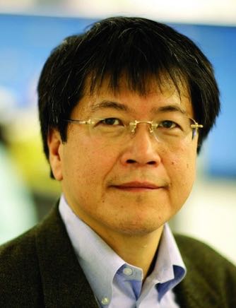
Martin YarmushKeynote Presentation

Hills and Valleys in the Landscape of Cell-Based BioMEMS and Tissue Engineering
Martin Yarmush, Founding Director of the Center for Engineering in Medicine, Massachusetts General Hospital and Harvard Medical School, United States of America

This presentation will cover some interesting advances in the fields of cellular BioMEMS and tissue engineering with an eye towards identifying important considerations that may help spur future meaningful discoveries and products. Also some discussion will focus on government-mandated programs and choices, artifactual results, and misleading terminology that have not been helpful in moving these fields forward.

09:30

Gabor ForgacsKeynote Presentation

Monitoring Biological Processes In Situ: Lab-in-the-Tissue
Gabor Forgacs, Professor, University of Missouri-Columbia; Scientific Founder, Organovo; CSO, Modern Meadow, United States of America

Engineering of 3D tissues and organs has seen spectacular progress in recent years. The technologies of 3D-printing and organ-­on-the chip have provided invaluable tools and structures for basic research, drug toxicity assays or disease models. In the case of drug toxicity studies the engineered construct is typically subjected to either known drugs or to new candidate drugs and metabolic functions are measured by detection of metabolites in the culture medium in which the engineered construct resides. This type of monitoring however, reveals only limited information about how the internal structure (e.g. cell-­cell interactions) of the engineered construct is effected and thus about the suitability of the tissue for further applications, such as its use in disease studies or ultimately for implantation. Here we propose to combine 3D printing and BioMEMS and related technologies to build Lab-in-the-­tissue devices for the in situ monitoring of metabolic and functional properties of engineered tissues and organ structures.

10:00

Mehmet TonerKeynote Presentation

Microfluidics to Isolate Single and Clusters of Rare Circulating Tumor Cells to Manage Cancer Patients
Mehmet Toner, Helen Andrus Benedict Professor of Biomedical Engineering, Massachusetts General Hospital (MGH), Harvard Medical School, and Harvard-MIT Division of Health Sciences and Technology, United States of America

Viable tumor-derived circulating tumor cells (CTCs) have been identified in peripheral blood from cancer patients and are not only the origin of intractable metastatic disease but also marker for early cancer. However, the ability to isolate CTCs has proven to be difficult due to the exceedingly low frequency of CTCs in circulation. As a result their clinical use until recently has been limited to prognosis with limited clinical utility.  More recently, we introduced several microfluidic methods to improve the sensitivity of rare event CTC isolation, a strategy that is particularly attractive because it can lead to efficient purification of viable CTCs from unprocessed whole blood. The micropost CTC-Chip (µpCTC-Chip) relies on laminar flow of blood cells through anti-EpCAM antibody-coated microposts, whereas the herringbone CTC-Chip (HbCTC-Chip) uses micro-vortices generated by herringbone-shaped grooves to efficiently direct cells toward antibody-coated surfaces. These antigen-dependent CTC isolation approaches, also called “positive selection”, led to the development of a third technology, which is tumor marker free (or antigen-independent) sorting of CTCs. We call this integrated microfluidic system the CTC-iChip, based on the inertial focusing strategy, which allows positioning of cells in a near-single file line, such that they can be precisely deflected using minimal magnetic force.

10:30

Coffee Break and Networking

11:00

Joshua EdelKeynote Presentation

Novel Strategies in Single Molecule Sensing
Joshua Edel, Professor, Imperial College London, United Kingdom

Analytical Sensors play a crucial role in today’s highly demanding exploration and development of new detection strategies. Whether it be medicine, biochemistry, bioengineering, or analytical chemistry the goals are essentially the same: 1) improve sensitivity, 2) maximize throughput, 3) and reduce the instrumental footprint. In order to address these key challenges, the analytical community has borrowed technologies and design philosophies which has been used by the semiconductor industry over the past 20 years. By doing so, key technological advances have been made which include the miniaturization of sensors and signal processing components which allows for the efficient detection of nanoscale object. One can imagine that by decreasing the dimensions of a sensor to a scale similar to that of a nanoscale object, the ultimate in sensitivity can potentially be achieved - the detection of single molecules. This talk highlights novel strategies for the detection of single molecules using nano-porous membranes.

11:30

Hsueh-Chia ChangKeynote Presentation

A Solid-State Nanopore microRNA Quantification Platform
Hsueh-Chia Chang, Bayer Professor of Chemical and Biomolecular Engineering, University of Notre Dame, Interim Chief Technology Officer, Aopia Biosciences, United States of America

We report a new highly selective (PCR-free) solid-state nanopore miRNA quantification platform for liquid biopsy and single-cell assays. A single ion-track nanopore in a PET membrane is asymmetrically etched into a conic geometry and is coated with a high-permittivity dielectric layer by Atomic Layer Deposition. The surface modified conic nanopore allows for high throughput molecular translocation (100 Hz vs 1 Hz for protein nanopores) and selective delay of single-stranded (ss) nucleic acids compared to their hybridized double-stranded (ds) duplex (mean translocation time of 100 ms vs 1 ms with 5% overlap in the two distributions). The delay is due to enhanced van der Waal attraction between exposed rings of the ss-nucleic acids, with delocalized electrons, with the high-permittivity coating. Individual translocation events can be recorded for a mixture of ss- and ds-nucleic acids numbering between 100 to 100,000.  Whether the translocating molecule of each event is an ss miRNA or its ds duplex can be discerned with 95% confidence.

12:00

Fiorenzo OmenettoDavid L. KaplanKeynote Presentation

Biomaterial Printing Frontiers
Fiorenzo Omenetto, Frank C. Doble Professor of Engineering, Associate Dean for Research, School of Engineering, Tufts University
David L. Kaplan, Stern Family Endowed Professor of Engineering, Professor & Chair -- Dept of Biomedical Engineering, Tufts University, United States of America

Biomaterials offer opportunities for devices that operate seamlessly at the interface of the biological and technological worlds.  Stringent requirements on material form and function are imposed when operating at the nanoscale or when interfacing such materials with microelectronic circuitry. Silk fibroin is a very attractive biopolymer for use as a polymorphic matrix for multiple material formats that are casted, printed, extruded, or molded. This opens opportunities for multi-functional, sustainable devices that leverage both the properties of the material and the  biological features that they can encompass.

12:30

Ron WeissKeynote Presentation

Synthetic Biology: From Parts to Modules to Therapeutic Systems
Ron Weiss, Director, MIT Synthetic Biology Center; Professor, Massachusetts Institute of Technology (MIT), United States of America

Synthetic biology is revolutionizing how we conceptualize and approach the engineering of biological systems. Recent advances in the field are allowing us to expand beyond the construction and analysis of small gene networks towards the implementation of complex multicellular systems with a variety of applications. In this talk I will describe our integrated computational / experimental approach to engineering complex behavior in a variety of cells, with a focus on mammalian cells. In our research, we appropriate design principles from electrical engineering and other established fields. These principles include abstraction, standardization, modularity, and computer aided design. But we also spend considerable effort towards understanding what makes synthetic biology different from all other existing engineering disciplines and discovering new design and construction rules that are effective for this unique discipline. We will briefly describe the implementation of genetic circuits and modules with finely-tuned digital and analog behavior and the use of artificial cell-cell communication to coordinate the behavior of cell populations. The first system to be presented is a genetic circuit that can detect and destroy specific cancer cells based on the presence or absence or specific biomarkers in the cell. We will also discuss preliminary experimental results for obtaining precise spatiotemporal control over stem cell differentiation for tissue engineering applications. We will conclude by discussing the design and preliminary results for creating an artificial tissue homeostasis system where genetically engineered stem cells maintain indefinitely a desired level of pancreatic beta cells despite attacks by the autoimmune response, relevant for diabetes.

13:00

Networking Lunch in the Exhibit Hall: Visit Exhibitors and Poster Viewing


Session Title: Emerging Themes in 3D-Bioprinting

14:00

Albert FolchKeynote Presentation

3D-Printing of Microfluidic Devices in Biocompatible Polymers
Albert Folch, Professor of Bioengineering, University of Washington, United States of America

The Folch lab's mission is to make microfluidic devices as easy to use as smartphones and make them easily avalable to clinicians in order to enable novel cancer diagnostics and therapies. However, biologists and clinicians typically do not have access to microfluidic technology because they do not have the engineering expertise or equipment required to fabricate and/or operate microfluidic devices. At present, microfluidic technology is universally based on the replica-molding and bonding of elastomers (such as poly(dimethyl siloxane) (PDMS)) or thermoplastics (such as poly(methyl methacrylate) (PMMA) or poly-styrene (PS)). Furthermore, the present commercialization path for microfluidic devices is usually restricted to high-volume applications in order to recover the large investment needed to develop the plastic molding processes. We are developing stereolithographic 3D-printing methods for the fabrication of microfluidic devices in order to make microfluidic technology readily available via the web to biomedical scientists. Until now, most available SL resins did not have all the favorable physicochemical properties of the above-named plastics (e.g., biocompatibility, transparency, elasticity, and gas permeability), so the performance of SL-printed devices was still inferior to that of equivalent PDMS devices. We have developed microfluidic devices by SL in two resins – low-MW PEG-DA and a 3D-printable form of PDMS – that have excellent transparency, cytocompatibility, and flexibility.

14:30

Jet-based Bioprinting: Implementation, Process Dynamics, and Process-Induced Cell Injury
Yong Huang, Professor, University of Florida, United States of America

Maskless jet-based (including laser- and inkjet-based) three-dimensional (3D) cell bioprinting is a revolutionary advance for printing arbitrary cell patterns as well as creating heterogeneous living constructs. Unfortunately, process-induced thermomechanical injury to cells as well as other biomaterials during printing still poses a significant challenge to ensuring satisfactory post-transfer cell viability. Using a representative laser bioprinting technology (laser-induced forward transfer) as a jet-based model system, we have been addressing the aforementioned printing-induced cell injury challenge by studying the process-induced cell thermomechanical loading during the cell droplet formation and landing processes and the post-transfer cell viability based on the process-induced thermomechanical loading. In this talk, the perspective of ongoing bioprinting research is first introduced. Then the modeling of the laser-induced cellular droplet formation and landing processes is discussed. The relationship between the mechanical loading information and the post-transfer cell injury/viability is further established through an apoptosis signaling pathway-based modeling approach. Finally, this talk shares some thoughts regarding basic scientific challenges during bioprinting.

15:00

Dong-Woo ChoKeynote Presentation

3D Cell Printing Technology with Tissue-Specific Bioink
Dong-Woo Cho, Professor, Pohang University of Science and Technology (POSTECH), Korea South

3D cell printing systems developed in our lab are introduced along with the high performance tissue specific bioinks also developed in our las. They are applied to construct several different kinds of organ chips as well as in-vitro models/orgnoids. In-vitro models for cardiac muscle, skeletal muscle, skin, and chips recapitulating airway, GBM, liver, etc are included.

15:30

CELLINKTechnology Spotlight:
Development of Bioinks for 3D Bioprinting of Soft Tissues
Hector Martínez, Chief Technology Officer, CELLINK

3D Bioprinting has gained attention in tissue engineering due to its ability to spatially control the placement of cells, biomaterials and biological molecules. The development of new hydrogel bioinks with good printability and bioactive properties has made it possible to 3D bioprint and accelerate the maturation of complex 3D tissue-like models. In this talk, we present our recent bioink development for different tissue engineering applications, such as brain, skin and bone tissues.

16:00

3D Bioprinting and Perfusion of Vascularized Tissues
David Kolesky, Research Scientist, Harvard University and The Wyss Institute for Bioloigcally Inspired Engineering, United States of America

Engineered thick living tissue constructs could enable new in vitro applications in 3D cell studies, drug screening, disease modeling, and, ultimately, therapeutic applications in regenerative medicine. We will highlight our recent efforts on concurrent patterning of cells and vasculature, along with new strategies to achieve active perfusion, long-term stability of thick living tissues (> 1cm thick), all of which are essential for creating a physiologically and therapeutically relevant tissue manufacturing method.  As a demonstration of complex architecture and function at a physiologically relevant size scales, we directly differentiate patterned hBM-MSCs towards the osteogenic lineage via the in situ delivery of various factors through a pervasive vascular network, illustrating control over the long-term growth and development of our printed tissue. With control over multicellular architecture, the chemo-mechanical microenvironment, and the ability to support thick, developing tissue for long time points, this method could serve as a platform for studying emergent biological functions in complex engineered micro-environments, and, may ultimately, find applications in vivo.

16:30

3D Printing Functional Materials & Devices
Michael McAlpine, Kuhrmeyer Family Chair Professor of Mechanical Engineering, University of Minnesota, United States of America

The development of methods for interfacing high performance functional devices with biology could impact regenerative medicine, smart prosthetics, and human-machine interfaces. Indeed, the ability to three-dimensionally interweave biological and functional materials could enable the creation of devices possessing unique geometries, properties, and functionalities. Yet, most high quality functional materials are two dimensional, hard and brittle, and require high crystallization temperatures for maximal performance. These properties render the corresponding devices incompatible with biology, which is three-dimensional, soft, stretchable, and temperature sensitive. We overcome these dichotomies by: 1) using 3D printing and scanning for customized, interwoven, anatomically accurate device architectures; 2) employing nanotechnology as an enabling route for overcoming mechanical discrepancies while retaining high performance; and 3) 3D printing a range of soft and nanoscale materials to enable the integration of a diverse palette of high quality functional nanomaterials with biology. 3D printing is a multi-scale platform, allowing for the incorporation of functional nanoscale inks, the printing of microscale features, and ultimately the creation of macroscale devices. This three-dimensional blending of functional materials and ‘living’ platforms may enable next-generation 3D printed devices.

17:00

James YooKeynote Presentation

Bioprinting for Translational Applications
James Yoo, Professor, Associate Director and Chief Scientific Officer, Wake Forest Institute for Regenerative Medicine, United States of America

Tissue engineering and regenerative medicine has emerged as an innovative scientific field that focuses on developing new approaches to repairing cells, tissues and organs. Over the years, various engineering strategies have been developed to build functional tissues and organs for clinical applications. However, challenges still exist in developing complex tissue systems. In recent years, 3D bioprinting has emerged as an innovative tool that enables rapid construction of complex 3D tissue structures with precision. This developing field promises to revolutionize the field of medicine addressing the dire need for tissues and organs suitable for surgical reconstruction. In this session novel and versatile approaches to building tissue structures using 3D printing technology will be discussed. Clinical perspectives unique to 3D printed structures will also be discussed.

17:30

Keith MurphyKeynote Presentation

3D-Bioprinted Human Liver Tissue For Chronic Liver Failure and Inborn Errors of Metabolism
Keith Murphy, CEO, Organovo, United States of America

Organovo has announced 3d bioprinted human liver tissue as the first transplantable tissue in its pipeline.  For patients in need of a liver transplant, no robust alternatives exist today.   Approximately 17,000 patients are on the U.S. liver transplant waiting list this year.  In addition, acute-on-chronic liver failure (“ACLF”), a recognized and distinct disease entity encompassing an acute deterioration of liver function in patients with liver disease, affects 150,000 patients annually in the United States.  Pediatric metabolic liver diseases represent another disease indication where a bioprinted liver tissue patch may show therapeutic benefits.  The combined total addressable market opportunity for treating these patients exceeds $3B. Organovo has performed preclinical studies in animal models showing engraftment, vascularization and sustained functionality of its bioprinted liver tissue, including stable detection of liver-specific proteins and metabolic enzymes.  With strong preclinical data in hand, Organovo is pursuing this compelling opportunity with a formal preclinical development program. Assuming development progresses according to its current expectations, Organovo intends to submit an Investigational New Drug (“IND”) application to the U.S. Food and Drug Administration (“FDA”) for its therapeutic liver tissue in three to five years.  As appropriate, Organovo will pursue breakthrough therapy designation, clinical development outside the United States, and other opportunities to accelerate time to market.

18:00

Networking Reception with Beer, Wine and Appetizers. Engage with Fellow Delegates and Enjoy Views of the Charles River and the Boston Skyline

19:30

Clase of Day 1 of the Conference

19:45

Dinner Short Course on Biofabrication, 3D-Printing & Microfluidics [Separate Registration Required]

Friday, 17 March 2017

07:00

Morning Coffee, Breakfast Pastries and Networking in the Exhibit Hall

07:30

Breakfast IP Briefing: The Intellectual Property Landscape of 3D-Bioprinting
Deborah Sterling, Director, Sterne, Kessler, Goldstein & Fox PLLC, United States of America
Stephanie Elmer, Associate, Sterne, Kessler, Goldstein & Fox PLLC, United States of America

A company seeking to make, use, or sell bioprinted organs and tissues must consider the patent landscape.  This talk will focus on ways to protect bioprinted organs and tissues, patent filings directed to bioprinted organs and tissues, certain exceptions to patent infringement, as well as the possibility of future litigation.


Session Title: Emerging Themes in Tissue Engineering

08:00

Scaffold-Free Biofabrication with the ‘Regenova Bio 3D Printer’
Nicanor Moldovan, Associate Research Professor, Departments of Biomedical Engineering and Ophthalmology, Indiana University-Purdue University Indianapolis, United States of America

Bioprinting is one of the most advanced forms of biofabrication. ‘Scaffold-free’ bioprinting relies only on the cells’ ability to reconstitute complex bio-similar tissues, without the need of an artificial material as bioprinting support (or ‘scaffold’). I will illustrate this concept with the case of ‘Regenova Bio 3D Printer’, commercialized in Japan by Cyfuse Biomedical K.K., and in US by its subsidiary Amuza, Inc. This technology is now available in our recently-established 3D Bioprinting Core facility that serves the researchers at our University and in Midwest USA.

08:30

Cardiovascular Tissue Engineering using 3D Printing Technology
Narutoshi Hibino, Assistant Professor, Cardiac Surgery, Johns Hopkins Hospital, United States of America

Cardiovascular disease is one of the leading causes of death worldwide despite the variety of medical, mechanical, and surgical strategies. We have developed novel 3D printing technologies that could change the practice of cardiovascular disease treatment, including patient specific 3D printed tissue engineered vascular graft and bio 3D printed cardiac tissue. I will discuss insights of these new 3D printing technology as well as challenges we need to overcome for future clinical application and commercialization.

09:00

Recent Advances in Micro-Liver Tissue Engineering
Berk Usta, Instructor in Surgery, Massachusetts General Hospital – Harvard Medical School – Shriners Hospitals for Children, United States of America

The liver performs many key functions such as serving as the metabolic hub of the body. Accordingly, the liver is the focal point of many investigations aimed at understanding an organism’s toxicological response to endogenous and exogenous challenges.  In this talk, I will review the recent advances in micro-tissue engineering by other groups and then focus on our own work at the Center for Engineering in Medicine at the Massachusetts General Hospital. Main subject areas will be our work on producing viable micro human/rat liver models, alternative biomaterials for micro-tissue engineering and finally our recent efforts towards recapitulation of physiological zonation of the liver.

09:30

Stimuli-Responsive Biomaterials for 3D Printing
Rachael Oldinski, Assistant Professor, Department of Mechanical Engineering, University of Vermont, United States of America

The development of stimuli-responsive, or ‘smart’ biomaterials is critical to the manipulative manufacturing of custom tissue engineering scaffolds and implants. This talk will focus on the modification of a seaweed derivative, alginate, for the design of sustainable materials for tissue engineering construct development using 3D printing technology.

10:00

Jeffrey MorganKeynote Presentation

A Modular Prefab Approach to Building Macrotissues
Jeffrey Morgan, Professor of Medical Science and Engineering, Brown University, United States of America

This presentation will focus on the use of micro-molds to direct the self-assembly of cells into large modular microtissues with complex geometries and a new instrument (BioP3) that picks, places and perfuses these modules as they are stacked and undergo fusion into a single macrotissue.

10:30

Coffee Break and Networking in the Exhibit Hall: Visit Exhibitors and Poster Viewing

11:00

Tissue Engineered Microenvironments For Studying Human Tumor Metastasis
Jungwoo Lee, Assistant Professor, University of Massachusetts-Amherst, United States of America

Disseminated tumor cells (DTCs) undergo varying periods of dormancy in ectopic tissue sites before developing overt metastatic tumors. Accumulating evidence suggests that metastatic dormancy and recurrence of DTCs is regulated by intrinsic genetic instability and close interaction with the surrounding microenvironment. Yet, the mechanism by which DTCs enter and escape dormancy remains largely uncertain due to the lack of model systems that can capture the initial activity of extremely rare DTCs. Here, we introduce a bioengineered approach to capture the critical events regulated by the extrinsic tissue microenvironments on DTCs with high experimental control and fidelity. We first developed human soluble factor enriched and vascularized microenvironments by subdermally implanting human bone marrow stromal cell preseeded scaffolds into immunodeficient NSG mice. Humanized tissue analogues recruited circulating human tumor cells released from physiologically relevant orthotopic xenograft tumors. Tail-vein delivery of human peripheral blood mononuclear cells further increased cellular complexity. Established human stromal-tumor-immune niches were serially transplanted into naïve NSG mice for continuous monitoring of metastatic tumor development. Our approach successfully recapitulated the heterogeneous phenotypes, dormant and aggressive, of DTCs and demonstrated human stromal and immune cell derived niche regulation.

11:30

Beyond Tissue Printing: Liquid Like Solids Support 3D Cell Science and Engineering
Thomas Angelini, Professor, Department of Mechanical and Aerospace Engineering, University of Florida, United States of America

Throughout the broad areas of tissue engineering, cellular therapeutics, and personalized medicine, there is an immediate need for research platforms with the facility and versatility of cell culture that rival or surpass the effectiveness of animal models, allowing biochemical, environmental, and mechanical parameters to be tuned precisely for each tissue, disease, patient and test condition. In this presentation I will describe a recently developed three-dimensional microenvironment for cell culture, as well as a 3D printing technique for creating micro-tissues within this 3D growth medium. The method leverages the fluid-solid jamming transition within liquid-like solids, which allows robotically controlled nozzles to create cellular structures, deposit molecules, and exchange fluids directly to suspended cell populations. This system enables the large scale, rapid generation of micro-tissues of reproducible size and geometry with access to nutrients or exogenously added bioactive molecules through unrestricted diffusion. This research is the focus of an ongoing collaboration between multiple laboratories from the college of engineering and the college of medicine at the University of Florida. The ultimate goal is to discover the chemical and physical environmental conditions necessary to create in vitro replicas of in vivo tissue groups for high-throughput drug screening, and implantation for tissue therapy or repair.

12:00

Elena BulanovaKeynote Presentation

Scalable Biofabrication and Systematic Characterization of Tissue Spheroids for Directed Tissue Self-Assembly Using Acoustic Waves
Elena Bulanova, Head of Cell Technologies, 3D Bioprinting Solutions Russia, Russian Federation

Tissue spheroids are gaining extensively their place in biofabrication as building blocks. We suggested a straightforward procedure for biofabrication and initial characterization of tissue spheroids with optimal controllable parameters prepared from different cell types employing non-adhesive technology. Applying different immortalized and primary cells we have demonstrated the reproducibility and scalability of spheroid generation, the strong dependency of ultimate spheroid diameter on initial cell seeding density and cell type. Additionally, the spheroids viability was shown to be governed by cell derivation. In our study we suggest a decision procedure to apply for any cell type one starts to work with to prepare a new type of tissue spheroids with predictable controllable optimal features suitable for high quality standards in biofabrication and drug discovery.

12:30

Ali KhademhosseiniKeynote Presentation

Nano- and Microfabricated Hydrogels for Regenerative Engineering
Ali Khademhosseini, Professor, Department of Medicine, Brigham and Women’s Hospital; Wyss Institute for Biologically Inspired Engineering, Harvard University, United States of America

Engineered materials that integrate advances in polymer chemistry, nanotechnology, and biological sciences have the potential to create powerful medical therapies. Our group aims to engineer tissue regenerative therapies using water-containing polymer networks, called hydrogels, that can regulate cell behavior. Specifically, we have developed photocrosslinkable hybrid hydrogels that combine natural biomolecules with nanoparticles to regulate the chemical, biological, mechanical and electrical properties of gels.  These functional scaffolds induce the differentiation of stem cells to desired cell types and direct the formation of vascularized heart or bone tissues. Since tissue function is highly dependent on architecture, we have also used microfabrication methods, such as microfluidics, photolithography, bioprinting, and molding, to regulate the architecture of these materials. We have employed these strategies to generate miniaturized tissues.  To create tissue complexity, we have also developed directed assembly techniques to compile small tissue modules into larger constructs. It is anticipated that such approaches will lead to the development of next-generation regenerative therapeutics and biomedical devices.

13:00

Networking Lunch in the Exhibit Hall: Visit Exhibitors and Engage with Your Fellow Delegates

13:30

Luncheon Presentation: 3D Tissue Printing for Clinical Cardiovascular Applications
Jonathan Butcher, Associate Professor, Biomedical Engineering, Cornell University, United States of America


Session Title: The Convergence of 3D-Bioprinting and Tissue Engineering Fields and Synthetic Biology Applications Development

14:00

3D Bioprinting of GelMA Scaffolds Triggers Mineral Deposition by Primary Human Osteoblasts
Christine McBeth, Senior Research Scientist, Fraunhofer Center for Manufacturing Innovation, United States of America

We discuss our bioprinter that can generate osseoinductive structures that are directly grafted to implants destined for orthopedic applications. We further describe our novel hybrid bioinks that are specifically designed to meet both the demands of bioprinting and downstream applications.

14:30

Toward Building Complex Biomaterial Matrix Microenvironments by Low-Voltage Continuous Electrospinning Patterning
Yan Yan Shery Huang, Professor of BioEngineering, University of Cambridge, United Kingdom

The creation of more complex tissue models in vitro points to the need for controlled assembly of artificial and biological material architectures in two dimensional (2D) and three dimensional (3D) space. Existing biomaterial fabrication techniques (e.g., 3D printing, electrospinning, and templating) fall short in building the complex combinations of chemical and structural elements, with limited feature resolution. Our recent development in low-voltage electrospinning patterning (LEP), and its combination with additive manufacturing, opens up new avenues in the creation of geometrically defined biomaterial matrices. This presentation will demonstrate LEP’s applications in fabricating bio-scaffolds, single-fibres with multi-scale morphologies, and the printing of microelectrodes onto flexible substrates.

15:00

Perfusion Directed 3D-Bone Mineralization
Pranav Soman, Associate Professor, Biomedical and Chemical Engineering, Syracuse University, United States of America

An inherent challenge in conventional tissue engineering strategies is the ability to efficiently deliver nutrients throughout the thickness of a complex, physiologically relevant biomimetic construct. In lieu of adequate interstitial perfusion, cellular viability and physiological function is compromised. In this work, we will present the creation of structurally supported, perfusable hydrogels capable of growing bone in user defined directions. Briefly, bone-like human osteosarcoma cells were encapsulated inside UV cross-linkable gelatin methacrylate (GelMA) hydrogels, and this cell-hydrogel mixture was casted onto a 3D printed poly(vinyl alcohol) (PVA) structure. PVA serves as a sacrificial material and was dissolved away to obtain hollow channels to facilitate the perfusion of media using a custom-made acrylonitrile butadiene styrene (ABS) bioreactor. Osteogenic media was perfused through the channels, and the radial zones of bone mineralization surrounding the channels were quantified.  This study demonstrates that user-defined 3D printed channels can be used to spatially control bone mineralization.

15:30

Modification of Bacterial Nano-cellulose with IKVAV Peptide for Tissue Engineering Applications
Luismar Porto, Associate Professor, Federal University of Santa Catarina (UFSC) Brazil , Brazil

The fabrication of suitable porous polymer scaffolds which mimic natural tissues is still one of the main challenges of tissue engineering. Bacterial nanocellulose (BNC) hydrogels possesses a nanofiber network that resembles the native extracellular matrix. The immobilization of bioactive molecules into its microstructure may add functionality and improves support for adhesion and proliferation of human cells. The main goal of this work was the functionalization of BNC hydrogel membrane with IKVAV peptide (BHMI) to enhance the biomaterial. BNC membranes were produced by Gluconacetobacter hansenii. BHMIs were prepared by oxidation reaction of BNC followed by chemical derivatization with EDC (1-ethyl-3-(3-dimethylaminopropyl) carbodiimide) and functionalization with IKVAV. Scanning electron microscopy imaging of BNC before oxidation showed a clear 3D nanofiber network structure; after IKVAV immobilization, granules of the peptide were formed on the nanofiber surface. Functionalization was confirmed by Fourier Transformed Infrared Spectroscopy. Human umbilical endothelial cells (HUVECs) were aseptically seeded on BHMI and, after 48 hours, the functionalized scaffold was able to promote HUVEC adhesion and proliferation. Cell alignment, spreading and endothelial tube organization was also observed. Our results showed that BNC-IKVAV hydrogels supports tubulogenesis in vitro showing that they are a very versatile biomaterial.

16:00

Harnessing the Inflammatory Response for Tissue Regeneration
Kara Spiller, Assistant Professor, Drexel University, United States of America

The inflammatory response plays a major role in the body’s response to injury, disease, or implantation of a biomaterial. When the inflammatory response functions normally, it can be a powerful force that promotes tissue repair and regeneration, but when it goes awry, disease takes hold and healing is impaired. The goal of the Biomaterials and Regenerative Medicine Laboratory at Drexel University is to understand the mechanisms by which the inflammatory response orchestrates successful tissue regeneration and to develop novel biomaterial strategies that apply these principles to situations in which tissue regeneration is impaired. In particular, we focus on the behavior of the macrophage, which can rapidly change behavior in response to environmental stimuli to promote inflammation (M1), tissue deposition (M2a), or remodeling (M2c). Through their dynamic phenotypic changes, macrophages function as major regulators of healing. Current emphasis is on tracking macrophage phenotype changes in the healing (or lack thereof) of human chronic diabetic foot ulcers, which holds potential to allow a personalized medicine approach to wound care. We will also discuss novel drug delivery strategies that harness macrophage behavior to promote tissue regeneration and healing in a diverse array of tissues.

16:30

3D Printing of Patient-Specific Brain Cancer Model
Hee-Gyeong Yi, Researcher, Pohang University of Science and Technology, Korea, Korea South

Demand for a patient-specific cancer model is critical to predicting the valuable clinical benefit of new drug combinations for personalized medicine. Since there are large numbers of drug candidates, the test solely relying on animals requires the prohibitively expensive and time-consuming procedures. Although the ex vivo cancer models are favorable to conduct high-throughput screening, their translational applications have been hindered by the inability to reflect the tumor micro-environment including three-dimensional (3D) extracellular matrix (ECM) and the cancer-associated cells. A challenge is to simultaneously mimic the dimensionality, complexity, and heterogeneity of the original environment. Here, we describe 3D printing technology for generating a highly biomimetic platform that replicates the heterogeneous tumor micro-environment and thereby can be a patient-specific model of human brain cancer. Our approach is demonstrated to have a promise to replicate the patient's brain cancer. We expect that our approach is probably applicable to other cancers.

17:00

Engineering Lungs for Transplantation: Harnessing the Regenerative Potential of Human Airway Stem Cells
Sarah Gilpin, Massachusetts General Hospital (MGH), Harvard Medical School, Instructor, Department of Surgery, United States of America

This presentation will discuss our progress toward bioengineering transplantable human lungs, and describe the use of tissue-derived airway stem cells for ex vivo lung regeneration.

17:30

Origami-Enabled Tissue Engineering
Carol Livermore, Associate Professor, Department of Mechanical and Industrial Engineering, Northeastern University, United States of America

Replicating liver structure in engineered tissue is challenging because of liver’s dependence on the effective diffusion of nutrients and metabolic byproducts and because of the massive parallelism of its fine structure.  Origami-based microfluidics offers a new paradigm for addressing these challenges. Folding offers a low-cost, rapid means of creating larger scale fluidic structure to mimic vasculature; co-folding of porous and nonporous materials permits diffusion between hepatocytes and “engineered sinusoids”; and directed cell assembly offers control of cell organization at smaller length scales for the creation of hierarchical architectures. This talk will present the enabling tools of origami tissue engineering and their demonstration through the design, fabrication, and biological characterization of origami liver tissue units.

18:00

Close of Day 2 of the Conference.