Other Track AgendasCirculating DNA, Circulating RNA, Circulating Tumor Cells | Exosomes and Microvesicles | Sample Preparation and Analysis |

Monday, 23 March 201508:00 | Conference Registration, Coffee, and Breakfast Pastries | |
Joint Plenary Session: Extracellular Vesicles and Extracellular Nucleic Acids as Circulating Biomarkers |
| | |
Session Chair: John Palma, Ph.D., Roche Molecular Systems |
| | 09:00 | 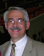 | Keynote Presentation Exosomes: From “Dust” to Multiplexed Diagnostic Biomarker
Doug Taylor, Chief Scientific Officer, Exosome Sciences Inc, United States of America
Exosomes are nano-sized vesicles released by all cell types and their protein and RNA cargo mirror the composition of their originating cells. In the case of cancer cells, the released exosomes exhibit proteins and RNA associated with the tumor and can be used as surrogates to define tumor type and stage and to predict therapeutic responses. While exosomes possess the FDA-defined features of ideal biomarkers and can be easily obtained and assessed, several obstacles have limited their clinical applications. The most critical barrier to their diagnostic application is the isolation of disease-specific exosomes and characterization their cargo. Emerging technologies will be discussed by the isolation of exosomes, associated specifically with cancers, allowing their use in diagnosis, patient stratification for therapy, monitoring therapeutic responses in real time and early identification of recurrence. These approaches target the unique properties and compositions of specific vesicle populations. The isolation of enriched pathology-specific vesicles enhances the signal to noise ratio and allows the identification of markers directly derived from the specific cell types. By correlating these circulating markers with the molecular characteristics and real-time clinical parameters, the use of circulating vesicles represents the ideal, multiplexed marker platform for clinical management. |
| 09:30 | 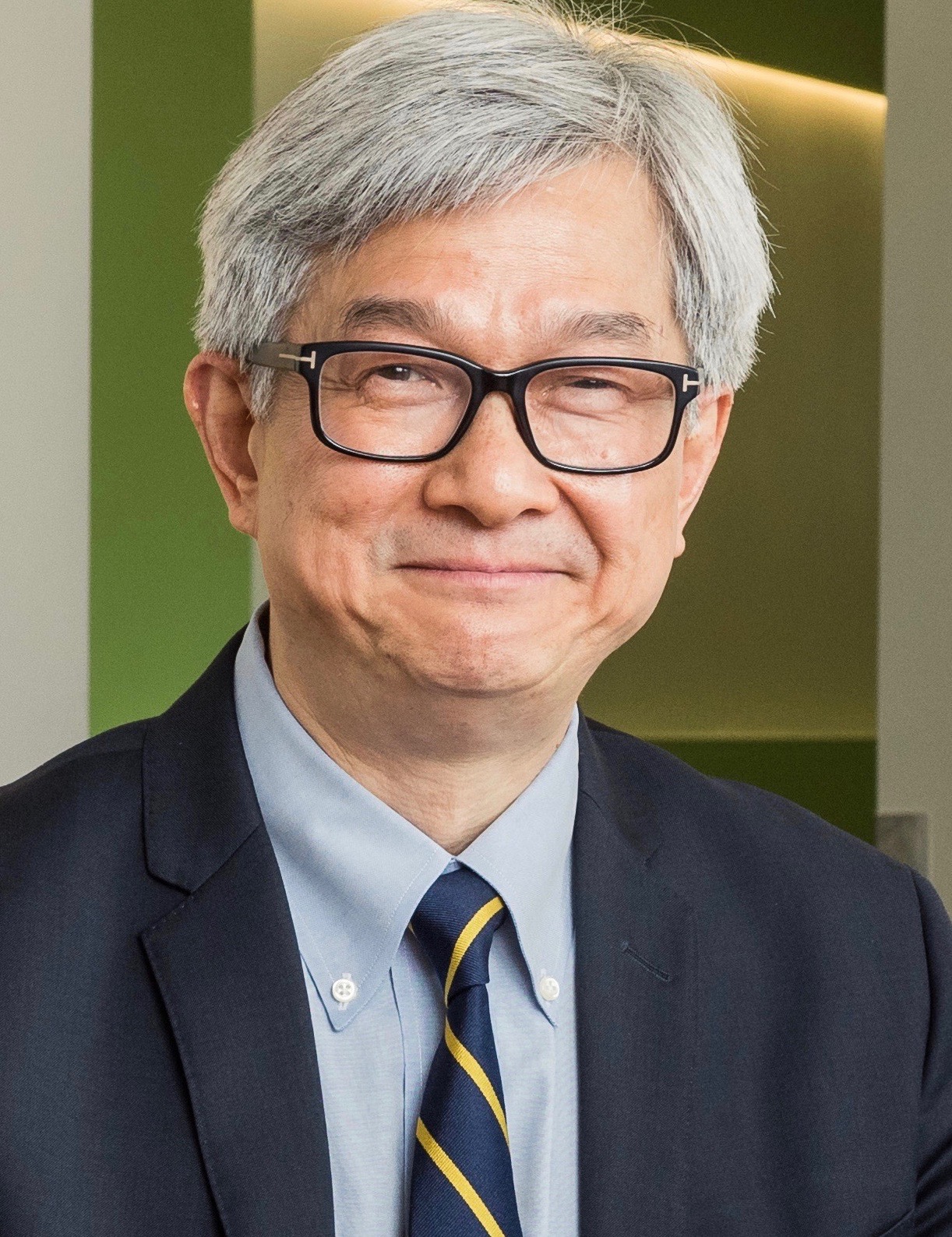 | Keynote Presentation Saliva-based Exosomes for Liquid Biopsies
David Wong, Felix and Mildred Yip Endowed Chair in Dentistry; Director for UCLA Center for Oral/Head & Neck Oncology Research, University of California-Los Angeles, United States of America
Constitutive activation of epithelial growth factor receptor (EGFR) is prevalent in epithelial cancers, particularly in non-small-cell lung carcinoma (NSCLC). EGFR mutation predicts sensitivity to EGFR-targeted therapy and mutation detection is mainly based on tissue biopsy, which is invasive, expensive and time-consuming. Non-invasive, real-time, point-of-care, inexpensive detection and monitoring of EGFR mutations in NSCLC patients is highly desirable. We developed a novel core technology, Electric Field-Induced Release and Measurement (EFIRM), relying on a multiplexible electrochemical sensor that can detect EGFR mutations directly from body fluids. EFIRM for EGFR mutation detection was established in vitro, and correlated with tumor size from xenografted mice. In clinical application, we demonstrated that EFIRM can detect EGFR mutations from saliva and serum of 22 NSCLC patients. And finally, a blinded test was performed on saliva from 40 NSCLC patient saliva samples. The receiver operating characteristic analysis indicated that EFIRM detected the exon 19 deletion with an area under the curve (AUC) of 0.94 and the L858R mutation with an AUC of 0.96. Our data indicate that EFIRM is effective, accurate, rapid, user-friendly, and cost effective for the detection of EGFR mutations in saliva of NSCLC patients. We termed this SAliva-Based EGFR (SABER) Mutation Detection for detection and monitoring EGFR mutations in saliva of NSCLC patients. |
| 10:00 | 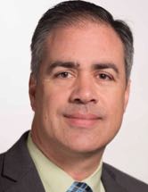 | Keynote Presentation Performance of a Blood-based Assay for the Detection of EGFR Mutations in NSCLC
John Palma, Chief Medical Officer, Roche Sequencing Solutions, United States of America
|
| 10:30 |  | Keynote Presentation Comprehensive Genomic Profiling in Clinical Oncology: Overcoming the Challenges
Vincent Miller, Chief Medical Officer, Foundation Medicine, United States of America
|
| 11:00 | Coffee Break, Visit Exhibitors and Networking | 11:30 |  | Keynote Presentation Extracellular RNA in Biofluids as Biomarkers of Disease
Xandra Breakefield, Professor, Mass General Hospital (MGH)/Harvard Medical School, United States of America
Most cells in the body release extracellular RNA (exRNA) within vesicles or in association with ribonucleoprotein/lipoprotein particles. This overview talk will consider the types of RNA released, the biofluids analyzed, and modes of evaluation of the quantity and sequence of exRNAs as a index of different disease states. Examples will be provided for analysis of exRNA from serum/plasma, urine and cerebral spinal fluid of cancer patients with a focus on discovery of novel non-coding RNAs and quantitation of rare, mutant RNAs. |
| 12:00 | 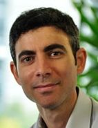  | Keynote Presentation Noninvasive Cancer Genomics and Exploring Potential Clinical Applications of Circulating Tumor DNA
Nitzan Rosenfeld, Senior Group Leader, Cancer Research UK and CSO, Inivata Ltd.
Davina Gale, Head of Molecular Diagnostics, Co-Founder, Inivata, United Kingdom
Novel biomarkers are required to accurately assess tumour burden and
response in cancer. Circulating tumour DNA (ctDNA), containing
tumour-specific DNA alterations, is present in patient plasma as a
fraction of the total cell-free DNA. The study of ctDNA levels and
mutational profile could potentially inform clinical decision making, be
used to monitor tumour dynamics, assess response to treatment and
identify mutations associated with acquired drug resistance. Inivata is
a clinical cancer genomics company focused on harnessing the emerging
potential of circulating DNA analysis to improve cancer testing and
treatment. Novel applications of ctDNA are enabled by Inivata’s
technology platform which includes its proprietary enhanced TAm-Seq™
technology, to allow the detection and analysis of genomic material from
a cancer patient’s cell-free DNA collected through routinely accessible
blood samples. This non-invasive approach – a liquid biopsy – offers a
revolution in how cancer is detected, monitored and treated. |
| 12:45 | Networking Lunch, Visit the Exhibitors and Poster Viewing | 14:00 |  Technology Spotlight: Technology Spotlight:
Beckman Coulter Cytoflex Violet SSC: An alternative to FSC PMT or Fluorescence in the Detection of Extracellular Vesicles
Vasilis Toxavidis, Resource Director of BIDMC/HSCI Flow Cytometry Core, Beth Israel Deaconess Medical Center, United States of America
John Tigges, Flow Cytometry Science Center Director, Beth Israel Deaconess Medical Center, United States of America
The interest in the identification and detection of submicron particles has increased in recent years. The ability to study them has been hindered by available techniques to measure particles at sizes below 1um. Flow Cytometry has become an important tool in EV research with instrumentation being developed to identify particles at the submicron level. Instrumentation such as cytometers optimized to improve light scattering collection and image cytometers. However, most equipment designed for the detection of EVs is expensive and complex. Hardware enhancements have focused around the development of the FSC PMT. While the FSC PMT enhancements have proven to enable the flow cytometer to detect particles <200nm in size, the instrumentation is not practical for all lab settings. In this comparison study, it has been shown that the Cytoflex is compatible to the results obtained from the AstriosEQ, NanoView, and Gallios. The ability to resolve and distinguish the populations as effectively as its counterparts, has proven the Cytoflex Violet SSC to be a viable alternative to the FSC PMT to detect EVs.
| |
Session Title: Exosomes and Microvesicles in Various Disease Classes -- Cancer, CNS, Cardiovascular Disease |
| | |
Session Chair: Kendall Van Keuren-Jensen, Ph.D., TGEN and Johan Skog, Ph.D., Exosome Diagnostics |
| | 14:30 |  | Keynote Presentation Exosome Biomarkers: Current State and Future Directions
Johan Skog, Chief Scientific Officer, Exosome Diagnostics Inc, United States of America
|
| 15:00 | RNA Signatures Associated with Brain Injury and Disease
Kendall Van Keuren-Jensen, Professor and Deputy Director, Translational Genomics Research Institute, United States of America
One of our goals has been to examine the extracellular RNA contents in cell-free biofluids to identify markers of brain injury and disease. We have examined multiple biofluid types from patients with head trauma and neurodegenerative disease. We discuss the utility of each biofluid in reliably reflecting injury and we will discuss common and specific RNAs that are altered by injury and disease. | 15:30 | Coffee Break, Networking, Exhibit and Poster Viewing | 16:00 | Roles of Mesenchymal Stem Cell–derived Exosomal Non-coding RNAs in the Oncogenic Potential of Glioma Stem Cells and in microRNA-based Therapy for Glioblastoma
Chaya Brodie, Professor, Henry Ford Hospital, United States of America
Mesenchymal stromal cells (MSCs) are multipotent stem cells that reside in various tissues such as bone marrow, adipose, placenta and umbilical cord. MSCs are able to cross the blood brain barrier and migrate to and track glioma. Endogenous MSCs derived from bone marrow and adipose tissues have been reported to migrate to tumor sites and promote EMT and metastasis. We explored the role of exosomes derived from adipose derived MSCs and found that administration of these MSCs or their exosomes increased the self-renewal, stemness markers, mesenhycmal transformation and the migration of glioma stem cells (GSCs). In contrast, cord and placenta-derived MSCs and their secreted exosomes exerted opposite effects and their intracranial administration decreased the tumor growth of GSC-derived xenografts and prolonged animal survival. miRNA sequencing and long non-coding RNA array analyses of these exosomes revealed a set of specific miRNAs and lncRNAs that were downregulated and acted as tumor suppressors in GSCs. Some of these miRNAs were delivered to GSCs following treatments with MSC-derived exosomes and exerted a strong inhibitory effect on the growth of GSC-derived xenograft. The role of exosome-derived miRNAs in the regulation of GSC oncogenic potential, and their clinical applications in stem cell-based glioma therapeutics will be discussed. | 16:30 | Large Oncosomes as a Novel Source of Circulating DNA and miRNA in Cancer
Dolores Di Vizio, Professor, Cedars Sinai Medical Center, United States of America
Our group and others have demonstrated that cancer cells release oncogenic cargo in extracellular vesicles (EVs). Transit of EVs through tissue spaces can alter the tumor microenvironment and elicit behavioral responses by cells exposed to them, such as cell motility, invasiveness and metastatic propensity. These discoveries point to a role in tumor evolution for a conserved and finely regulated biological process that allows intercellular transfer of bioactive proteins, nucleic acids and lipids in the form of pre-assembled plasma membrane structures. Several approaches toward development of a “liquid biopsy” for cancer have been attempted. It has been known for years that both DNA and RNA can be detected in the circulation, and that circulating, cell-free (cf) DNA is more abundant in cancer patients than in controls, and can be an indicator of resistance to therapeutic regimens. Circulating DNA is also present in EVs derived from normal and tumor cells. The possibility that EVs preserve the stability of extracellular nucleic acid in the bloodstream is stimulating efforts to use EV fraction(s) in blood as a non-invasive source of personalized markers of disease aggressiveness, and as a means of following cancer progression and regression in real time. Our team recently reported that silencing of the gene encoding the cytoskeletal regulator Diaphanous related formin-3, DIAPH3, in tumor cells results in a transition to a rapid migratory phenotype characterized by dynamic membrane perturbations and increased metastatic potential. We also discovered that DIAPH3 silencing results in the export of large (1-10 µm diameter) bioactive EVs (large oncosomes) that originate from the shedding of bulky membrane protrusions from the plasma membrane. We have demonstrated that the abundance of large oncosomes in the circulation and in tissues correlate with advanced disease in mouse models and human subjects. | 17:00 | Small Volume Biofluid Analysis: Challenges and Solutions for Working with Extracellular and Exosomal RNAs
Shlomit Kenigsberg, Senior Researcher, Create Fertility Centre, Canada
Small volumes of bodily fluids are a vital source for clinical diagnostics, providing important information on tissues and organs. These fluids contain an assortment of biomarkers, including protein complexes, extracellular and vesicular RNAs, each carrying a distinct signature from the point of origin, and potentially cargo for downstream cellular processes. However, many times these samples are limited in volume and precious to get, therefor the analysis of these fluids is inherently challenging. Firstly, these fluids come in extremely small volumes, and thus, only allow for a limited number of tests. Secondly, these fluids contain a large amount of abundant proteins, such as albumin and immunoglobulins and complement factors, which can mask and contaminate the proteomic analyses. Furthermore, extracellular RNAs are not easily separated from the intra-vesicular RNAs and thus, might skew the transcriptome results. Lastly, these fluids can contain a heterogeneous population of extracellular vesicles that are difficult to parse. In this review, I will discuss the sample processing of Cerebrospinal fluid (CSF), synovial fluid, lacrimal fluid, nipple discharge (breast cancer), aspirates from cysts, cervical mucus, uterine cavity aspirates and tubal fluids, with a focus on the challenges we face with our results from follicular and, and embryo culture media, providing some solutions to study these fluids.
| |
Session Title: Engineering of Exosomes as Delivery Vehicles |
| | 17:30 | Exosome Mediated Delivery of Therapeutic Oligonucleotides for Treatment of Neurodegenerative Disorders
Anastasia Khvorova, Professor, RNA Therapeutics Institute, University of Massachusetts Medical School, United States of America
Oligonucleotide therapeutics is a new class of drugs, the clinical utility of which has been limited by inefficient tissue distribution and cellular uptake. Through our research, we have developed a novel methodology that enables the loading of hydrophobically modified oligonucleotides (hsiRNA) into exosomes. These hsiRNAs show efficient cellular uptake in vitro as well as broad brain distribution and in vivo efficacy. Exosome-formulated oligonucleotide therapeutics might be a solution for the development of novel therapeutics for the treatment of neurodegenerative disorders. | 18:00 | Using Extracellular Vesicles to Enhance Gene Delivery Properties of a Virus Vector
Casey Maguire, Assistant Professor of Neurology, Harvard Medical School, United States of America
We have previously shown that extracellular vesicles (EVs) can enhance the in vivo gene delivery efficacy of adeno-associated virus (AAV) vectors, the first commercialized gene therapy product in the western world.
Here we will provide an overview of our hybrid EV-AAV technology and provide current data on its use as a therapeutic to treat CNS disease. | 18:30 | Networking Reception: Enjoy Premium Beers, California Wines, and Appetizers with Your Colleagues with a Beautiful View of Boston and The Charles River | 19:45 | Close of Day 1 of the Conference |
Tuesday, 24 March 201507:30 | Morning Coffee and Breakfast Pastries | 08:00 | Breakfast Briefing: Tissue Print Technologies for the Preparation of High Quality Human Biospecimens: A Practical and Cost Effective Approach to Obtaining Snap-Frozen Tissue for Molecular Biomarker Analysis
Sandra Gaston, Director, Molecular Biomarkers Research Laboratory; Scientific Director, Tufts Medical Center Biorepository, Tufts Medical Center, United States of America
Although technologies that support the use of FFPE specimens for molecular studies have advanced rapidly in recent years, high quality snap-frozen tissue is still required for many research applications. We have developed a set of innovative tissue-print technologies that support the analysis of RNA, DNA and protein biomarkers in human biopsies and surgical specimens without compromising the tissue for pathology diagnosis. My presentation will highlight the results of the adoption of tissue print technologies by our multi-center prostate cancer collaborative research group. This practical and cost-effective approach has allowed us to obtain snap frozen “touch prep” samples from prostate needle biopsies, dramatically expanding the range of patients and samples available for molecular genetic analysis. | |
Session Title: Methodologies and Approaches for the Study of Exosomes and Microvesicles and their Utility as Circulating Biomarkers |
| | |
Session Chairs: Leonora Balaj, Ph.D., MGH/Harvard Medical School and Dolores Di Vizio, M.D., Ph.D., Cedars Sinai Los Angeles |
| | 08:30 | Nanoplasmonic Exosome Analysis
Hyungsoon Im, Associate Professor, Center for Systems Biology, Mass General Hospital (MGH)/Harvard Medical School, United States of America
This presentation will review the recently developed exosome analysis platform, nPLEX (nano-plasmonic exosome) technology. The sensing sensing is based on transmission surface plasmon resonance (SPR) through periodic nanohole arrays. Target-specific exosome binding on the array causes significant SPR signal changes, which enables sensitive and fast detection of exosomes. We applied the first generation nPLEX system to detect exosomes collected from ovarian cancer patients. The analyses not only identified cancer-specific exosomes, but also differentiated treatment responses to standard ovarian cancer therapies. | 09:00 | Microfluidic Technology for Analysis of Plasma-Derived Exosomes
Yong Zeng, Associate Professor, University of Florida, United States of America
This presentation will discuss an integrated microfluidic system that streamlines and expedites immuno-isolation, surface phenotyping, and intravesicular protein analysis of tumor-derived circulating exosomes from cancer patients. Microfluidics has evolved from initially a scale-dependent chemical analysis technology towards a versatile platform for quantitative analysis of molecular and cellular events in complex biological systems. We have developed microfluidics-based biotechnologies and assays for quantitative measurement of circulating cancer biomarkers, such as glycoproteins and exosomes. This presentation will discuss an integrated microfluidic system that streamlines and expedites immuno-isolation, surface phenotyping, and intravesicular protein analysis of tumor-derived circulating exosomes from cancer patients. | 09:30 | Exosome-Associated RNA in Diagnosis and Management of Cancer
Cicek Gercel-Taylor, Clinical Research Director, Exosome Sciences Inc., United States of America
Overview of nucleic acid diagnostics will be presented with specific emphasis on RNA including the recent developments in the cancer arena. RNA characterization approaches will be summarized. Current developments in the utility of RNA in clinic and their future potential will be discussed. | 10:00 | Coffee Break, Networking with Exhibitors, and Poster Viewing | 10:30 | The Role of Extracellular Vesicle-derived Micro-calcifications in Atherosclerotic Plaque Vulnerability
Elena Aikawa, Associate Professor of Medicine, Brigham and Women’s Hospital, Harvard Medical School, United States of America
Cardiovascular calcification, a growing burden in Westernized countries, is not only a risk factor for cardiovascular events, but may itself contribute to cardiovascular risk. Research into treatment of cardiovascular calcification is lacking, as shown by clinical trials that have failed to demonstrate the reduction of calcific aortic stenosis. Hence the need to elucidate the pathways that contribute to cardiovascular calcification and to develop new therapeutic strategies to prevent or reverse calcification has driven our research investigations. This presentation will discuss studies that have used molecular imaging methods to advance knowledge of cardiovascular calcification, focusing in particular on the alternative mechanisms of vascular calcification via the release of calcifying matrix vesicles. | 11:00 |  Technology Spotlight: Technology Spotlight:
Complete Next-Generation Gene Expression Workflow for microRNAs from Bodily Fluids, Extracellular Vesicles and Small Sample Input - Sample Preservation, RNA Extraction, Amplification and Detection
Bernard Lam, Senior Research Scientist, Norgen Biotek Corporation
The tremendous advancements in our basic understanding of cancer biology at the molecular level have led to the increased use of molecules such as DNA, RNA and microRNA (miRNA) as a biomarker for diagnosis and prognosis of various diseases. In particular, much research has focused on detecting biomarkers in bodily fluids such as plasma, serum, urine or saliva, as they are either readily available as clinical samples or they can be easily obtained using non-invasive approaches. The characteristics of RNA present in bodily fluids are quite different from traditional samples such as cells and tissues. First, many of the RNAs in bodily fluids are either free-circulating or are within extra-cellular vesicles such as exosomes. These RNAs are generally small in size - either mRNA of less than 1000 nts or miRNAs. More importantly, the RNA present in bodily fluids is usually of very low abundance and this presents challenges for detection and studies using many next-generation gene expression technologies which require a large amount of RNA template, sometimes at the microgram level.
| 11:30 | Urinary Exosomes
Leileata Russo, Research Professor, Mass General Hospital (MGH)/Harvard Medical School, United States of America
This presentation will detail our work to date on understanding urinary exosomes and the RNA species they contain. Particular emphasis will be made regarding urinary exosomes as a diagnostic platform. | 12:00 | Networking Lunch and Exhibit, Poster Viewing | 13:00 |  Technology Spotlight: Technology Spotlight:
Isolation of Exosomes, Other Extracellular Vesicles and Vesicular RNA
Martin Schlumpberger, Associate Director, Scientific Applications, QIAGEN
A new affinity-based spin column procedure has been developed for highly specific isolation of exosomes and other extracellular vesicles (EVs) from serum or plasma, as well as for vesicular RNA content (exoEasy and exoRNeasy protocols, respectively). Results demonstrate that vesicular RNA (incl. long RNA transcripts and miRNA) can be separated from non-vesicular RNA. Fully intact long RNAs as well as miRNAs can be recovered from EVs and successfully profiled. In addition, the influence of different sample collection, handling and processing devices and workflows on RNA representation has been tested.
The applications presented here are for molecular biology use only. Not for use in diagnostic procedures.
| |
Session Title: Exosomes and Microvesicles for the Development of Biofluid Biopsies |
| | |
Session Chair: Shlomit Kenigsberg, Ph.D., MBA, University of Toronto |
| | 13:30 | Defining the Role of Tumor-secreted Exosomes in Metastasis and as Surrogate Markers of DNA Mutations
Héctor Peinado Selgas, Head of Microenvironment and Metastasis Group, Spanish National Cancer Research Centre (CNIO), Weill Medical College of Cornell University, United States of America
Cancer is a systemic disease, while most of the research effort has been focused on analyzing the primary tumor, there is a lack of information on how the tumor microenvironment influences metastasis and metastatic niche formation. The importance of the microenvironment in cancer formation, metastasis and treatment is now fully acknowledged. Prominent roles for resident cells, such as fibroblasts, bone marrow-derived cells, soluble factors, ECM molecules and secreted vesicles have been established. We have recently found a novel role for tumor-secreted exosomes promoting a pro-metastatic phenotype of bone marrow progenitor cells through in a process called ‘education of the tumor microenvironment’. In addition, in collaboration with Drs. David Lyden and Jacqueline Bromberg, we have found that double stranded DNA is secreted in tumor exosomes and reflects the mutational status of parental tumor cells. The analysis of cargo and DNA in plasma-circulating exosomes in cancer patients demonstrated that they contain specific signatures and mutations being critical factors in diagnosis and prognosis. Our studies suggest that specific molecules shed on tumor exosomes dictate the behavior of the tumor microenvironment favoring metastasis. | 14:00 | Circulating microRNAs as Biomarkers in Multiple Sclerosis
Roopali Gandhi, Assistant Professor in Neurology, Head of MS Biomarkers, Brigham and Women's Hospital/Harvard Medical School, United States of America
A major challenge in MS is to develop immune biomarkers that will allow a better understanding of an individual Multiple Sclerosis patient including response to therapy and disease stage. Stable expression of miRNA in plasma and serum, makes them ideal potential immune biomarker. We found that the expression of miRNAs could be used to differentiate MS patients from control and other neurologic diseases and also to find differences among various clinical groups of MS patients.
| 14:30 | Extracellular Hsp90 and Exosome Release
Daniel Jay, Professor, Tufts University, United States of America
We identified extracellular Hsp90 (eHsp90) as pro-invasive using a FALI-based functional proteomic screen for proteins required for cancer invasiveness and showed that Hsp90 helps in the activation of Matrix Metalloproteinase-2 (MMP2) to facilitate breast cancer invasion. Since then, many pro-invasive proteins activated by eHsp90 for different cancers have been identified by several labs suggesting that eHsp90 may serve as an activation hub for cancer invasion. We showed that Hsp90 is released from cancer cells via exosomes and these exosomes enhance invasion in an Hsp-90 dependent manner while Barrott et al. (2013) showed that eHsp90 facilitates uptake of tethered Hsp90 inhibitors suggesting that Hsp90 may be trafficked back into cells. Together, these observations led us postulate that eHsp90 may act in exosome trafficking. We tested this notion by treating MDA-MB-231 breast cancer cells in culture with the Hsp90 inhibitors ganetespib or STA-12-7191 (a biotinylated and thus impermeant derivative of ganetespib) and measured the release of exosomes and eHsp90 from these cells. We had recently shown that STA-12-7191 inhibits cell migration of cancer cells and is 5-fold less toxic to normal cells than ganetespib. Both of these drug treatments resulted in a 50% reduction in exosomes as assessed by protein concentration and Hsp90 compared to no drug controls indicating that inhibition of Hsp90 is involved in exosome release. These findings support a functional role for Hsp90 in exosome release from cancer cells, a new and important process for the communication of tumors with their extracellular milieu that is known to enhance invasion and metastasis. The mechanisms of exosome trafficking are poorly understood and these studies suggest that Hsp90 is part of this mechanism.
| 15:00 | Coffee Break, Networking, Exhibit and Poster Viewing | 15:30 | The Role of Red Blood Cell-derived Microparticles in Cardiac Remodeling
Ionita Ghiran, Assistant Professor of Medicine, Beth Israel Deaconess Medical Center, United States of America
Advances in the management of acute myocardial infarction (MI), notably the extensive use of reperfusion therapies have led to a significant decline in early mortality. However, surviving patients with myocardial damage continue to suffer substantial morbidity and mortality from its sequelae. The adverse mechanical remodeling that occurs subsequent to an MI, hallmarks of which include fibrosis and hypertrophy, underlies the development of heart failure (HF). The intimate mechanisms responsible for progression towards HF are not currently well understood. During normal and inflammatory conditions, such as MI, complement activation targets the plasma membrane of nearby cells, especially red blood cells (RBCs), generating large quantities of EVs. We have found that: (i) circulating RBC-derived EVs reach a maximum early in the morning hours, and (ii) RBC-derived EVs are enriched in miR-451, a regulator of mTOR and AMPK pathways. Moreover, our co-culture experiments shown that RBC-derived EVs efficiently fuse with and transfer the EV miRNA content to both endothelial and cardiac cells. Following the fusion of the RBC-derived EVs to endothelial cells, the RBC-derived Ago-2:miRNA-451 complex effectively targeted several mRNAs species such as, ATF-2, CAV-2 and S1RP-2, all of which contain miR451 recognition site. The net result was a significant down-regulation of the levels of targeted mRNA following 12 hours post-EVs incubation. Thus, our in vitro results suggest a novel mechanisms responsible for cardiac remodeling, specifically the unforeseen role of circulating RBCs and RBC-derived EVs in this process. | 16:00 | Poster Awards and Presentations from Poster Award Winners
Leonora Balaj, Instructor in Neurosurgery, Mass General Hospital (MGH)/Harvard Medical School, United States of America
Dr. Leonora Balaj, Mass General Hospital (MGH)/Harvard Medical School will Announce The Top 3 Poster Award Winners.
Topics Covered in Posters
1. Exosomes and Microvesicles as Biomarker Classes
2. Engineering Designer Exosomes and Microvesicles
3. Exosomes and Microvesicles as Delivery Vehicles
4. DNA, RNA, and Proteins in Exosomes and Microvesicles
| 17:00 | Close of Day 2 of the Conference |
|


 Add to Calendar ▼2015-03-23 00:00:002015-03-24 00:00:00Europe/LondonExosomes and MicrovesiclesSELECTBIOenquiries@selectbiosciences.com