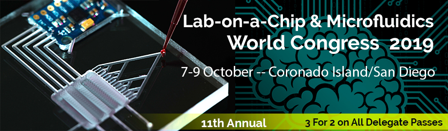
Co-Located Conference AgendasMicrofluidics & Flow Chemistry 2019 | Point-of-Care Diagnostics, Global Health & Biosensors 2019 | Single Cell Analysis Summit 2019 |  Other Track Agendas Other Track AgendasLab-on-a-Chip & Microfluidics 2019: Companies, Emerging Technologies & Commercialization Track "B" | Lab-on-a-Chip & Microfluidics 2019: Emerging Themes, Technologies and Applications Track "A" |

Monday, 7 October 201913:00 | Conference Registration and Conference Materials Pick-Up | |
Session Title: Opening Plenary Session -- Emerging Themes and Trends in Microfluidics and Lab-on-a-Chip, circa 2019 |
| | |
Plenary Session Venue: Coronado Ballrooms C & D |
| | 13:40 |  | Conference Chair Conference Chaiperson Opening Remarks and Welcome to the Attendees
Leanna Levine, Founder & CEO, ALine, Inc., United States of America
|
| 13:50 |  | Conference Chair Conference Chaiperson Opening Remarks and Welcome to the Attendees
Amy Adelberger, Founder and CEO, Global Impact Advisors, United States of America
|
| 14:00 |  | Keynote Presentation Microfluidic Methods for Single-Cell Manipulation and -Omics Analyses
Aaron Wheeler, Canada Research Chair of Bioanalytical Chemistry, University of Toronto, Canada
|
| 14:30 |  | Keynote Presentation Polymer-based Nanosensors for Single-Molecule Sequencing
Steve Soper, Foundation Distinguished Professor, Director, Center of BioModular Multi-Scale System for Precision Medicine, The University of Kansas, United States of America
We are generating a single-molecule DNA/RNA sequencing platform that can acquire sequencing information with high accuracy (>95%) at unprecedented throughputs (106 bases s-1). The technology employs high density arrays of nanosensors that read the identity of individual mononucleotides from their characteristic flight-time through a 2-dimensional (2D) nanochannel (~50 nm in width and depth; >10 µm in length) fabricated in a thermoplastic via nanoimprint lithography (NIL). The mononucleotides are generated from an intact DNA fragment using a highly processive exonuclease, which is covalently anchored to a plastic solid support contained within a bioreactor that sequentially feeds mononucleotides into the 2D nanochannel. The identity of the mononucleotides is deduced from a molecular-dependent flight-time through the 2D nanochannel. The flight time is read in a label-free fashion by measuring current transients (i.e., resistive pulse sensing) induced by a single mononucleotide when it travels through a constriction with molecular dimensions (<5 nm in effective diameter) poised at the input/output ends of the flight tube. In this presentation, our efforts on building these nanosensors using NIL in thermoplastics will be discussed and the detection of single molecules using electrical transduction with their identity deduced from the associated flight time provided. Also, surface modifications of thermoplastics for the immobilization of biologics, such as exonucleases, will be discussed as well as the activity of biological enzymes when immobilized to a plastic support. Finally, information on the manipulation of single DNA molecules using nanofluidic circuits will be presented that takes advantage of forming unique nanoscale features to shape electric fields for DNA manipulation and serves as the operational basis of the nanosensing platform. |
| 15:00 |  | Keynote Presentation Approaches to Wearable Microfluidic Sensor Devices for Well-Being and Personalized Healthcare
Martyn Boutelle, Professor of Biomedical Sensors Engineering, Imperial College London, United Kingdom
A new generation of portable, wearable biomedical devices is becoming possible through recent advances in microfluidic technologies, microelectronics, sensors and biosensors. Key to this integration is the phenomenal growth in mobile computing power provided by current tablets and mobile phones. Through miniaturization and carefully engineered smart designs we can embed computer control and analytical best practice into portable even wearable devices that are able to compensate for shortcomings such as falling performance. These hybrid microfluidic systems appear to their target users as simple stable systems that tell them what they want to know. My group specializes in designing and building such microfluidic systems to meet the needs of acute critical care medicine. Key molecular markers are measured using both optical and electrochemical sensors and biosensors. We then work with clinical care teams to show proof of concept of the real-time continuous chemical information that microfluidic systems can produce. Our ultimate goal is that such systems can be used to monitor patients and guide therapy in a patient-specific, personalized way. |
| 15:30 | Coffee Break and Networking | 16:15 |  | Keynote Presentation Rapid Diagnostics of Antibiotic Resistance
Rustem Ismagilov, Ethel Wilson Bowles and Robert Bowles Professor of Chemistry and Chemical Engineering, California Institute of Technology, United States of America
Rapid antibiotic susceptibility testing (AST) is critical for
determining appropriate treatments and for enabling global antibiotic
stewardship. Phenotypic AST is the gold standard, but these tests are
unacceptably slow, requiring pre-culture steps. Genotypic AST methods
are not sufficiently general to replace the gold standard (especially in
Gram-negative organisms). Our lab uses microfluidic digital
single-molecule counting of pathogen-specific RNA and DNA to perform
phenotypic AST directly from clinical samples in as fast as 30 min.
Further, we have demonstrated refinements of this approach that can
enable rapid AST in high-priority organisms such as Neisseria
gonorrhoeae and Carbapenem-resistant Enterobacteriaceae (CRE). |
| 16:45 |  | Keynote Presentation Optical and Electrical Single Molecule Analysis With Nanopore Optofluidic Devices
Holger Schmidt, Narinder Kapany Professor of Electrical Engineering, University of California-Santa Cruz, United States of America
In recent years, single molecule analysis has gained increasing importance for next-generation medical diagnostics. Among the leading approaches are optical fluorescence detection and electrical analysis of ionic current across nanopores. I will discuss an integrated platform that can combine both approaches in a single chip. Design and applications of these electro-optofluidic chips will be discussed. These include multiplex detection of multiple molecular biomarkers, dual-mode detection of single viruses, and feedback controlled gating of a nanopore for on-demand delivery and selection of biomolecules for chip-based analysis. The advanced level of electrical, optical, and fluidic integration is ideal for point-of-care applications. |
| 17:15 |  | Keynote Presentation The Difficult First Step for POC Diagnostics
Paul Yager, Professor, Department of Bioengineering, University of Washington, United States of America
A diagnostic process, no matter how long, begins with a first step.
Whether to guide treatment of an individual’s infection, or to control
the outbreak of a pandemic, there is an urgent need for low-cost rapid
diagnostic devices capable of identifying the cause of infectious
disease that work wherever the person is, not just in a centralized
laboratory. “Ubiquitous diagnostics” can bring the best diagnostic
capabilities to homes, physicians’ office laboratories and pharmacies in
the developed world, or to places in the developing world where nothing
is available now. However, the greatest challenge is often not
detection of the analyte(s), but the preparation for the sample for that
detection process. Samples differ in volumes, the concentration of the
analyte, and the presence of components that can interfere with analyte
detection. We have been working to develop simple devices for nucleic
acid amplification tests (NAATs) for the presence of Chlamydia and
gonorrhea, Mycobacterium tuberculosis and HIV. To enable them we have
created a suite of stand-alone or integrated sample-handling components
that can process blood, urine or swabs from different cavities in the
body: the goal in all cases is to create a few microliters of a fluid
that is both concentrated and “clean” enough that it can be introduced
automatically into a sensitive NAAT device of our own design. We will
show recent progress in reducing such processes to a few user-friendly
steps appropriate for untrained users. |
| 17:45 |  | Keynote Presentation Cell-based Point-of-Care Oncology Tool (POCOT) For Precision Medicine
John McDevitt, Chair, Department Biomaterials, New York University College of Dentistry Bioengineering Institute, United States of America
The U.S. health care spending in 2016 reached $3.3 trillion or $10,348
per person. Health spending now accounts for 17.9% the US Gross Domestic
Product. Chronic diseases like cardiac and cancer account for the
majority of the costs in this area. Further, contributing to the $2.8B
total costs for each new pharmaceutical is the cumbersome decade long
approval process. These somber statistics seem to be offset by the
promise of cutting edge biomarker developments with 157,000 biomarker
papers published in last decade, yet only ~1 biomarker per year gets
approved by the FDA. Likewise, the true potential for precision medicine
is tampered by an infrastructure and incentive structure that slows the
arrival of the benefits of precision medicine. To overcome these
limitations, transformative new tools are desperately needed to enable a
new era of medicine through research, technology, and policies that
empower patients, researchers, and providers to work together toward
development of individualized treatment. This talk features the
development, optimization and validation of the first cell-based
point-of-care oncology tool (POCOT) for precision medicine. Using
single-cell data collected non-invasively from cytology samples of
prospectively recruited patients with gold-standard-confirmed diagnoses,
a series of predictive models were developed and validated resulting in
a “continuous numerical risk score”. Model development consisted of:
(1) training binary classification models for each diagnostic class
pair, (2) pairwise coupling to obtain diagnostic class probabilities,
and (3) a weighted aggregation to obtain a final risk score on a
continuous scale. Diagnostic accuracy based on optimized cutpoints for
the validation dataset ranged from 76% for Benign lesions, to 82.4% for
Dysplastic, 89.6% for Malignant, and 97.6% for Normal controls. The
weighted aggregation of pairwise probabilities into a continuous
variable was associated with a minor decrease of 6.4% for overall
accuracy and demonstrated a strong positive relationship with diagnostic
severity (Pearson’s coefficient = 0.805 for validation dataset).
Frequencies of 5 cellular phenotypes recorded for 3 different ranges of
the numeric index score agreed with common trends observed in
conventional cytopathology. Finally, a simulation demonstrated that the
numeric index can respond to changes in as few as 1% of the cells within
a cytology sample, which is a necessary requirement for future
evaluations of its utility for lesion monitoring. The first cell-based
point-of-care oncology tool for precision medicine is demonstared in the
context of a continuous numeric index for potentially malignant oral
disorders. These efforts result in a sensitive, accurate, and
non-invasive method for enabling monitoring of these lesions. Continuous
quantitative severity indices devoid of human subjectivity and
categorization bias could have significant implications in a variety of
medical decision making situations where microscopic evaluations and
grading of biopsy samples by pathologists currently determine
diagnostic, prognostic and treatment decisions. As such, this numeric
index has the potential to monitor disease progression, recurrence, and
the need for therapeutic intervention. |
| 18:15 |  | Keynote Presentation Enzyme-based Bioelectronic Wearable Sensing Devices
Joseph Wang, Distinguished Professor, SAIC Endowed Chair, University of California-San Diego, United States of America
Wearable bioelectronic devices rely on oxidoreductase enzymes and have already demonstrated considerable promise for on-body applications ranging from highly selective non-invasive biomarker monitoring to epidermal energy harvesting. Critical to such progress is the judicious design of the enzyme-electronic interface, along with flexible platforms with mechanical properties similar to those of biological tissues. Such devices require special attention to the enzyme-electronic interface and to several considerations related to wearable applications, such as mechanical properties (flexibility and stretchability), operational stability in different biofluids and under changing conditions (e.g., pH, temperature), biofouling, selectivity, and low target concentrations. Keeping these requirements in mind, our group has pioneered a variety of wearable biocatalytic sensors and biofuel cells devices. By leveraging the advantages of biocatalysis, electrochemistry, and flexible electronics, and addressing key challenges, wearable bioelectronic devices could have a tremendous impact on diverse biomedical, fitness and defense fields. |
| 18:45 | Networking Dinner in the Exhibit Hall with Beer and Wine. Meet the Exhibitors and Network with Colleagues | 20:00 | Close of Day 1 of the Conference |
Tuesday, 8 October 201908:00 | Conference Registration, Materials Pick-Up, Morning Coffee and Pastries in the Exhibit Hall | |
Session Title: Technology Trends in Microfluidics and Lab-on-a-Chip |
| | |
Venue: Coronado Ballroom C |
| | 09:00 | | 09:30 | Parallel Flow Cytometry Made Simple
Steven Graves, Professor, University of New Mexico; President & CEO, BennuBio Inc., United States of America
Cell killing pressure and turbulence created by high linear flow velocities, as well as the stochastic arrival of particles, inherently limit any single stream flow cytometer to approximately 50,000 events per second and flow rates of about 250 microliters to a milliliter per minute. As particle size increases, these limitations worsen due to increased turbulence in the required larger flow channels, which thereby limits the achievable linear velocities and reduces analytical rate to hundreds of particles per second. These limitations prevent flow cytometry from being used efficiently in applications that range from extremely rare cell analysis to the use of large multicellular systems in pharmaceutical discovery. Therefore, there have been many efforts to create parallel flow cytometers, which can increase analytical and volumetric rates through the use of multiple streams. However, such systems have been plagued by complexity due to the use of multiple channels, optical paths, and laser sources that make long term operation extremely difficult. Here we present a highly parallel flow cytometer that uses a multinode acoustic standing wave, a line focused laser, and a high speed sCMOS camera to create a single optical path, single laser, and single detector system that will be able to detect up to 16 colors of emitted/scattered light from up to 4 excitation lasers. | 10:00 |  | Keynote Presentation Fast, Cartridge-Ready Sample Prep Using Off-the-Shelf Components
Richard Chasen Spero, CEO, Redbud Labs, United States of America
Sample-to-answer products are catching fire in the diagnostics and
research markets, but microfluidic sample prep remains a major
challenge. Methods that are ubiquitous at the bench, such as magnetic
beads and centrifugation, translate poorly to cartridge formats.
Meanwhile, microfluidic sample prep methods are generally proprietary
and challenging to customize. We demonstrate microfluidic sample prep
using commercially available components to deliver rapid affinity
purification of target analytes. The result is an open platform for
cartridge-ready sample prep that can deliver for lab-on-a-chip
developers what magnetic beads enable at the bench. |
| 10:30 | Morning Coffee Break and Networking in the Exhibit Hall | 11:15 |  Reagent Management: Between Wet & Dry Reagent Management: Between Wet & Dry
Branston Williams, Director of Product Management, IDEX Health & Science
Having a comprehensive reagent handling plan is pivotal in the development of a successful consumables platform. Understanding the interdependencies of assay workflow, reagent manufacturing, consumable design, and cartridge assembly, creates a cost-effective and scalable product. This presentation will review technologies and capabilities to store wet and dry reagents, load reagents and manage on-card reagents through reconstitution, mixing, and precisely dispensing. A reliable and robust reagent handling setup requires collaboration throughout the value stream; from reagent manufacturing through to consumable assembly. IDEX Health & Science will share broad fluidic expertise and a set of functional tools to support a systems-based view of lab-on-a-chip and point-of-care reagent handling.
| 11:45 | 3D-Printed Microfluidics For Automation of Large-Library Molecular Selection Against Cancer Targets
Noah Malmstadt, Professor, Mork Family Dept. of Chemical Engineering & Materials Science, University of Southern California, United States of America
Diagnosing and treating cancer requires having a reliable set of affinity reagents that can specifically and strongly bind to cancer-related protein targets. These reagents are the necessary molecular tools that will enable next-generation technologies for studying, diagnosing, and fighting cancer. Current approaches to producing such reagents, however, are unreliable, expensive, and slow. mRNA display is a molecular selection technology that is uniquely capable of searching libraries of more than a trillion unique compounds to develop ultrahigh affinity reagents against cancer-relevant targets. While this technology has an impressive demonstrated track record of producing such reagents, it has so far been limited to the laboratory scale.
We have built a microfluidic platform that automates the key steps of mRNA display, allowing it to be deployed in a high-throughput fashion. The system is based on modular components manufactured by 3D printing. This manufacturing approach minimizes the cost of the system, allows for rapid design iterations, and facilitates complex fluid routing in three dimensions. The modular architecture isolates the various steps of mRNA display into distinct physical and logical locations. These steps include selection of mRNA display library members that bind to a target, photoligation of the encoded mRNA library to puromycin constructs for peptide generation, sample optical interrogation for quantification of results, and various oligonucleotide processing reactions. Physical isolation allows for modules that are sensitive to surface adhesion to be disposable while the rest of the system can be reused. We have demonstrated the performance of this modular microfluidic system in the selection of molecules for binding multiple cancer-related target proteins. This approach will eventually facilitate the use of mRNA display by non-expert uses: an laboratory that can produce a cancer target protein will be able to simply input this protein into the system and in a matter of hours generate a molecule that binds the target with extraordinarily high affinity. | 12:15 | Advanced 3D Printing for Microfluidics
Gregory Nordin, Professor, Brigham Young University, United States of America
While there is great interest in 3D printing for microfluidic device fabrication, the challenge has been to achieve feature sizes that are in the truly microfluidic regime (<100 µm). The fundamental problem is that commercial tools and materials, which excel in many other application areas, have not been developed to address the unique needs of microfluidic device fabrication. Consequently, we have created our own stereolithographic 3D printer and materials that are specifically tailored to meet these needs. We show that flow channels as small as 18 µm x 20 µm can be reliably fabricated, as well as compact active elements such as valves and pumps. With these capabilities, we demonstrate highly integrated 3D printed microfluidic devices that measure only a few millimeters on a side, and that integrate separate chip-to-world interfaces through high density interconnects (up to 88 interconnects per square mm) that are directly 3D printed as part of a device chip. These advances open the door to 3D printing as a replacement for expensive cleanroom fabrication processes, with the additional advantage of fast (30 minute), parallel fabrication of many devices in a single print run due to their small size. | 12:45 | Networking Lunch in the Exhibit Hall, Exhibits and Poster Viewing | |
Session Title: Exploring the Applications of Microfluidics and Lab-on-a-Chip Technologies |
| | 13:30 | Microfluidic Sorting of Sperm For Applications in Assisted Reproductive Technologies
Utkan Demirci, Professor, Stanford University School of Medicine, United States of America
Micro- and nano-scale technologies can have a significant impact on medicine and biology in the areas of cell manipulation, diagnostics and monitoring. At the convergence of these new technologies and biology, we research for enabling solutions to real-world problems at the clinic. Emerging nano-scale and microfluidic technologies integrated with biology offer innovative possibilities for creating intelligent, mobile medical lab-chip devices that could transform diagnostics and monitoring, tissue engineering and regenerative medicine. Male infertility is a reproductive disease, and existing clinical solutions for this condition often involve long and cumbersome sperm sorting methods, including preprocessing and centrifugation-based steps. These methods also fall short when sorting for sperm free of reactive oxygen species, DNA damage, and epigenetic aberrations. Existing platforms suffer from structural complexities, i.e., pumps or chemoattractants, setting insurmountable barriers to clinical adoption. Inspired by the natural filter-like capabilities of the female reproductive tract for sperm selection, a model-driven design—featuring pillar arrays that efficiently and noninvasively isolate highly-motile and morphologically normal sperm, with lower epigenetic global methylation, from raw semen—is presented. The microfluidic sperm sorters that we created, such as the Simple Periodic ARray for Trapping And isolatioN (SPARTAN), modulate the directional persistence of sperm, increasing the spatial separation between progressive and non-progressive motile sperm populations. They lead to results within an unprecedentedly short 10-minute assay time. With over 99% motility of sorted sperm, a 5-fold improvement in morphology, 3-fold increase in nuclear maturity, and 2–4-fold enhancement in DNA integrity, SPARTAN offers to standardize sperm selection while eliminating operator-to-operator variations, centrifugation, and flow. Some of these innovative microfluidic devices have been translated into FDA approved and CE-marked products, where they have been widely used by fertility clinics around the world to serve patients, leading to an estimated 10,000+ live births globally. | 14:00 |  | Keynote Presentation Free-Surface Microfluidics and SERS or High Performance Sample Capture and Analysis
Carl Meinhart, Professor, University of California-Santa Barbara, United States of America
Nearly all microfluidic devices to date consist of some type of fully-enclosed microfluidic channel. The concept of ‘free-surface’ microfluidics has been pioneered at UCSB during the past several years, where at least one surface of the microchannel is exposed to the surrounding air. Surface tension is a dominating force at the micron scale, which can be used to control effectively fluid motion. There are a number of distinct advantages to the free surface microfluidic architecture. For example, the free surface provides a highly effective mechanism for capturing certain low-density vapor molecules. This mechanism is a key component (in combination with surface-enhanced Raman spectroscopy, i.e. SERS) of a novel explosives vapor detection platform, which is capable of sub part-per-billion sensitivity with high specificity. |
| 14:30 | Microfluidics Trapping Systems For Cell Engineering and Assays
Lidong Qin, Professor and CPRIT Scholar, Houston Methodist Research Institute, United States of America
Cellular and molecular assays, especially in the study of phenotype-genotype correlations at the single-cell level, are critical for the understanding of intratumor heterogeneity and identification of cancer phenotype-related genes and new cell subsets, and assist in cancer prevention, diagnosis, and therapy. Traditional technologies for single-cell manipulation and analysis are often limited by operational complexity, limited efficiency, and/or low-throughout. Integrated microfluidics devices have become a robust technique for single-cell manipulation. By the rational design of microfluidics platforms, we can achieve rapid and high-throughput cellular and molecular assays, including (1) single cell based analysis such as Block-Cell-Printing for live single-cell printing, Single-Cell Pipette for convenient single-cell isolation, and yeast chips for high-throughput analysis of yeast replicative aging, (2) double cells based assay such as vertical cell pairing for high-resolution imaging of the immunological synapse, and (3) CRISPR/Cas9 based genome editing for hard-to-transfect cells. | 15:00 |  “Putting the Lid on Microfluidics” – A Review of Cover Layer Bonding and Sealing Techniques for Microfluidic Lab-on-Chip Devices “Putting the Lid on Microfluidics” – A Review of Cover Layer Bonding and Sealing Techniques for Microfluidic Lab-on-Chip Devices
John Town, SVP Technology, TECHNICOLOR PRECISION BIODEVICES
Technicolor brings over 100 years of technical innovation across a broad range of disciplines to the microfluidic consumable manufacturing space. This presentation provides a review of current and emerging techniques used in bonding polymers to create functional microfluidic lab-on-chip devices. The review includes a discussion of material considerations, performance attributes and manufacturing processes.
| 15:30 | Why Considering the Impact of Cartridge Architecture Matters for the Final Product Cost of Goods
Leanna Levine, Founder & CEO, ALine, Inc., United States of America
As microfluidics-based products mature and move toward the consumer market, the drive toward a lower cost of goods for the instrument, in addition to a low-cost consumable, requires consideration of the overall cartridge architecture and the instrument interface early in the development program. For example, choices in the valve design and actuation method can lead to the instrument, rather than the assay protocol, driving the cartridge design. In this presentation, we will review examples and strategies that have been implemented in development programs, and provide context on the impact of different choices on the system, including impact on the size of the microfluidics-based consumable, and overall system complexity. | 16:00 | Afternoon Coffee Break and Networking in the Exhibit Hall | 16:30 |  | Keynote Presentation Circulatory System on a Chip -- From in vitro to in vivo, From Single Cell to Microphysiological Systems
Abraham Lee, Chancellor’s Professor, Biomedical Engineering & Director, Center for Advanced Design & Manufacturing of Integrated Microfluidics, University of California-Irvine, United States of America
The circulatory system is a critical physiological process of the human body that maintains homeostasis by balancing biological parameters by the delivery and removal of nutrients/waste and fighting off invading pathogens. Through the advancement of microfluidics technologies, we have enabled the automation of biological fluids delivery through physiological vasculature networks that mimic the physiological circulation of the human body. The critical bottleneck is to engineer the microenvironment for the formation of 3D tissues and organs and to also pump and perfuse the tissue vascular network for on-chip microcirculation. On the other hand, microfluidics play an important role in the recent advances in liquid biopsy, an emerging technique that analyzes biological samples such as blood for the detection of biomolecules or cells that are indicative of disease or physiological state. Specifically, liquid biopsy has become a promising technology to isolate and target rare cells such as circulating tumor cells (CTCs) in body fluids thanks to many of these microfluidic cell sorting techniques. This advent of microfluidic liquid biopsy provides an in vitro snap shot into the patient’s physiological status via the in vivo circulation that enables one to monitor disease state and progression for diagnosis and prognosis. A key bottleneck is to identify the critical subpopulation of cells, often at single cell resolution among billions of cells in circulation. Along with the aforementioned in vitro on-chip perfused vascularized tissue platforms, these two technologies go hand-in-hand to connect in vitro screening to in vivo screening with great potential in the development of personalized medicine. Ultimately this is the microfluidic maintenance of physiological equilibrium, or ‘microfluidic homeostasis.' |
| 17:00 |  | Keynote Presentation A Mobile Healthcare System Based on Smartphone and Smart Lab-on-a-Chip using Microchannel Capillary Flow Assay (MCFA)
Chong Ahn, Distinguished University Research Professor, Mitchell P. Kartalia Chair Professor of BioMEMS, University of Cincinnati, United States of America
A new mobile healthcare system using smartphone and smart lab-on-a-chip for the rapid and high sensitive detection of malaria biomarker, Plasmodium falciparum Histidine Rich Protein 2 (PfHRP2), has been developed and characterized in this work. Chemiluminescence-based sandwich enzyme linked immunosorbent assay (ELISA) for PfHRP2 as the target antigen was performed on the lab-on-a-chip for high sensitive microchannel-based capillary flow assay (MCFA). A limit of detection (LoD) of 1.2 ng/ml of PfHRP2 was achieved, which is considered enough for the diagnostics of possible malaria infection. A smartphone based self-powered portable analyzer was also developed for optical detection. An optical detection system, which can interface with a smartphone, has been developed for the chemiluminescence detection. The results obtained from this work envisages a new stand-alone mobile healthcare system for the rapid diagnosis of infectious diseases using a smartphone. |
| 17:30 | Automated Liquid Handling with Microfluidic Droplet-on-Demand Arrays
Elliot Hui, Associate Professor of Biomedical Engineering, University of California-Irvine, United States of America
Microfluidic emulsion droplets have proven to be capable vessels for nanoliter biochemical reactions. However, liquid handling in droplets has remained much less flexible than conventional pipetting into well plates. We present a droplet-on-demand dispenser array that can apportion arbitrary combinations of reagents into individual droplets. The dispensers draw from a standard-format array of wells and employ a pneumatic digital logic control system to enable binary addressing of large dispenser arrays. Droplets are tracked by machine vision as they wind past individual dispenser nozzles, allowing dispenser actuation to be fully automated. We envision the miniaturization of a wide variety of automated liquid handling protocols including high throughput screening applications. | 18:00 | Networking Reception in the Exhibit Hall with Beer and Wine -- Network with the Exhibitors and Engage with Speakers and Attendees | 19:00 | Close of Day 2 of the Conference |
Wednesday, 9 October 201907:00 | Morning Coffee, Pastries and Networking in the Exhibit Hall | |
Session Title: Emerging Technologies, Applications and Late-Breaking Development in Microfluidics, Lab-on-a-Chip and Associated Ecosystem |
| | |
Venue: Coronado Ballroom C |
| | 08:00 | Design Automation of Low-cost Microfluidic Droplet Generators
Ali Lashkaripour, Researcher, CIDAR Lab, Boston University, Boston University, United States of America
Low-cost micromilling in thermoplastics and machine learning techniques were exploited to build a large experimental dataset and learn from it. This allowed for accurate performance prediction and design automation of microfluidic droplet generators, in terms of droplet diameter, generation rate, and formation regime. | 08:30 | Isolation, Discrimination and Transportation of Individual Specific Chromosomes From Single Mammalian Cells in a Microfluidic Channel
Hidehiro Oana, Associate Professor, The University of Tokyo, Japan
Chromosomes were isolated from single mammalian cells in a microfluidic channel. Then, in the microfluidic channel, X chromosomes were fluorescently labeled by utilizing fluorescence-tagged dCas9 probes and discriminated. After that, capture/transportation of the X chromosomes were performed using optical tweezers. | 09:00 |  | Keynote Presentation Droplet Emulsion Generation based on Electrokinetics: Digital PCR and Cell Encapsulation
Hsueh-Chia Chang, Bayer Professor of Chemical and Biomolecular Engineering, University of Notre Dame, Interim Chief Technology Officer, Aopia Biosciences, United States of America
We report a new microdroplet generation technology that uses AC electric field rather than hydrodynamic shear to generate droplets. Intricate flow-rate tuning of both phases is hence unnecessary and massive parallelization is now possible because of the absence of hydrodynamic cross-talk. The bulky and expensive precision micropump is replaced by a simple and miniature AC source that can be driven by a battery. With a less viscous oil phase and a properly tuned AC frequency, we are able to eliminate electro-coalescence and Rayleigh fission. Monodispersed droplets with CV less than 3% and with a size range from 10 to 100 microns can be generated at a high throughput of nearly 1000 Hz per nozzle. Moreover, the size of the droplets can be easily adjusted by simply tuning the voltage or the frequency. One hundred to one million droplets and, with parallelization, more than 1 billion droplets can be generated in 30 minutes. This large range allows high-dynamic range digital PCR and conformal cell encapsulation. |
| 09:30 | Precision Assembly by All-Aqueous Interfaces
Anderson Shum, Professor, Department of Mechanical Engineering; Director, Advanced Biomedical Instrumentation Centre, University of Hong Kong, Hong Kong
Biomedical applications, such as tissue engineering and drug delivery, require controlled assembly of biomolecules, cells and small molecules. These have been achieved either by a synthetic route, or using a directing substrate. Aqueous-aqueous Interfaces, which can be formed by inducing phase separation of aqueous solution containing two or more water-soluble additives, that can be salt, polymer and biomacromolecules, such as proteins and DNAs. Their bio- and cyto-compatibility can be ensured by choosing the appropriate additives, making these all-aqueous systems particularly appealing for applications that require precise and mild handling of delicate molecules and cells. In this work, we propose the use of the interface formed between two immiscible aqueous phases. These interfaces are selective to different molecules, as they are permeable to water-soluble additives yet exhibit preferential solubility through partitioning of the additive molecules based on this affinity to the aqueous phases. We show how these interfaces can be used to control the assembly of macromolecules, as well as cells into membranes and capsules that can be used for biomedical applications that require precise configuration of the constituent components. | 10:00 | Morning Coffee Break and Networking in the Exhibit Hall | 10:30 |  Tools for Ensuring Successful Outcomes in Microfluidic Experiments Tools for Ensuring Successful Outcomes in Microfluidic Experiments
Thomas Corso, Chief Technical Officer, CorSolutions
The challenges of designing and developing microfluidic assays are frequently overlooked, which can negatively impact the robustness, repeatability and reliability of the outcome. However if the microfluidic researcher has the correct tools available, these subtleties and nuances can be understood and often circumvented. These essential tools for troubleshooting problematic microfluidic experiments will be discussed including flow meters, pressure meters, and closed-loop feedback control pumps. Additionally, common pitfalls and ways to avoid them will be examined, such as leaky connections, pulsatile flows, and presence of bubbles. Finally ways of designing microfluidic experiments to minimize the possibility of issues developing will be considered.
| 11:00 | Dielectrophoresis-Assisted Creation of Cell Aggregates for Lab-On-A Chip Applications
Jonathan Cottet, Researcher, EPFL, Switzerland
A microfluidic platform allowing dielectrophoresis-assisted formation of cell aggregates of controlled size and composition under flow conditions is presented. The created aggregate proved to be stable after 5 minutes of cell-cell contact. | 11:30 |  | Keynote Presentation Improving Sustainability in the Lab-On-a-Chip Field: Environmentally-Friendly Device Prototyping and Manufacturing
Maiwenn Kersaudy-Kerhoas, Professor of Microfluidic Engineering, Heriot-Watt University, United Kingdom
The use of single-use, disposable medical equipment has increased the amount of medical waste produced and the advent of point-of-care diagnostics in lab-on-chip format is likely to add further volume. Current materials used for the manufacture of these devices are derived from petroleum sources and are, therefore, unsustainable. In addition, disposal of contaminated plastics necessitates combustion to reduce infection risk, which has, depending on material composition, and incineration capacity, an undesirable environmental impact. To address these issues, we have developed several sustainable approaches for the rapid prototyping of single-use point-of-care cartridges, including use recycled PMMA or laminated PLA. Parameters such as optical quality, cost, and translation to mass-market products will be discussed. These techniques present exciting opportunities for immediate and future sustainable solutions in the LOAC field. |
| 12:00 |  Slot Die Coating Techniques for Microfluidics and Biotechnology Applications Slot Die Coating Techniques for Microfluidics and Biotechnology Applications
Greg Gibson, Chief Technology Officer, nTact
Slot Die Coating is a proven method for the deposition of a variety of liquid chemistries onto glass, silicon, metal and plastic substrates, for the development and production of a broad range of products. Historically used in industries such as flat panel display and photovoltaic manufacturing, the coating method is now gaining acceptance for biotechnology and pharmaceutical applications. Advantages of this technique include high uniformity, low material waste, and compatibility with a wide range of coating materials. Applications and process results will be discussed, including selective coating methods used for depositing an array of rectangular shapes on a single substrate or multiple substrates.
| 12:30 | Networking Lunch in the Exhibit Hall, Exhibits and Poster Viewing | 13:30 | Poster Awards | 13:45 |  Surface Preparation for Microfluidic and Biomedical Devices Surface Preparation for Microfluidic and Biomedical Devices
Bill Moffat, Founder & CTO, Yield Engineering Systems, Inc. (YES)
This presentation traces YES’s experience in modifying surfaces for microfluidics and biomedical devices, from the company’s work on surface tension modification using silane chemistry in the semiconductor industry, to plasma surface modification in the semiconductor industry, to extensive silane chemistry modification in the microfluidic and biotech industry, to combination plasma and silane deposition equipment for both industries.
| 14:15 |  | Keynote Presentation A Fully Automated Lab-on-a-Chip Platform For Biological Applications
Zeynep Altintas, Head of Biosensors and Receptor Development Group, Technical University of Berlin, Germany
Our custom-designed microfludic sensor provides rapid, sensitive and specific diagnostic methods. It is capable of detecting carcinogenic toxins, various bacteria species and their DNAs, and serves as a promising tool for pathogen detection in environmental and medical samples. |
| 14:45 |  | Keynote Presentation Quantum Diagnostics: From Single-Cells to Single-Molecules
Dino Di Carlo, Armond and Elena Hairapetian Chair in Engineering and Medicine, Professor and Vice Chair of Bioengineering, University of California-Los Angeles, United States of America
The ultimate limits of diagnostics in biology are the “quantum” units that convey information, e.g. single nucleic acids, proteins, and cells. Microfluidics has emerged as a powerful tool to compartmentalize single cells and molecules into sub-nanoliter droplets as individual bioreactors to enable sensitive detection and analysis down to this quantum limit. However, the current systems for quantum diagnostics have not been widely adopted, partly due to the requirement of specialized instruments and microfluidic chips to generate uniform droplets and perform adequate manipulations. I will discuss the platforms we are developing to fractionate volumes in simplified, instrument-free ways using 3D-shaped microparticles. Each “lab-on-a-particle” can be analyzed using widely available flow cytometers. These new lab-on-a-particle reagents eliminate the need for specialized new equipment for microfluidic compartmentalization and readout and promise to democratize single-molecule and single-cell technologies. |
| 15:15 | A Minimized Valveless Electromagnetic Micropump for the Microfluidics Actuation On Organ-on-a-Chip
Shengli Mi, Associate Professor, Biomanufacturing Engineering Laboratory, Tsinghua University-Shenzhen, China
In recent years, organ chips have become an ideal alternative for physiologic, pathologic studies and drug screening. In order to simulate the physiological environment in vivo more realistically, micropumps are often needed as a power source to achieve dynamic culture in organ chips. In this study, we present a valveless electromagnetic micropump which can be used in organ chips. The fluid flow is actuated by the vibration of PDMS membrane through a varying magnetic field. This micropump uses a nozzle/diffuser structure instead of valves, which makes the structure quite simple. By reducing the volume of coil and magnet, the volume of the electromagnetic micropump is minimized so that it can be integrated on a microfluidic chip. The micropump and the chip can be portably packaged by using a small signal generation module and dry battery to supply the coil with square wave, reducing the dependence on external devices. In addition, multiple electromagnetic micropumps can be integrated on a single chip. These micropumps can be connected in series or in parallel, which can achieve complicated flow conditions and simulate the fluid flow in human body more realistically in different types of organ chips. The flow rate of the microfluidics was measured to characterize the actuating performance of the micropump. We established the dynamic co-culture of liver model and the breast cancer model on the chip, with medium actuated by the electromagnetic micropump. Cell viability, albumin and IL-6 were analyzed. Compared with the static control group, the results indicated that dynamic co-culture actuated by the electromagnetic micropump was beneficial for the growth and functions of cells. |
|


 Add to Calendar ▼2019-10-07 00:00:002019-10-09 00:00:00Europe/LondonLab-on-a-Chip and Microfluidics 2019: Emerging Themes, Technologies and Applications Track "A"SELECTBIOenquiries@selectbiosciences.com