07:30 | Morning Coffee and Breakfast Pastries |
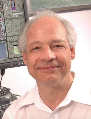
08:00 | Breakfast Briefing: Measuring Neutrophil Phenotype in Sepsis from a Single Drop of Blood
Daniel Irimia, Associate Professor, Surgery Department, Massachusetts General Hospital (MGH), Shriners Burns Hospital, and Harvard Medical School, United States of America
The absolute neutrophil count (ANC) is today one of the most common blood tests used in the clinic for evaluating the risk of infections in patients. However, this test implicitly assumes that all patients’ neutrophils are normal and it does not account for possible alterations of their functions. Such alterations occur in patients after burn injury and can significantly reduce the ability of the body to fight infections, even when neutrophils counts are in the normal range. To estimate the risk for infections more accurately, we designed microfluidic devices that measure neutrophil migration with higher precision than ever before. To circumvent the challenging logistics of neutrophil isolation in the clinic, we designed these devices to work directly with one droplet of blood. Using these devices, we identified a set of phenotype markers for the early detection and monitoring of sepsis in patients after major burn injuries. |
|
Session Title: Next Generation Sequencing (NGS) as a Means to Interrogate Biomarker Cargo -- How to Incorporate into Practice? |
| |
|
Session Chair: Jamie Platt, PhD, Geneuity (Molecular Pathology Laboratory Network, Inc.) |
| |
08:30 | Surveying the RNA-Seq Field
Joshua Levin, Senior Group Leader, Research Scientist, Stanley Center for Psychiatric Research, Klarman Cell Observatory, The Broad Institute of MIT and Harvard, United States of America
RNA-seq allows us to comprehensively characterize the transcripts present in a biological sample. This talk will focus on the “basics” of RNA-Seq as well as how to approach the selection of an appropriate method for specialized RNA-Seq. An overview of key breakthroughs will also be presented. |
09:00 |  | Keynote Presentation Getting Back to Basics: Sample Preparation Considerations for Advanced Sequencing Technologies
Jamie Platt, Vice President, Geneuity (Molecular Pathology Laboratory Network, Inc.), United States of America
Advanced sequencing technologies like NGS have truly transformed genomic and gene-based research and clinical diagnostics. As tools and technologies in advanced sequencing rapidly emerge, the potential for greater sensitivity, increased depth, and greater breadth grow. Within a clinical diagnostic or clinical trial setting, understanding the limitations of the sample and sample prep methods are key to overall system performance. Critical sample preparation considerations will be discussed, both within the context of clinical studies, assay validation, and the clinical diagnostic setting. |
|
09:30 |  | Keynote Presentation Nanotrap Nanoparticles for Discovery and Measurement of Previously Invisible Low Abundance Biomarkers
Lance Liotta, Co Director, George Mason University, United States of America
Unresolved challenges in the biomarker discovery and measurement field are 1) Very low concentration of early disease biomarkers 100 fold below the detection limit of biomarker discovery and measurement platforms, 2) Candidate body fluid biomarkers are masked by a billion fold excess quantities of resident proteins such as immunoglobulin and albumin, and 3) Degradation and perishability of candidate biomarkers ex vivo following clinical sample collection, shipping and storage. We created a new class of versatile multifunctional nanotechnology that addresses all of these challenges in one step. The technology is novel porous, buoyant, core-shell hydrogel nanoparticles containing novel high affinity reactive chemical baits that harvests biomarkers in body fluids. Upon contact with the sample, the suspended nanoparticles immediately affinity-sequester target biomarkers inside the particles, exclude albumin, fully protect the biomarkers from degradation (even at elevated temperatures), and massively concentrate the sequestered biomarkers into a small volume. The technology can dramatically (demonstrated up to 10,000 fold) improve the lower limits of detection and the precision of: a) mass spectrometry (MS) biomarker discovery, b) quantitation by multiple reaction monitoring (MRM), or c) quantification by any clinical grade immunoassay. The technology has been applied to biomarker discovery and high precision measurement of clinical analytes in all classes of body fluids including urine, saliva, sweat, and blood. |
|
10:00 | Coffee Break, Networking, Exhibit and Poster Viewing |
10:30 | Standardizing High-Throughput Sequencing of Extracellular RNA from Human Plasma
Yaoyu Wang, Associate Director, Dana-Farber Cancer Institute, United States of America
Extracellular vesicles have been shown to regulate intercellular signaling by transmitting RNA materials such as mRNA, microRNA and snRNA. This phenomenon implies extracellular RNA (exRNA) may partially reflect cellular content within the human body and may show disease specific variation. As such, exRNA profiles have great potential as disease biomarkers. While high-throughput sequencing technology offers a potentially sensitive means to characterize and quantify exRNA, there is a lack of a understanding of the efficacy and reliability of commercial kits for extracellular RNA sequencing. Here, we aim to optimize protocols for sequencing extracellular RNA from human samples. |
11:00 | Increasing Sensitivity of Next Generation Sequencing-based Transcriptome Profiling by Selectively Depleting Abundant RNAs
Daniela Munafo, Applications and Product Development Scientist, New England Biolabs, Inc., United States of America
Next Generation Sequencing has become the method of choice in research and clinical diagnostics for transcriptome profiling and biomarker discovery. However, whole-transcriptome sequencing is challenging due to the large dynamic range of transcript expression within a total RNA sample. Highly expressed transcripts with minimal biological interest can dominate readouts, masking detection of more informative lower abundant transcripts. I’ll present a method to enrich for RNAs of interest by eliminating unwanted RNAs before sequencing. This method is based on hybridization of probes to the targeted RNA and subsequent enzymatic degradation of the selected RNAs. This method removes abundant RNAs (such us cytoplasmic and mitochondrial ribosomal RNA and hemoglobin transcripts from derivate blood samples) while retaining coding and non-coding RNAs. The reduction of abundant transcripts for RNA-Seq studies significantly increases the ability to detect true biological variations that could not be detected in non-depleted samples. |
11:30 |  Technology Spotlight: Technology Spotlight:
Enriching Nucleic Acids for Next-Generation Sequencing Analyses of SNPs, CNVs, Gene Fusions and More
Steve Kain, Director, Product Marketing, NuGEN Technologies
The novel Single Primer Enrichment Technology (SPET) and how it differs from existing target enrichment methods will be described. Sensitive variant detection from genomic DNA derived from fresh and FFPE tissues using 344 cancer-related genes will be demonstrated as well as utilization of SPET as a rapid, cost-effective screening tool for discovery of novel fusions and detection of known fusions with a panel of 500 cancer genes implicated in fusions events.
|
12:00 | Networking Lunch, Visit the Exhibitors and Poster Viewing |
|
Session Title: Sample Preparation from Various Different Clinically-Relevant Specimens and Biorepositories -- Methodologies and Approaches for Archiving and Interrogating Clinical Samples |
| |
13:30 | Computational Analysis of Exosomal Small-RNA-seq
Robert Kitchen, Research Associate, Yale University, United States of America
Introduction to analysis of small RNAs by RNA-sequencing, including best practices for mitigating nuisance contaminants and special considerations for profiling extra-cellular RNAs (exRNAs). Based on these best practices, we present a software package for the analysis of RNA-seq data obtained from both cellular and extracellular small-RNA preparations. We extend beyond existing analysis methods used to assess cytosolic micro-RNAs (miRNAs) to specifically address experimental issues that may arise in exRNA profiling, such as variable contamination of ribosomal RNAs, the presence of endogenous non-miRNA small-RNAs, and the presence of exogenous small-RNA molecules derived from a variety of plant, bacteria, and viral species. |
14:00 | Sample-to-Result Molecular Diagnostics in an Easy-to-Use Disposable Test
Barry Lutz, Assistant Professor, University Of Washington, United States of America
Infectious diseases are a leading cause of death in the developing world. Even when treatment is available, misdiagnosis leads to ineffective treatment, waste of scarce medications, and increased pressure for drug resistance. While these diseases can be diagnosed in centralized laboratories, these assays require trained operators to carry out multi-step assay protocols, expensive and fragile instrumentation, and cold storage conditions for many reagents. At the University of Washington we are developing rapid immunoassays and nucleic acid tests designed for minimal-resource settings, whether it be a pharmacy, an outdoor clinic, or your home. DARPA is supporting a project to develop a fully-disposable test for multiplexed detection of DNA and RNA that is simple enough for home use. All steps from sample preparation to visual readout are carried out automatically, and there is no need for external equipment or sample handling by the user (other than swabbing their nose). Data collection by a cell phone could allow transmission of results to a healthcare provider and medical record. Project goals include commercializable tests as well as broad platform capabilities for future tests. I will present the concept and progress as a means for sample-to-result testing outside conventional labs, and perhaps even in your home. |
14:30 | Pre-Analytical and Analytical Variables Impacting Sample Quality and Assay Development
Veronique Neumeister, Laboratory Director of Specialized Translational Services Lab, Yale University School of Medicine, United States of America
Accurate assessment of tissue biomarkers in the research and the clinical setting is of utmost importance in order to develop reproducible and reliable assays and support the increasing efforts towards individualized molecular targeted therapy of cancer patients. However, tissue handling and processing are not always tightly controlled and pre-analytical variables can significantly alter tissue quality and impact companion diagnostic testing. This presentation will focus on the effects of pre-analytical and analytical variables in the diagnostic and research setting. Methodology and importance of quality control, optimization and standardization of tissue processing, biomarker validation and development of useful laboratory tests for the clinical setting will be discussed.
|
15:00 | Coffee Break and Networking |
15:30 | The Physics and Applications of Nanoelectronic Biosensors
Mark Reed, Professor, Yale University, United States of America
Nanoscale electronic devices have the potential to achieve exquisite sensitivity as sensors for the direct detection of molecular interactions, thereby decreasing diagnostics costs and enabling previously impossible sensing in disparate field environments. Semiconducting nanowire-field effect transistors (NW-FETs) hold particular promise, though contemporary NW approaches are inadequate for realistic applications and integrated assays. We present here an integrated nanodevice biosensor approach that is compatible with CMOS technology, has achieved unprecedented sensitivity, and simultaneously facilitates system-scale integration of nanosensors. These approaches enable a wide range of label-free biochemical and macromolecule sensing applications, such as specific protein and complementary DNA recognition assays, and specific macromolecule interactions at femtomolar concentrations. Critical limitations of nanowire sensors are the Debye screening limitation and the lack of internal calibration for analyte quantification, which has prevented their use in clinical applications and physiologically relevant solutions. We will present approaches that solve these longstanding problems, which demonstrates the detection at clinically important concentrations of biomarkers from whole blood samples, integrated assays of cancer biomarkers, and the use of these as a quantitative tool for drug design and discovery, including binding kinetics, chirality detection, enzyme detection, and activity. |
16:30 | Fit-For-Purpose Biospecimens: Matchmaking in the Biorepository
Sherilyn Sawyer, Director, BWH/Harvard Cohorts Biorepository, Brigham and Women's Hospital/Harvard Medical School, United States of America
Matchmaking: searching for a match that works together, creates a complimentary pair, and results in a strong result. For biorepositories, this means locating or collecting biospecimens that are the best match to the research question and assay modality at hand. While some specimens can be collected with the needs of a current research question and analysis platform in mind, a large percentage of research questions cannot be addressed by this “just in time inventory” approach due to limitations in available case numbers, requirements for outcome measures, or the need to build upon prior research results within the same sample set. This presentation will examine the use of pilot experimentation to test the match between specimen and analysis to ensure fit-for-purpose use of prospectively collected biospecimens. |
17:00 | Next Generation BioSpecimen Sciences: Sample-Centered Precision Medicine
Michael Roehrl, Director, UHN Program in BioSpecimen Sciences, University Health Network, University of Toronto, Canada
The deep omic analysis of human samples has become the centerpiece of cutting-edge precision medicine. We will discuss the role of biospecimen sciences and the innovative role of modern pathology in this context, including specific examples from the UHN Program in BioSpecimen Sciences, one of the largest academic programs of its kind. |
17:30 | Advanced Diagnostic and Sample Preparation Platform for Bio-surveillance
Kamlesh Patel, R&D Advanced System Engineering and Deployment Manager, Sandia National Laboratories, United States of America
Emerging infectious diseases present a profound threat to global health, economic development, and political stability, and therefore represent a significant national security concern for the U.S. The key to preventing an outbreak before it goes global is to establish a biosurveillance network that effectively reaches even the most remote regions and provides a network-integrated, location-appropriate diagnostic capability. At present, the two main factors that prevent the extension of biosurveillance activities beyond centralized laboratory facilities are the lack of a deployable rapid-response diagnostic platform and a method to safely and consistently process infected samples in the field for analysis. We report on our work to develop a forward-deployed sample preparation and diagnostic platform to safely and consistently process infected clinical samples in the field for analysis as part of a comprehensive biosurveillance strategy. Small-volume blood samples are processed on a digital microfluidic hub platform with integrated modules to 1) extract and process RNA and 2) stabilize and format RNA into a cDNA intermediate for either real-time multiplex PCR analysis or lab-based next generation sequencing. |
18:00 | Isolation of Dilute Pathogens from Blood
Alexis Sauer-Budge, Biotechnology Managing Scientist, Exponent, Inc., Adjunct Research Assistant Professor, Biomedical Engineering Dept, Boston University, United States of America
Traditionally, bacterial pathogens in the blood have been identified using culture-based methods that can take several days to obtain results. This can lead to physicians making treatment decisions based on an incomplete diagnosis contributing to patient morbidity. To decrease diagnosis time, we are developing a novel sample preparation device for isolating and concentrating dilute bacteria from blood. Though commercial kits exist for the removal of blood from these samples, they typically capture only DNA, thereby necessitating the use of blood culture for antimicrobial testing. Here, we report a novel, scaled-up sample preparation protocol carried out in a new microbial concentration device. The process can efficiently lyse 10 mL of bacteremic blood while maintaining the microorganisms’ viability. This talk will present the methodology and examples of integration with downstream detection technologies, including PCR, SERS, and standard culture. Our sample preparation protocol holds great promise for the rapid diagnosis of bacteremia directly from a primary sample. |
18:30 | Close of Day 2 of the Conference |


